Phytochemical investigation of the folk medicinal plant Argyreia acuta Lour. has led to the isolation of a pair of new 3-alkylated coumarin enantiomers, (±)-acutamarin [(±)-1] and 3 known structurally related coumarins (2-4), along with 4 known flavonoids (5-8). The structures of these compounds were elucidated on the basis of extensive spectroscopic (including one-dimensional and two-dimensional nuclear magnetic resonance) analyses and by comparison of their spectral data with those reported in the literature. The absolute configurations of ( − )-1 and ( + )-1 were proposed by comparison of experimental and calculated electronic circular dichroism data. Compounds 5 and 7 exhibited in vitro anti-inflammatory activity by inhibiting the production of nitric oxide with IC50 values of 24.54 ± 0.36 μM and 10.60 ± 0.15 μM, respectively.
The genus Argyreia (Convolvulaceae) comprises approximately 90 species, which are mainly distributed in south and southeast Asia.1 Plants of this genus are known to have broad biological properties, such as cytotoxic,2 anti-inflammatory,3 hepatoprotective,4 and neuroprotective5 activities. Among them, A acuta Lour., locally known as “Goudahao,” is a perennial liana or climbing vine growing in southwest mainland China and has been used as a folk herbal medicine for the treatment of inflammatory diseases, including acute or chronic bronchitis, cough, sore throat, and mouth ulcers.6 Previous phytochemical and biological investigations of this plant have resulted in the isolation of a series of resin glycosides with α-glucosidase inhibitory activity.7-10 As part of a program to discover new agents with potential in the treatment of inflammatory diseases, our attention was drawn to the ethyl acetate extract of A acuta, which exhibited inhibitory activity on nitric oxide (NO) production in lipopolysaccharide (LPS)-induced RAW264.7 macrophage cells. Further fractionation led to the isolation of a new 3-alkylated coumarin, acutamarin (1), together with 7 known compounds, scopoletin (2), umbelliferone (3), scopolin (4), (2R,3R)-7-O-methyl-dihydrokaempferol (7-O-methylaromadendrin) (5), 4′-7-di-O-methyl-kaempferol (6), 7-O-methyl-kaempferol (rhamnocitrin) (7), and 3′-7-di-O-methyl-quercetin (rhamnazin) (8) (Figure 1). The details of the isolation, structure elucidation, and biological activity of these metabolites are reported herein.
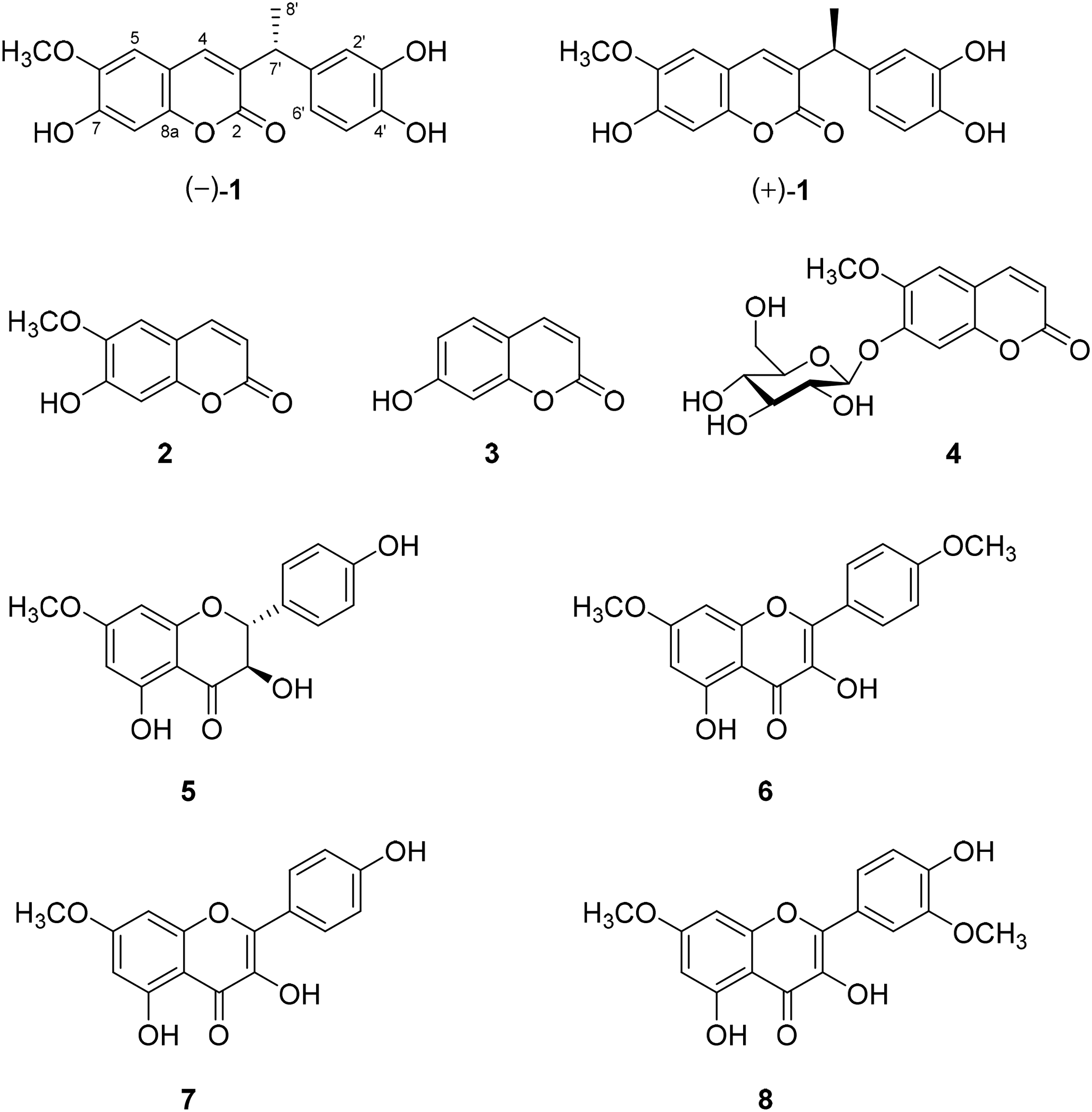
Structures of compounds 1 to 8.
Results and Discussion
Compound 1 was obtained as a yellow, amorphous solid. It was assigned the molecular formula C18H16O6 (11 degrees of unsaturation) on the basis of its HRESIMS data (m/z 351.0842 [M + Na]+). The 1H and 13C NMR spectra of 1 displayed signals for 2 methyl groups including 1 O-methyl, 1 methine, 14 aromatic/olefinic carbons (6 of which were protonated), and 1 carboxyl carbon (δC 164.1). These data accounted for all the NMR resonances for 1 and agreed with the molecular formula C18H16O6, except for 3 exchangeable protons. In the aromatic region of the 1H NMR spectrum, 3 singlets at δH 7.57 (H-4), 7.07 (H-5), and 6.74 (H-8) were consistent with a 36,7-trisubstituted coumarin core,11,12 which was supported by heteronuclear multiple bond correlation (HMBC) cross-peaks from H-4 to C-2 (δC 164.1)/C-5 (δC 109.7)/C-8a (δC 150.0), from H-5 to C-4 (δC 140.1)/C-7 (δC 151.9)/C-8a (δC 150.0), and from H-8 to C-4a (δC 113.0)/C-6 (δC 147.0) (Figure 2). HMBC from the protons of the oxygenated methyl (δH 3.88, 6-OCH3) to C-6 (δC 147.0) located the methoxy group at C-6, which was confirmed by the NOESY correlation of H-5 (δH 7.07) with 6-OCH3. In turn, an ABX-type spin system comprised of the signals at δH 6.71 (H-2′, d, J = 2.0 Hz), 6.70 (H-5′, d, J = 8.1, Hz), and 6.61 (H-6′, dd, J = 8.1, 2.1 Hz) revealed the presence of a 1-substituted 3,4-dihydroxyphenyl moiety, and relevant HMBC cross-peaks further supported this assignment (Figure 2). Finally, key HMBCs from Me-8′ with C-7′/C-1′/C-3 and from H-7′ to C-2/C-4/C-2′/C-6′ indicated that C-3, C-1′, and C-8′ are all connected to the methine C-7′. On the basis of these data, the planar structure of 1 was established, as shown in Figure 1.
Key heteronuclear multiple bond correlations (HMBCs) of compound 1.
Compound 1 has 1 chiral center at C-7′, but is optically inactive, suggesting that it was a racemic mixture. This is consistent with the transparent CD curve. Subsequent chiral resolution of 1 was performed using a Daicel Chiralpak IC type chiral HPLC column, leading to the separation of (–)-1 and ( + )-1 in a ratio of approximately 1:1. As expected, both (–)-1 and ( + )-1 showed opposite optical rotations and mirror-like Cotton effects in the electronic circular dichroic (ECD) curves (Figure 3). To determine absolute configurations of the enantiomers, ECD calculations were carried out at the CAM-B3LYP/def2-TZVP level in methanol using the PCM model. Results (Figure 3) indicated that the calculated ECD spectrum of (R)-1 fitted well with the ECD measurement value of (–)-1, so that the absolute configurations of compounds (–)-1 and ( + )-1 were established to be R and S, respectively. Therefore, compounds (–)-1 and ( + )-1 were assigned as (R)-3-(1-(3,4-dihydroxyphenyl)ethyl)-7-hydroxy-6-methoxycoumarin and (S)-3-(1-(3,4-dihydroxyphenyl)ethyl)-7-hydroxy-6-methoxycoumarin, respectively. They were named ( − )-acutamarin [(-)-1] and ( + )-acutamarin [( + )-1] after the plant origin.
Comparison of the measured electronic circular dichroism (ECD) spectrum for 1 with the CAM-B3LYP/def2-TZVP/PCM(MeOH) calculated curve for (R)-isomer. σ = 0.30 eV, shift = 14 nm.
Three known structurally related coumarins, scopoletin (2),13 umbelliferone (3),14 and scopolin (4),13 together with 4 known flavonoids, (2R,3R)-7-O-methyl-dihydrokaempferol (7-O-methylaromadendrin) (5),15 4′-7-di-O-methyl-kaempferol (6),16 7-O-methyl-kaempferol (rhamnocitrin) (7),15 and 3′-7-di-O-methyl-quercetin (rhamnazin) (8),17 were also isolated and identified by spectral data (Supplemental Tables S2, S3 and S4) in comparison with those reported in literatures.
Compound 1 is a new member of the 3-alkylated coumarin family, which has valuable structural motifs with a wide range of applications in the chemical and pharmaceutical industries.18-20 To our knowledge, 3-(3,4-dihydroxybenzyl)-6,7-dihydroxycoumarin isolated from Onychium japonicum is the closest naturally occurring precedent.21 In addition, it should be noted that 1 shares the same structural core with warfarin (Supplemental Figure S1), a famous synthetic 3-alkylated coumarin used as an anticoagulant drug. However, no inhibitory activity was observed for 1 in the evaluation of in vitro anti-platelet aggregation activity, suggesting that the substitution pattern of the core structure is critical for the potent anticoagulant effect.
All isolated compounds were tested for in vitro anti-inflammatory activities by measuring the NO production in LPS-stimulated RAW264.7 mouse macrophages and the results are shown in Table 1. Among them, only compounds 5 and 7 exhibited inhibition of NO production with IC50 values of 24.54 ± 0.36 μM and 10.60 ± 0.15 μM, respectively. L-NMMA was used as a positive control and presented an IC50 value of 39.82 ± 1.92 μM.
The values represent mean ± SD from triplicate tests.
Positive control.
In summary, compound 1, a new racemic 3-alkylated coumarin, 3 known coumarin analogues (2-4), and 4 known flavonoids (5-8) were isolated from the folk medicinal plant A acuta. Compound 1 was further resolved on a chiral column to yield ( − )-1 and ( + )-1 whose absolute configurations were elucidated by comparison of the observed and calculated ECDs. The new naturally occurring 3-alkylated coumarins [( − )-1 and ( + )-1] identified in this research expand the chemical space of coumarins. Among these isolates, compounds 5 and 7 showed inhibitory activities against NO production in activated macrophages. The findings may provide a clue for understanding the anti-inflammatory mechanism of A acuta.
Experimental
General
Optical rotations were obtained on a Jasco P-1020 Automatic Digital Polarimeter (JASCO), UV measurements on a Shimadzu UV-2700 (Shimadzu), and ECD data with a Jasco J-810 CD spectrometer (JASCO). 1H NMR (500 MHz), 13C NMR (125 MHz), and 2D NMR spectra were recorded on a Bruker AV-600 instrument (Bruker) (CD3OD, δH 3.30, δC 49.0). ESIMS data were obtained on an MDS SCIEX API 2000 LC/MS instrument (SCIEX), and HRESIMS data on an Agilent G6230 TOF mass spectrometer (Agilent). Semipreparative HPLC was performed with an HPLC system equipped with a Shimadzu LC-20AR pump (Shimadzu) using a YMC-pack ODS-A column (5 μm, 10 × 250 mm, YMC). Chiral resolution was performed using a Daicel Chiralpak IC type chiral HPLC column (3 μm, 4.6 × 250 mm, Daicel). For column chromatography, silica gel 60 (100 - 200 mesh, Qingdao Marine Chemical Ltd) and YMC ODS (75 μm, YMC) were used. TLC was performed using HSGF254 silica gel plates (Yantai Jiangyou Silica Gel Development Co. Ltd).
Plant Material
The whole herbs of A acuta Lour were collected from Nanning City (22°45′N, 108°26′E), Guangxi Zhuang Autonomous Region, China in June 2017, and identified by Prof. Songji Wei of College of Pharmacy, Guangxi University of Traditional Chinese Medicine, where a voucher specimen (No. YPC-001) is kept at the College of Pharmacy, Guangxi University of Traditional Chinese Medicine.
Extraction and Isolation
The air-dried, powdered aerial parts of A acuta (15.0 kg) were extracted with 95% ethanol (35 L, 72 h), followed by 75% ethanol (2 × 35 L, each 72 h). A suspension of the concentrated ethanol extract (1.3 kg) in H2O (3 L) was partitioned in sequence with light petroleum, EtOAc and n-butanol (saturated with H2O), each 3 × 3 L, to give fractions soluble in light petroleum (60 g), EtOAc (200 g), n-butanol (500 g), and water (480 g), after evaporation under reduced pressure at 45°C. The EtOAc-soluble portion (200 g) was subjected to Si gel CC, eluted with a light petroleum/EtOAc gradient system, to afford fractions A to I. Fraction A (4.6 g) was chromatographed on a silica gel column, eluted with step gradient mixtures of light petroleum/EtOAc, followed by recrystallization with methanol to afford compound 6 (1.0 mg) as yellow needles. Fraction C (2.4 g) was subjected to CC on silica gel, eluted with a light petroleum/EtOAc gradient, and Sephadex LH-20, eluted with light petroleum − CHCl3−CH3OH (1:1:1), followed by RP-18 semipreparative HPLC, delivered with CH3OH − H2O (11:9) to give 2 (4.0 mg; tR = 10.5 min), 7 (4.5 mg; tR = 27.0 min), and 8 (6.3 mg; tR = 29.9 min). Similarly, fractions E (4.5 g), G (1.0 g), and H (7.5 g) were fractionated on a Sephadex LH-20 column (light petroleum − CHCl3−CH3OH 1:1:1), followed by separation by RP-18 semipreparative HPLC, to yield 5 (1.7 mg; CH3OH − H2O 7:3, tR = 12.5 min) and 3 (4.3 mg; CH3OH − H2O 2:8, tR = 22.5 min) from fraction E, and 1 (2.8 mg; CH3OH − H2O 4:6, tR = 19.3 min) from fraction G, and 4 (5.0 mg; CH3OH − H2O 2:8, tR = 11.5 min) from fraction H.
Chiral Separation of 1
Compound 1 (2.8 mg) was optically separated over a Daicel Chiralpak IC type chiral HPLC column using n-hexane/isopropanol (85:15) as eluent at a flow rate of 1 mL/min, yielding (‒)-1 (1.1 mg; tR = 52.2 min) and ( + )-1 (1.0 mg; tR = 64.6 min).
The anti-inflammatory activity of compounds 1 to 8 was evaluated by measuring inhibitory effects on NO production in LPS-induced RAW 264.7 macrophage cells. Briefly, RAW264.7 cells were seeded in 96-well plates and stimulated with 1 μg/mL of LPS. Test compounds (final concentration 50 μM) were added to 96-well plates at the same time. After overnight incubation, NO production was evaluated by measuring the absorbance at 570 nm. MTS was added to the remaining medium to detect cell viability to eliminate the compounds’ toxic effect on cells. L-NMMA was used as a positive control.
Anti-Platelet Aggregation Activity Assay
The anti-ADP-induced rabbit platelet aggregation assays were carried out as in the previous report.23
Supplemental Material
sj-docx-1-npx-10.1177_1934578X211044563 - Supplemental material for Chemical Constituents of the Folk Medicinal Plant Argyreia acuta Lour. and Their Anti-Inflammatory Activity
Supplemental material, sj-docx-1-npx-10.1177_1934578X211044563 for Chemical Constituents of the Folk Medicinal Plant Argyreia acuta Lour. and Their Anti-Inflammatory Activity by Yunqing Li, Xiaoyong Zhu, Liuyan Mo, Cailan Liu, Jun Li, Bing Li, Jianhua Wei, Guangfeng Liao and Rumei Lu in Natural Product Communications
Footnotes
Acknowledgments
The authors thank Qinghua Kong of Kunming Institute of Botany, Chinese Academy of Sciences for the biological assays.
Declaration of Conflicting Interests
The authors declared no potential conflicts of interest with respect to the research, authorship, and/or publication of this article.
Funding
This work was supported by the Guangxi Science and Technology Base and Talent Special Project (AD20159007), the BasicResearch Ability Enhancement Project for Young and Middle-aged Teachers in Colleges and Universities of Guangxi (2020KY07038), theDoctoral Research Start-up Fund of Guangxi University of Chinese Medicine (2019BS011), the Guangxi First-class discipline (2018XK034) andthe Innovation Project of Graduate Education of Guangxi University of Chinese Medicine (YCSZ2020023).
Ethical Approval
Not applicable, because this article does not contain any studies with human or animal subjects.
Informed Consent
Not applicable, because this article does not contain any studies with human or animal subjects.
Trial Registration
Not applicable, because this article does not contain any clinical trials.
ORCID iDs
Guangfeng Liao
Rumei Lu
Supplemental Material
Supplemental material for this article is available online.
References
1.
TofernBKalogaMWitteLHartmannTEichE. Occurrence of loline alkaloids in Argyreia mollis (Convolvulaceae). Phytochemistry. 1999;51(8):1177‐1180. doi: 10.1016/s0031-9422(99)00121-1
2.
SharmaBDhamijaIKumarSChaudharyH. In vitro and in vivo evaluation of antitumor activity of methanolic extract of Argyreia nervosa leaves on Ehrlich ascites carcinoma. Bangladesh J Pharmacol. 2015;10(2):399‐408. doi: 10.3329/ bjp.v10i2.22334
3.
LalanBKHirayRSGhonganeBB. Evaluation of analgesic and anti-inflammatory activity of extract of Holoptelea integrifolia and Argyreia speciosa in animal models. J Clin Diagn Res. 2015;9(7):FF01‐FF04. doi: 10.7860/jcdr/2015/ 12059.6200
KashyapPRamHShuklaSDKumarS. Scopoletin: antiamyloidogenic, anticholinesterase, and neuroprotective potential of a natural compound present in Argyreia speciosa roots by in vitro and in silico study. Neurosci Insights. 2020;15:1–10. doi: 10.1177/2633105520937693
6.
ZhangRWeiJTangCNongBLuGWangY. Analysis on quality control of Argyreia acuta Lour. Med Plant. 2020;11(6):42‐51. doi: 10.19600/j.enki.issn2152-3924.2020.06.009
7.
HuJ-YWuX-HYinY-QPanJ-TYuB-W. Three resin glycosides isolated from Argyreia acuta, including two isomers. Chin J Nat Med. 2018;16(5):354‐357. doi: 10.1016/s1875-5364(18)30067-0
8.
YuB-WSunJ-JPanJ-T, et al. Four pentasaccharide resin glycosides from Argyreia acuta. Molecules. 2017;22(3):440. doi: 10.3390/molecules22030440
9.
WangLYanY-SCuiH-HYinY-QPanJ-TYuB-W. Three new resin glycosides compounds from Argyreia acuta and their alpha-glucosidase inhibitory activity. Nat Prod Res. 2017;31(5):537‐542. doi: 10.1080/14786419.2016.1201669
10.
YinY-QPanJ-TYuB-WCuiH-HYanY-SChenY-F. Two pentasaccharide resin glycosides from Argyreia acuta. Nat Prod Res. 2016;30(1):20‐24. doi: 10.1080/14786419.2015.1030739
11.
ZhaoDGZhouAYDuZYZhangYZhangKMaYY. Coumarins with alpha-glucosidase and alpha-amylase inhibitory activities from the flower of Edgeworthia gardneri. Fitoterapia. 2015;107:122‐127. doi: 10.1016/j.fitote. 2015.10.012
12.
ShiXJYeGATangWJZhaoWM. A new coumarin and carbazole alkaloid from Clausena vestita D. D. TAO. Helv Chim Acta. 2010;93(5):985‐990. doi: 10.1002/ hlca.200900337
13.
SibandaSNdenguBMultariGPompiVGaleffiC. A coumarin glucoside from Xeromphis obovata. Phytochem Lett. 1989;28(5):1550‐1552. doi: 10.1016/S0031-9422(00)97791-4
14.
SinghRSinghBSinghSKumarNKumarSAroraS. Umbelliferone—an antioxidant isolated from Acacia nilotica (L.) Willd. ex. Del. Food Chem. 2010;120(3):825‐830. doi: 10.1016/j.foodchem.2009.11.022
15.
ZhangXFHungTMPhuongPT, et al. Anti-inflammatory activity of flavonoids from Populus davidiana. Arch Pharmacal Res. 2006;29(12):1102‐1108. doi: 10.1007/bf02969299
El-GhalyEMShaheenURagabEEl-hilaAAAbd-AllahM.R. Bioactive constituents of Pulicaria jaubertii: a promising antihypertensive activity. Pharmacogn. J. 2016;8:81‐86. doi: 10.5530/pj.2016.1.18
18.
JafarpourFDarvishmollaMAzaddoostNMohagheghF. Direct C-3 alkylation of coumarins via decarboxylative coupling with carboxylic acids. New J Chem. 2019;43(24):9328‐9332. doi: 10.1039/C8NJ06410E
19.
MirandaLDIcelo-ÁvilaERentería-GómezÁPilaMMarreroJG. AC-3-selective direct alkylation of coumarins by using a microwave-assisted xanthate-based radical reaction. Eur J Org Chem. 2015;2015(19):4098‐4101. doi: 10.1002/ejoc.201500322
20.
BanerjeeASantraSKKhatunNAliWPatelBK. Oxidant controlled regioselective mono- and di-functionalization reactions of coumarins. Chem Commun. 2015;51(84):15422‐15425. doi: 10.1039/C5CC06200D
21.
LiMCYaoZTakaishiYTangSADuanHQ. Isolation of novel phenolic compounds with multidrug resistance (MDR) reversal properties from Onychium japonicum. Chem Biodivers. 2011;8(6):1112‐1120. doi: 10.1002/cbdv.201000224
22.
TaoJMorikawaTAndoSMatsudaHYoshikawaM. Bioactive constituents from Chinese natural medicines. XI. Inhibitors on NO production and degranulation in RBL-2H3 from Rubia yunnanensis: structures of rubianosides II, III, and IV, rubianol-G, and rubianthraquinone. Chem Pharm Bull.2003;51(6):654‐662. doi: 10.1248/cpb.51.654
23.
XieYZhangJLiuWXieNFengFQuW. New urushiols with platelet aggregation inhibitory activities from resin of Toxicodendron vernicifluum. Fitoterapia. 2016;112:38‐44. doi: 10.1016/j.fitote.2016.05.001
Supplementary Material
Please find the following supplemental material available below.
For Open Access articles published under a Creative Commons License, all supplemental material carries the same license as the article it is associated with.
For non-Open Access articles published, all supplemental material carries a non-exclusive license, and permission requests for re-use of supplemental material or any part of supplemental material shall be sent directly to the copyright owner as specified in the copyright notice associated with the article.