Two new glycosides, periplanosides A (1) and B (2), 3 compounds reported from a natural source for the first time (3 − 5), and 6 known compounds 6 − 11 were isolated from the ethanol extract of Periplaneta americana (Linnaeus). Their structures, including absolute configurations, were unambiguously identified by comprehensive spectroscopic and chemical methods. Compound 3 is a racemate whose enantiomers were purified by chiral high-performance liquid chromatography . The biological evaluation results showed that compound 7 (0 − 20 μM) did not affect the viability of RAW264.7 cells and could effectively inhibit the production of interleukin-6 stimulated by lipopolysaccharide in a concentration-dependent manner, indicating the potential to develop novel agents against inflammation-related diseases.
Periplaneta americana (Linnaeus) is a medicinal insect traditionally used for the treatment of stomach diseases, ulcers, and burns.1 Pharmacological studies showed that P americana could promote tissue repair and regeneration,2 and possess anticancer and anti-inflammatory activities as well.3 “Kangfuxinye,” with P americana as the only ingredient, is a well-known drug in the Chinese market to treat various ulcers and wounds.4 Chemical investigations of the insect have disclosed the presence of amino acids,5 cyclic dipeptides,6 sex pheromones,6 carnosine,7 and isocoumarin.7 We are interested in small-molecule compounds of P americana and our previous studies have identified the existence of some phenolic derivatives,8 dopamine derivatives,9 and dihydrocoumarin glucosides.10 Continuing with our study of P americana, the present research records the isolation of 11 small molecules, including 2 new compounds, periplanoside A (1) and B (2), 3 compounds (3 − 5) which were isolated from a natural source for the first time, and 6 known compounds (6 − 11) (Figure 1).
Chemical formulas of compounds 1−11.
Results and Discussion
Compound 1 was obtained as a yellowish gum. Its molecular formula was determined as C16H17NO8 on the basis of its high-resolution electrospray ionization mass spectrometry (HRESIMS) ion at m/z 374.0843 [M + Na]+ (calcd for C16H17NO8Na, 374.0850), and 13C nuclear magnetic resonance (NMR) and distortionless enhancement by polarization transfer (DEPT) spectra, with the molecular formula indicating 9° of unsaturation. The 1H and 13C NMR spectroscopic data (Table 1) showed the presence of a glucose moiety in the structure. Its β-form was indicated by the anomeric proton (δH 5.16, d, J = 7.8 Hz, H-1′). The 13C NMR and DEPT spectra contained 11 carbon resonances attributed to 5 sp2 methines and 5 quaternary carbons. The 1H–1H COSY spectrum (Figure 2) showed the correlations of H-3/H-4, H-5/H-6 and H-6/H-7. The HMBC spectrum presented the correlations of H-3/C-2, C-11, H-4/C-2, C-9, C-10, H-5/C-4, C-10, C-9, H-6/C-8, C-10, and H-7/ C-5, C-8, C-9. Considering these data and the downfield chemical shifts of C-2 (δC 148.7) and C-9 (δC 140.2), the molecular composition, and the requirement of unsaturation degrees, it can be found that 1 contains a quinoline ring with a carboxylic acid group at C-2. The connection of glucose with the aglycone was supported by the HMBC observation of H-1′/C-8 (δC 154.4). As a result, the planar structure of 1 was deduced. As for the sugar moiety of 1, the configuration was determined by acid hydrolysis followed by derivatization and comparison with a reference standard. Given that the standard retention times (min) of D-glucose and L-glucose derivatives were 12.43 and 14.04, respectively, and the retention time (min) of derivative 1 was 12.25, the glucose of 1 was determined as the D-form. Therefore, the structure of 1 was finally determined and named as periplanoside A.
The key COSY () and HMBC () correlations of compounds 1−4.
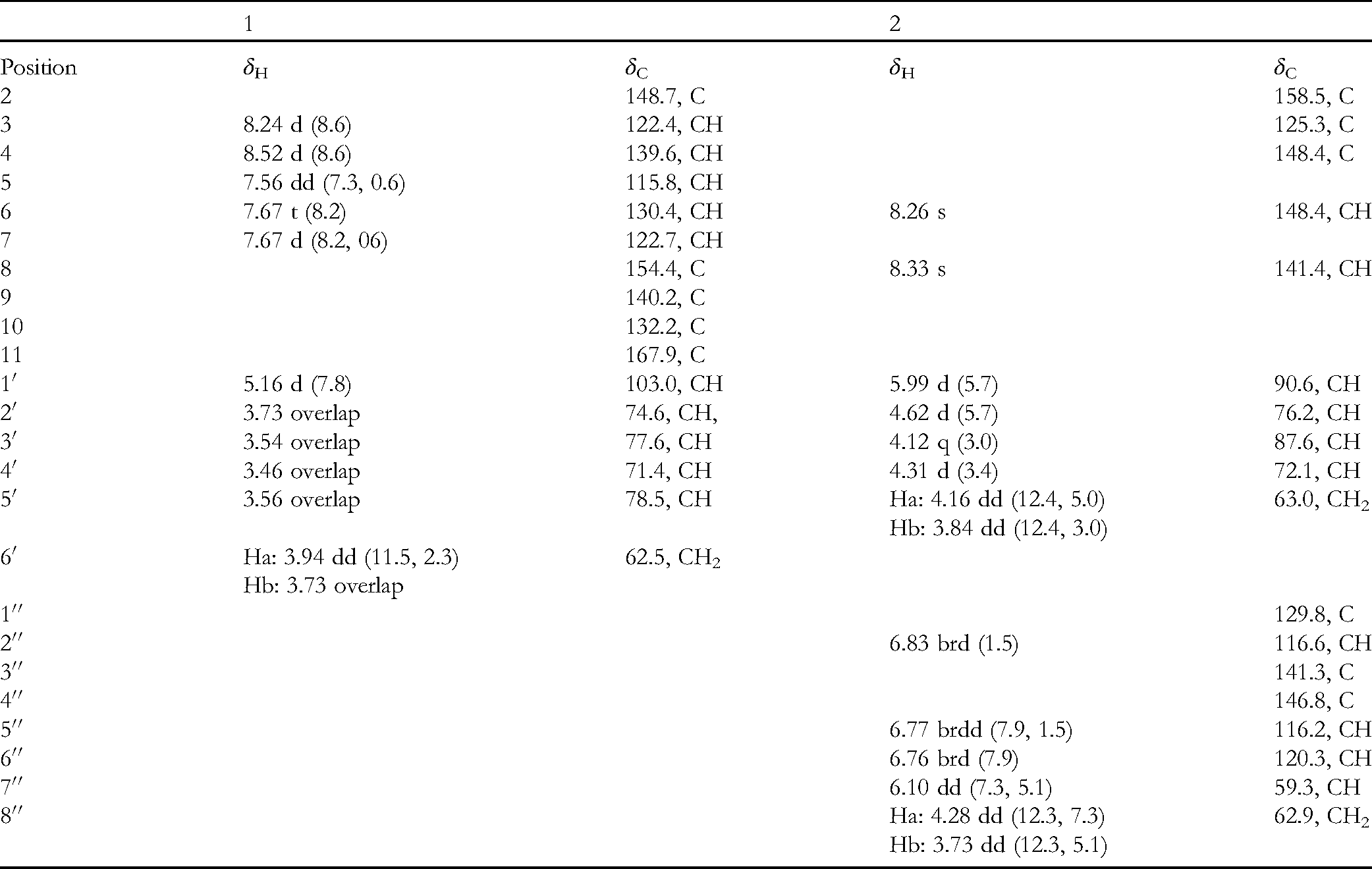
1H (600 MHz) and 13C Nuclear Magnetic Resonance (NMR) (150 MHz) Spectroscopic Data of 1 and 2 in Methanol-d4 (δ in ppm, J in Hz).
1
2
Position
δH
δC
δH
δC
2
148.7, C
158.5, C
3
8.24 d (8.6)
122.4, CH
125.3, C
4
8.52 d (8.6)
139.6, CH
148.4, C
5
7.56 dd (7.3, 0.6)
115.8, CH
6
7.67 t (8.2)
130.4, CH
8.26 s
148.4, CH
7
7.67 d (8.2, 06)
122.7, CH
8
154.4, C
8.33 s
141.4, CH
9
140.2, C
10
132.2, C
11
167.9, C
1′
5.16 d (7.8)
103.0, CH
5.99 d (5.7)
90.6, CH
2′
3.73 overlap
74.6, CH,
4.62 d (5.7)
76.2, CH
3′
3.54 overlap
77.6, CH
4.12 q (3.0)
87.6, CH
4′
3.46 overlap
71.4, CH
4.31 d (3.4)
72.1, CH
5′
3.56 overlap
78.5, CH
Ha: 4.16 dd (12.4, 5.0)
63.0, CH2
Hb: 3.84 dd (12.4, 3.0)
6′
Ha: 3.94 dd (11.5, 2.3)
62.5, CH2
Hb: 3.73 overlap
1′′
129.8, C
2′′
6.83 brd (1.5)
116.6, CH
3′′
141.3, C
4′′
146.8, C
5′′
6.77 brdd (7.9, 1.5)
116.2, CH
6′′
6.76 brd (7.9)
120.3, CH
7′′
6.10 dd (7.3, 5.1)
59.3, CH
8′′
Ha: 4.28 dd (12.3, 7.3)
62.9, CH2
Hb: 3.73 dd (12.3, 5.1)
Compound 2 was also obtained as a yellowish gum. Its molecular formula was determined as C18H20N4O8 on the basis of its HRESIMS ion at m/z 443.1175 [M + Na]+ (calcd for C18H20N4O8Na, 443.1170), indicating 11° of unsaturation. The 1H NMR spectrum of compound 2 (Table 1) showed two signals at δH 8.33 (s, 1H) and δH 8.26 (s, 1H), an ABX (1,2,4- or 1,3,4-trisubstituted) spin system (δH 6.76 [brd, J = 7.9 Hz, 1H], δH 6.83 [brd, J = 1.5 Hz, 1H], and δH 6.77 [brdd, J = 7.9 and 1.5 Hz, 1H]). Besides, there are signals at δH 6.10 (dd, J = 7.3, 5.1 Hz, 1H, H-7''), δH 5.99 (d, J = 5.7 Hz, 1H, H-1'), and five characteristics of the presence of a ribose. The 13C NMR and DEPT spectra of 2 contained 18 carbon resonances attributed to 2 methylenes, 10 methines, and 6 quaternary carbons with a carbonyl. These diagnostic signals, in consideration of the previously isolated compounds from insects,11 prompted us to conclude the presence of a guanosine moiety. In addition, the other signals were identified as a phenylethyl alcohol group, evidenced from the observation of an ABX spin system, 1H–1H COSY correlation of H-7''/H-8'', and HMBC correlation of H-7''/C-1'', C-2'', and C-6''. The chemical shifts of C-3'' (δC 141.3) and C-4'' (δC 146.8) indicated that they were oxygenated. The phenylethyl alcohol group was connected with guanosine via C-7''−N-5, supported by the HMBC correlations of H-7''/C-4 and C-6. Thus far, the planar structure of 2 was deduced. As for the sugar moiety of 2, its configuration was determined by acid hydrolysis followed by derivatization and comparison with the reference standards. The standard retention times (min) of D-ribose and L-ribose derivatives were 17.10 and 13.30, respectively. The retention time (min) of derivative 2 was 17.00, indicating that the ribose was the D-form. However, it is challenging to assign the configuration at C-7''. To clarify this, electronic circular dichroism (ECD) calculations were utilized. The results showed that the ECD spectrum of 7′′R agreed well with the experimental CD of 2 (Figure 3), leading to the unambiguous assignment of the absolute configuration at C-7′′. The structure of 2 was determined and named as periplanoside B.
Comparison of B3LYP/6-311 g (d, p) calculated electronic circular dichroism (ECD) spectrum for (7’’R)-2 with the experimental spectrum of 2 in MeOH. σ = 0.3 eV, shift = +21 nm.
Compound 3 has a molecular formula of C11H11NO4 by analysis of its positive HRESIMS ion at m/z 244.0581 [M + Na]+ (calcd for C11H11NO4Na, 244.0580), with 7° of unsaturation. The 1H NMR spectrum of 3 (Table 2) showed a 1,2,3-trisubstituted benzene ring (δH 5.84 [dd, J = 7.5, 1.4 Hz, H-5], δH 5.93 [t, 6.35, H-6], and δH 5.88 [dd, J = 6.5, 1.4 Hz, H-7]). The 13C NMR and DEPT spectra displayed 1 methyl group, 1 methylene, 4 methines, and 5 quaternary carbons (2 carbonyl groups). The NMR data of 3 were similar to those of jineol-8-sulfate,12 which is a quinoline derivative. Thus, it could be inferred that 3 was an analog of jineol-8-sulfate. The differences between 3 and jineol-8-sulfate were that compound 3 lacked a sulfonic acid group and 1 hydroxyl group, but had 2 more carbonyl groups, according to the chemical shifts of C-2 (δC 167.5) and C-11 (δC 172.1). In addition, the 1H–1H COSY correlations of H2-3/H-4 and the HMBC correlations (Figure 2) of H-4/C-2, C-5, H2-3/C-10, and C-11 were sufficient to prove that the two carbonyl groups were located on C-2 and C-4, respectively. Further, the HMBC correlation of H-12/C-11 (δC 172.1) suggested that the methoxy was located at C-11. Chiral high-performance liquid chromatography (HPLC) analysis indicated that 3 was a racemic mixture, which was further separated by chiral HPLC to afford two enantiomers (3a and 3b). The absolute configurations of these 2 enantiomers were determined to be 4R for 3a and 4S for 3b by comparing the calculated ECD curves with the experimental CD spectra (Figure 4). After searching the database, it was found that 3 was commercially available, but no literature was found. Therefore, compound 3, as a naturally occurring substance, is reported for the first time. Here, we tentatively named it as methyl 8-hydroxy-2-oxo-1,2,3,4-tetrahydroquinoline-4-carboxylate.
Comparison of B3LYP/6-311 g (d, p) calculated electronic circular dichroism (ECD) spectrum for (4R)-3 with the experimental spectra of 3a and 3b in MeOH. σ = 0.3 eV, shift = +24 nm.
1H and 13C Nuclear Magnetic Resonance (NMR) Spectroscopic Data of Compounds 3 − 5 (δ in ppm, J in Hz).
Compound 4 has a molecular formula of C8H17NO2 by analysis of its HRESIMS ion at m/z 182.1150 [M + Na]+ (calcd for C8H17NO2Na, 182.1150), with 1° of unsaturation. The 1H NMR spectrum of 4 (Table 2) exhibited 1 methyl (0.91, t, J = 7.2 Hz, H-6) and 6 sp3 methylenes (2 hetero-atom bearing). The 13C NMR and DEPT spectra displayed resonances for 8 carbons including 1 methyl, 6 methylenes (1 oxygenated), and 1 amide carbonyl. The 1H–1H COSY spectrum (Figure 2) showed correlations of H-2/H-3/H-4/H-5/H-6 and H-1'/H-2', suggesting 2 spin systems, which were assembled via HMBC correlations (Figure 2) of H-2, H-3, and H-1'/C-1 (δc 176.6). Apart from the above fragment, the remaining signal corresponded to an amino group on the basis of the chemical composition of 4 and the chemical shift of C-1' (δC 42.9). Therefore, the structure of 4 was determined. Compound 4 had been previously synthesized,13 but this was the first record of it as a natural product.
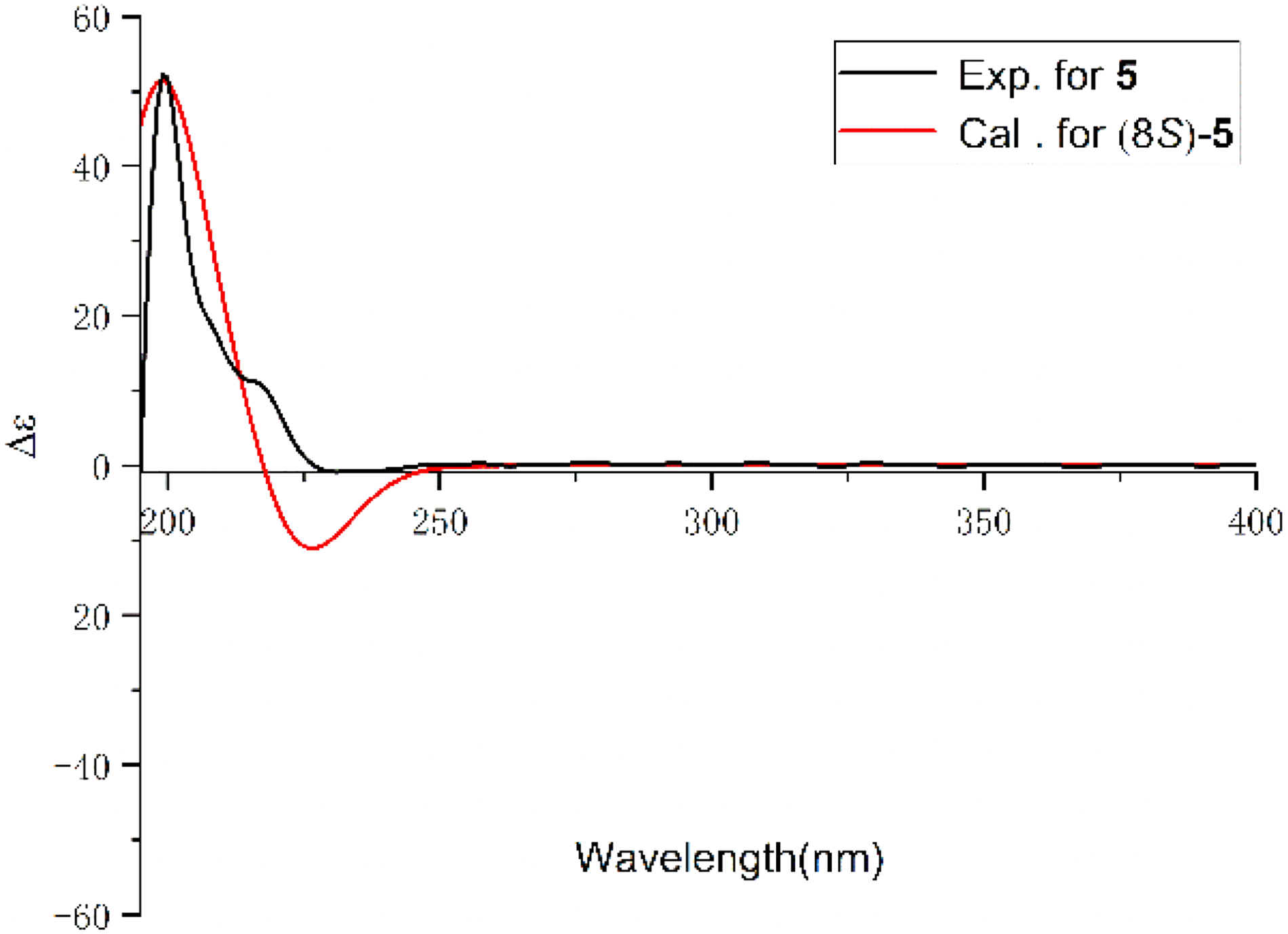
Compound 5 was isolated as white solid and its molecular formula was determined as C10H11NO3. The NMR data of 5 were in accordance with those of a compound previously synthesized by Martínez-Gudiño.14 However, it was characterized from a natural source for the first time. The absolute configuration of 5 was assigned as 8S by comparing the experimental CD curve with the calculated one in Figure 5.
Comparison of B3LYP/6-311 g(d,p) calculated electronic circular dichroism (ECD) spectrum for (8S)-5 with the experimental spectrum of 5 in MeOH. σ = 0.2 eV, shift = +15 nm.
By comparing their spectroscopic data with those in the literature, 6 known compounds were identified as trans-4-hydroxy-2-nonenal (6),15 (1S,3S)-1-methyl-1,2,3,4-tetrahydro-β-carboline-3-carboxylic acid (7),16 6-(2-formyl-1-pyrrolyl)-L-norleucin (8),17 (±)-1,2,3,4-tetrahydro-2-oxo-4-quinolinecarboxylic acid (9),18 phenylalanine (10),19 and proline (11).20
As illustrated in Figure 6, the cell counting kit-8 (CCK-8) assay showed that compound 7 did not affect the viability of RAW264.7 cells when using any of the examined concentrations. The enzyme-linked immunosorbent assay (ELISA) assay results showed that compound 7 could effectively suppress the production of interleukin-6 (IL-6) in RAW264.7 cells stimulated by lipopolysaccharide (LPS) in a concentration-dependent manner. Thus, the present data suggested that compound 7 might act as a lead compound for the development of novel agents against diseases resulting from pathogenic expression or activation of IL-6.
A: RAW264.7 cell proliferation in response to compound 7 at different doses assayed by the CCK-8 assay. B: Compound 7 suppressed LPS-induced IL-6 expression in RAW 264.7 cells. The cells were pretreated with different concentrations of compound 7 for 2 h and then stimulated with 1 μg/mL LPS for 4 h. Culture media were collected in order to measure IL-6 concentrations using an ELISA kit. Data represent mean ± SEM values of three experiments. *P<0.05, and **P<0.01 compared with LPS alone.
Conclusions
In the present study, 2 new glycosides, periplanosides A and B, 3 known, but new as natural products, and 6 known compounds were characterized. Compound 7 was shown to effectively suppress the production of IL-6 in RAW264.7 cells stimulated by LPS in a concentration-dependent manner. Therefore, the present data suggest that compound 7 possesses potential for the treatment of diseases resulting from pathogenic expression or activation of IL-6.
Experimental
General
HRESIMS were collected by a Shimazu LC-20AD AB SCIEX triple TOF X500R mass spectrometry (MS) spectrometer (Shimadzu Corporation). Ultraviolet (UV) and circular dichroism (CD) spectra were measured on a Jasco J-815 CD spectrometer (JASCO), and optical rotations on an Anton Paar MCP-100 digital polarimeter. MCI gel CHP 20P (75-150 μm, Mitsubishi Chemical Industries), macroporous adsorbents (Rohm Haas Amberlite TM XAD 16N) , RP-18 (40-60μm; Daiso Co.), YMC gel ODS-A-HG (40-60 μm; YMC Co), and Sephadex LH-20 (Amersham Biosciences) were used for column chromatography. For semipreparative HPLC, a Saipuruisi chromatograph with a YMC-Pack ODS-A column (25 mm × 10 mm, i.d., 5 μm) was used. For preparative HPLC, a Chuangxin-Tongheng Chromatograph was used, equipped with a Thermo Hypersil GOLD-C18 column (250 × 21.2 mm, i.d., 5 μm). Racemic compounds were purified by chiral HPLC on a Daicel Chiralpak AD-H column (250 mm × 4.6 mm, i.d., 5 μm), a Daicel Chiralpak IC column (250 mm × 4.6 mm, i.d., 5 μm) or a Phenomenex column (OOG-4762-E0 LUX i-Amylose-1, 250 mm × 4.6 mm, i.d., 5 μm). NMR spectra were recorded on either a Bruker AV-500 or an AV-600 spectrometer (Bruker) with tetramethylsilane (TMS) as an internal standard.
Insect Material
P americana was bought from the Breeding Base of He-Yuan-Mei-Lian in Midu County, Dali Autonomous Prefecture, Yunnan Province, PR China, in December 2017. A voucher specimen (CHYX-0627) was deposited at the School of Pharmaceutical Sciences, Shenzhen University, PR China.
Extraction and Isolation
Dried and powdered P americana (50.0 kg) were extracted with 70% EtOH to afford a crude extract. This was suspended in water and then extracted with an equal volume of ethyl acetate, three times, to obtain ethyl acetate and water extracts, respectively. The water-soluble extract was divided into 6 parts (Fraction [Fr.] A–Fraction F) using macroporous adsorbent resin and eluting with a gradient of aqueous ethanol (1%-100%). Fr. A (1.0 kg) was divided into 10 parts (Fr. A.1-Fr. A.10) by using a MCI gel CHP 20P column and eluting with a gradient of aqueous MeOH (3%-100%). Fr. A.6 (7.0 g) was gel filtered over Sephadex LH-20 (aqueous MeOH, 65%) to yield Fr. A.6.1 to Fr. A.6.4. Fr. A.6.2 (3.0 g) was applied to a RP-18 column and eluted with a gradient of aqueous MeOH (2%-100%) to produce 4 fractions (Fr. A.6.2.1-Fr. A.6.2.4). Fr. A.6.2.2 (650.0 mg) was gel filtered over Sephadex LH-20 (MeOH) to give Fr. A.6.2.1 to Fr. A.6.2.4. Fr.A.6.2.2 (200.0 mg) was purified by preparative HPLC (aqueous MeOH, 3%-100%, flow rate: 8 mL/min) to obtain Fr. A.6.2.2.1 to Fr. A.6.2.2.4. Fr. A.6.2.2.2 (18.0 mg) was separated by semipreparative HPLC (aqueous ACN, 11%) to obtain compound 1 (1.2 mg, tR = 18.4 min, flow rate: 3 mL/min). Fr. A.6.2.3 (100.0 mg) was purified by preparative HPLC (aqueous MeOH, 3%-100%, flow rate: 8 mL/min) to obtain Fr. A.6.2.3.1 to Fr. A.6.2.2.3. Fr. 6.2.3.1 (15.0 mg) was separated by semipreparative HPLC (aqueous MeOH, 11%) to obtain compound 2 (2.3 mg, tR = 16.3 min, flow rate: 3 mL/min) and compound 10 (8.0 mg, tR = 7 min, flow rate: 3 mL/min). Fr. A.6.2.1 (300.0 mg) was subjected to preparative HPLC (aqueous MeOH, 3%-100%, flow rate: 8 mL/min) to provide 5 portions (Fr. A.6.2.1.1-Fr. A.6.2.1.5). Fr. A6.2.1.5 (70.0 mg) was separated by semipreparative HPLC (aqueous MeOH, 40%) to yield compounds 4 (8.0 mg, tR = 18.6 min, flow rate: 3 mL/min) and 5 (4.0 mg, tR = 22.3 min, flow rate: 3 mL/min). Fr. A.6.1 (3.7 g) was subjected to an RP-18 column (aqueous MeOH, 3%-100%) to produce 3 portions (Fr. A.6.1.1–Fr. A.6.1.3). Among them, Fr. A.6.1.3 (600.0 mg) was separated by Sephadex LH-20 (MeOH) to yield 4 fractions (Fr. A.6.1.3.1-Fr. A.6.1.3.4). Fr. A.6.1.3.4 (30.0 mg) was further treated by HPLC (aqueous MeOH, 11%) to afford compound 6 (3.0 mg, tR = 19.2 min, flow rate: 3 mL/min). Fr. A.6.1 (3.7 g) was fractionated on an MCI gel CHP 20P column eluted with a gradient of aqueous MeOH (5%-100%) to provide 3 parts (Fr. A.6.1.1-Fr. A.6.1.3). Fr. A.6.1.3 (502.0 mg) was chromatographed over Sephadex LH-20 (MeOH) to produce 5 portions (Fr. A.6.1.3.1-Fr. A.6.1.3.5). Among them, Fr. A.6.1.3.5 was submitted to semipreparative HPLC (aqueous MeOH, 27%), followed by semipreparative HPLC (aqueous ACN, 12%) to afford compound 11 (3.6 mg, tR = 31.0 min, flow rate: 3 mL/min). Fr. B (200.0 g) was divided into 5 parts (Fr. B.1–Fr. B.5) by using an MCI gel CHP 20P column eluted with a gradient of aqueous MeOH (3%-100%). Then, Fr. B.3 (30.0 g) was divided into 9 portions (Fr. B.3.1-Fr. B.3.9) by RP-18 (aqueous MeOH, 5%-100%). Fr. B.3.8 (1.9 g) was gel filtered through Sephadex LH-20 (MeOH) to give Fr. B.3.8.1 to Fr. B.3.8.5. Fr. B.3.8.5 (450.0 mg) was subjected to preparative HPLC (aqueous MeOH, 3%-100%, flow rate: 8 mL/min) to provide eight portions (Fr. B.3.8.5.1-Fr. B.3.8.5.8). Purification of Fr. B.3.8.5.1 (96.0 mg) with aqueous ACN (16%) afforded compound 3 (3.0 mg, tR = 16.6 min, flow rate: 3 mL/min). Fr. B.3.4 (3.7 g) was separated on a Sephadex LH-20 column (MeOH) to give Fr. B. 3.4.1 to Fr. B.3.4.3, and Fr. B.3.4.3 (1.5 g) on a RP-18 column (aqueous MeOH, 3%-100%) to produce 5 portions (Fr. B.3.4.3.1-Fr. B.3.4.3.5). Among them, Fr. B.3.4.3.3 (700.0 mg) was submitted to a Sephadex LH-20 column (MeOH) to yield 4 fractions (Fr. B.3.4.3.3.1-Fr. B.3.4.3.3.4). Fr. B.3.4.3.3.4 (40.0 mg), after semipreparative HPLC (aqueous MeOH, 33%), afforded compound 7 (5.0 mg, tR = 14.1 min, flow rate: 3 mL/min). Fr. B.3.4.3.3 (70.0 mg), following semipreparative HPLC (aqueous ACN, 22%), afforded compounds 8 (3.0 mg, tR = 16.6 min, flow rate: 3 mL/min) and 9 (8.0 mg, tR = 22.4 min, flow rate: 3 mL/min).
Compound Characterization Data
Periplanoside A (1): yellowish gum; UV (MeOH) λmax (logε) 203 (4.10), 248 (4.12) nm; ([α]D20−18.5 [c 0.05, MeOH]); HRESIMS (positive) m/z 374.0843 [M + Na]+ (calcd for C16H17NO8Na, 374.0850); for 1H and 13C NMR data, see Table 1.
Periplanoside B (2): yellowish gum; UV (MeOH) λmax (logε) 203 (4.60), 225 (4.08), 244 (4.03), 263 (3.80), 280 (3.85) nm; ([α]D20 + 17.3 [c 0.05, MeOH]; CD [MeOH] Δε207+ 2.58, Δε231−1.89, Δε271 + 1.72, Δε284+ 2.68); HRESIMS (positive) m/z 443.1175 [M + Na]+ (calcd for C18H20N4O8Na, 443.1170); for 1H and 13C NMR data, see Table 1.
N-(Hexanoyl)ethanolamine (4): yellowish gum; UV (MeOH) λmax (logε) 200 (3.60), 254 (2.55) nm; ([α]D20 + 3.33 [c 0.09, MeOH]}; HRESIMS (positive) m/z 182.1150 [M + Na]+ (calcd for C8H17NO2Na, 182.1150); for 1H and 13C NMR data, see Table 2.
N-Formylphenylalanine (5): white solid; UV (MeOH) λmax (logε) 196 (4.05) nm; ([α]D20 + 37.9 [c 0.04, MeOH]; CD [MeOH] Δε199+ 8.24, Δε213 + 1.89, Δε218 + 1.63, Δε233−0.13); HRESIMS (positive) m/z 216.0636 [M + Na]+ (calcd for C11H11NO4Na, 216.0630); for 1H and 13C NMR data, see Table 2.
Acid Hydrolysis and Preparation of Sugar Derivatives of Compounds 1 and 2
Compounds 1 and 2 (each 0.5 mg) were separately dissolved in 6 N HCl (0.8 mL) and heated at 65 °C for 1.5 h. L-cysteine methyl ester in 1 mL pyridine was added to the concentrated mixture and heated at 65 ° for 1 h. Using the same method, ribose (D/L) and glucose (D/L) were also derivatized by using L-cysteine methyl ester.11,21 Then, 2-methylphenyl isothiocyanate was added to the reaction mixtures and heated at 65 °C for 1 h. The reaction mixtures were concentrated and analyzed using chiral HPLC (Daicel Chiralpak IC column [250 mm × 4.6 mm, i.d., 5 μm]); n-hexane/ethanol, 80:20; flow rate: 1 mL/min; compound 1, D-glucose, and L-glucose) with a UV detector and RP-18 HPLC (YMC-Pack ODS-A column, [250 mm × 4.6 mm, i.d., 5 μm]; MeOH/H2O [0.05% CF3COOH], 40:60; flow rate: 1 mL/min; compound 2, D-ribose, and L-ribose). The standard retention times (min) of the derivatives were as follows: D-glucose (12.43), L-glucose (14.04), D-ribose (17.10), and L-ribose (13.30). By comparing the retention times in our experiment with these standards, both glucose in compound 1 and ribose in compound 2 were determined to be in the D-form (see Supplemental Material).
Biological Evaluation
To obtain enough cells for analysis, a mouse macrophage line of RAW264.7 (Procell Life Science & Technology Co. ), was cultured in high-glucose Dulbecco’s modified Eagle’s medium (DMEM) (C11995500BT, Gibco), which was supplemented with 100 U/mL penicillin, 100 μg/mL streptomycin, and 10% fetal bovine serum (2094468CP, Gibco) in a humidified environment containing 5% CO2 at 37 °C. To examine the cytotoxic effect of compounds on RAW264.7 cells, 2 × 10⁴ cells/mL were seeded into 96-well plates with DMEM for overnight culture. Then, the cells were treated with either dimethyl sulfoxide (DMSO) or various concentrations of compounds for 24 h, following by the addition of 10 μL CCK-8 (Beyotime) into each well for 1 h at 37 °C. The absorbance of each well was recorded at 450 nm using a microplate reader (BioTek). To investigate whether compounds possess biological activities, different concentrations of compounds were used to pretreat RAW264.7 cells (2 × 10⁴ cells/mL, 100 μL medium/well) for 2 h followed by stimulation with LPS (1 μg/mL) for another 4 h. According to the instructions from the manufacturer, the collected culture supernatant from each well was assayed with an IL-6 ELISA Kit (Jiangsu Yutong Biotech Co. Ltd).
Supplemental Material
sj-docx-1-npx-10.1177_1934578X211033180 - Supplemental material for Small Molecule Constituents of Periplaneta americana and Their IL-6 Inhibitory Activities
Supplemental material, sj-docx-1-npx-10.1177_1934578X211033180 for Small Molecule Constituents of Periplaneta americana and Their IL-6 Inhibitory Activities by Hua-Sheng Zhang, Yong-Ming Yan, Dai-Wei Wang, Qing Lv, Yong-Xian Cheng and Shu-Mei Wang in Natural Product Communications
Footnotes
Declaration of Conflicting Interests
The author(s) declared no potential conflicts of interest with respect to the research, authorship, and/or publication of this article.
Funding
The author(s) disclosed receipt of the following financial support for the research, authorship, and/or publication of this article: This work was supported by the Guangdong Key Laboratory for Functional Substances in Medicinal Edible Resources and Healthcare Products (grant number 2021B1212040015, 81525026, 81773603, JCYJ20200109114225087, 2020B1515120051, JCYJ20170412110504956, and 86000000210).
ORCID iD
Yong-Ming Yan
Supplemental Material
Supplementary material relating to this article is available online.
References
1.
LiSZGangmuB. The Great Pharmacopoeia. The People’s Health Publishing House; 1981.
2.
HeZCPengFSongLY, et al.Review on investigations related to chemical constituents and biological activities of Periplaneta americana. China J Chin Mater Med. 2007;32:2326-2331.
3.
LiWDuanLFHeGQShenZQYangHQLiangYP. Periplaneta americana extract effects on experimental liver fibrosis. Lishizhen Med Mater Med Res. 2010;21:2520-2522. doi: cnki:sun:wsxb.0.1959-03-012
4.
LuoYSGaoMTMaFFLiuGMZhangCG. Research advances in pharmacological action and clinical application of Periplaneta americana. J Anhui Univ Nat Sci. 2012;40:5933-5935, 5942. doi: 10.13989/j.cnki.0517-6611.2012.10.073
5.
TangJJZhangLJiangLP, et al.Dopamine derivatives from the insect Polyrhachis dives as inhibitors of ROCK1/2 and stimulators of neural stem cell proliferation. Tetrahedron. 2014;70:8852-8857. doi: 10.1016/j.tet.2014.09.095
6.
WangXLXiangBLiYMGengFNChengYX. Water soluble compounds from Periplaneta americana and their angiogenesis activity. J Nat Prod. 2017;29:2004-2009. doi:10.16333/j.1001-6880.2017.12.002
7.
ZhuHJYanYMTuZC, et al.Compounds from Polyphaga plancyi and their inhibitory activities against JAK3 and DDR1 kinases. Fitoterapia. 2016;114:163-167. doi:10.1016/j.fitote.2016.09.005
8.
BaiHFLiYPQinFY, et al.Periplanetols A − F, phenolic compounds from Periplaneta americana with potent COX-2 inhibitory activity. Fitoterapia. 2020;143:104589. doi:10.1016/j.fitote.2020.104589
YanYMXiangBZhuHJ, et al.N-containing compounds from Periplaneta americana and their activities against wound healing. J Asian Nat Prod Res. 2019;21(2):93-102. doi:10.1080/10286020.2018.1450392
11.
YangYXLuoQHouB, et al.Periplanosides A–C: new insect-derived dihydroisocoumarin glucosides from Periplaneta americana stimulating collagen production in human dermal fibroblasts. J Asian Nat Prod Res. 2015;17(10):988-995. doi:10.1080/10286020.2015.1047771
12.
LeeJIKulkarniRKimMAHwangJSNaMKBaeJS. Antithrombotic and antiplatelet activities of small-molecule alkaloids from Scolopendra subspinipes mutilans. Sci Rep. 2016;6:1-12. doi:10.1038/srep21956
13.
TsujiKKawaiYKatYOsawaT. Formation of N-(hexanoyl)ethanolamine, a novel phosphatidylethanolamine adduct, during the oxidation of erythrocyte membrane and low-density lipoprotein. Biochem Biophys Res Commun. 2003;306(3):706-711. doi:10.1016/S0006-291X(03)01038-6
14.
Martínez-GudiñoGPérez-RojasNATrujillo-SerratoJJMora-PérezYSuárez-CastilloORMorales-RíosMS. Mechanistic study of reversible solid-state melt isomerization of 2-oxindoles to 2-quinolinones and its occurrence in a mass spectrometer. J Mol Struct. 2019;1175:828-835. doi:10.1016/j.molstruc.2018.08.056
15.
MuLHJuQFLiuPHuYZhaoHX. Chemical constituents of the roots of Pottsia laxiflora. Chem Nat Compd. 2013;48(6):1004-1007. doi:10.1007/s10600-013-0450-2
16.
WangXLiuRYangYZhangM. Isolation, purification and identification of antioxidants in an aqueous aged garlic extract. Food Chem. 2015;187:37-43. doi:10.1016/j.foodchem.2015.03.109
17.
HellwigMHenleT. Formyline, a new glycation compound from the reaction of lysine and 3-deoxypentosone. Eur Food Res Technol. 2010;230(6):903-914. doi:10.1007/s00217-010-1237-3
18.
FayolAHoussemanCSunXJanvierPBienaymeHZhuJ. Synthesis of α-isocyano-α-alkyl (aryl) acetamides and their use in the multicomponent synthesis of 5-aminooxazole, pyrrolo [3, 4–b] pyridin-5-one and 4, 5, 6, 7-tetrahydrofuro [2, 3-c] pyridine. Synthesis (Mass).2005;01:161-165. doi:10.1055/s-2004-831225
19.
MaJJTangJSGaoHHongKZhouGXYaoXS. Secondary metabolites from marine actinomycete Streptomyces. Chin J Mar. Drugs. 2010;29(2):6-9. doi:10.13400/j.cnki.cjmd.2010.02.011
20.
PengYLiuYH. Chemical constituents of starfish Asterina pectinifera from Yellow Sea. Chin J Pharm. 2011;46(24):1883-1885. doi:10.13400/j.cnki.cjmd.2010.02.011
21.
TanakaTNakashimaTUedaTTomiiKKounoI. Facile discrimination of aldose enantiomers by reversed-phase HPLC. Chem Pharm Bull. 2007;55:899-901. doi:10.1248/cpb.55.899
Supplementary Material
Please find the following supplemental material available below.
For Open Access articles published under a Creative Commons License, all supplemental material carries the same license as the article it is associated with.
For non-Open Access articles published, all supplemental material carries a non-exclusive license, and permission requests for re-use of supplemental material or any part of supplemental material shall be sent directly to the copyright owner as specified in the copyright notice associated with the article.


 ) correlations of compounds