Three new naphthoquinones, namely 5,8-dihydroxy-7′-isopropyl-furan ring-1″-prenyl-naphthoquinone (1), 5,8-dihydroxy-7′-propenyl-furan ring-1″-isopropanol-naphthoquinone (2), and 5,8-dihydroxy-7′-isopropanol-furan ring-1″-(5″-hydroxyphenyl)-naphthoquinone (3), along with 10 known naphthoquinone derivatives (4-13) were isolated from Cucumis bisexualis for the first time. All the compounds were elucidated on the basis of spectroscopic data analysis and chemical evidence. Compounds (1-13) were evaluated for their hepatoprotective activites on human L-O2 cells. Among them, compounds 1, 4, and 8 exhibited significant promotion effects on the proliferation of L-O2 cells.
Cucumis bisexualis belonging to the Cucurbitaceae family is not only used as a favorite wild fruit but also served for an important folk medicine distributed in China.1 Up to now, there are only 2 reports on the studies of the chemical components and biological activities of C. bisexualis. In 2017, it was found that its extract could reduce blood glucose in diabetic mice and enhance the antioxidant capacity of type 2 diabetic mice.2 Preliminary studies of our group have found that the coumarin derivatives from C. bisexualis possessed hepatoprotective activities.3 As part of our continuous phytochemical investigations on the bioactive isolates from C. bisexualis based on the previous research, this prompted us to perform a bioassay-guided investigation of C. bisexualis in order to evaluate its further hepatoprotective activities. We report here on the isolation and structural elucidation of 3 new naphthoquinones (1-3), together with 10 known naphthoquinone derivatives (4-13) that were isolated from the active fraction (EtOAc layer) of C. bisexualis for the first time (Figure 1). Moreover, the hepatoprotective activities of compounds (1-13) were evaluated in cell proliferation inhibition test and hepatoprotective test on CCl 4-induced injury of human L-O2 cells by 3-(4,5-dimethyl-2-thiazolyl)-2,5-diphenyl-2-H-tetrazolium bromide (MTT) assay. It was found that compounds 1, 4, and 8 (μM) exhibited important hepatoprotective activities. These research results may guide the search for new natural products with hepatoprotective attributes.
Structures of compounds 1 to 13.
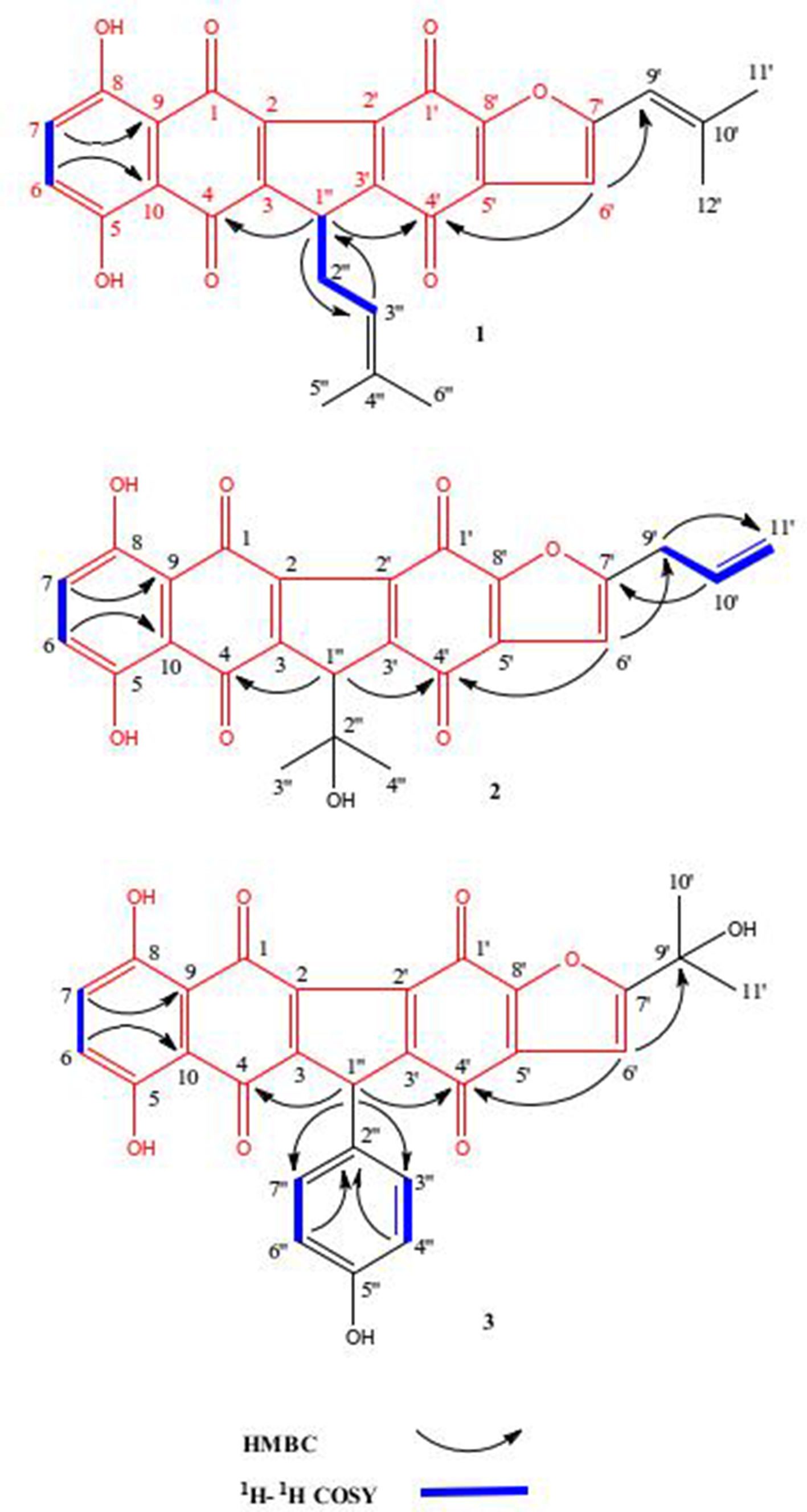
Compound 1 was obtained as a pale yellow powder. The molecular formula C28H22O7 was established by the presence of a deprotonated molecular ion [M−H]− at m/z 469.1275 (calcd. for C28H21O7, 469.1287) in the high resolution-electrospray ionization-mass spectrometry (HR-ESI-MS) data, which requires 18 degrees of unsaturation. The UV spectrum showed absorptions at λmax 210, 232, 280, and 431 nm, suggesting the presence of the naphthoquinone skeleton in compound 1.4 Its infrared (IR) spectrum showed the presence of hydroxyl (3438 cm−1), carbonyl (1736 cm−1), aromatic ring (1617 cm−1), and methyl (1376 cm−1) functionalities. The 1H nuclear magnetic resonance (NMR) data of compound 1 showed that a typical prenyl (δH 2.23, 2H, d, J = 7.2 Hz, H-2″; δH 5.24, 1H, t, J = 7.2, 1.5 Hz, H-3″; δH 1.81, 3H, s, 5″-CH3; δH 1.63, 3H, s, 6″-CH3) was connected to C-1″ by the heteronuclear multiple bond correlation (HMBC) correlations of H-3″ to C-1″, H-1″ to C-3″ (Figure 2). Moreover, an isobutene group (δH 6.20, 1H, s, H-9′; δH 1.77, 6H, s, 11″/12″-CH3) was assigned to C-7′ according to the HMBC correlations of H-6′ to C-8′, H-8′ to C-6′ (Figure 2). Two typical broad single peaks (δH 7.27, 2H, brs, H-6/7) were found in the low field of 1H NMR spectrum, which also confirmed that the 1,2-anthraquinone unit was in compound 1.
Key HMBC and 1H-1H COSY correlations of compounds 1 to 3.
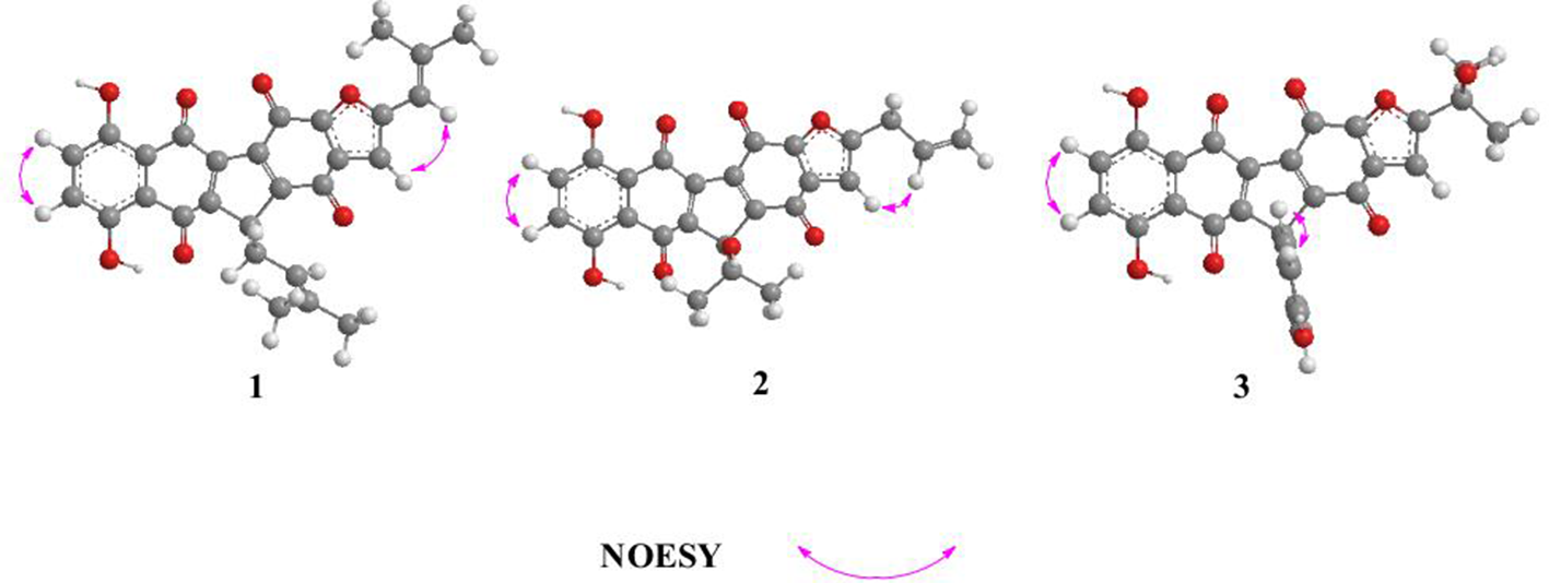
The other fragments of compound 1 were determined by the HMBC correlations of H-6 to C-10, H-7 to C-9, H-6′ to C-4′/9′ (Figure 2), the 1H-1H correlation spectroscopy (COSY) correlations of H-6 to H-7, H-1″ to H-2″, H-2″ to H-3″ (Figure 2), and the nuclear overhauser enhancement spectroscopy (NOESY) correlations of H-6 to H-7, H-6′ to H-8′ (Figure 3). Consequently, compound 1 was elucidated as 5,8-dihydroxy-7′-isopropyl-furan ring-1″-prenyl-naphthoquinone.
Key NOESY correlations of compounds 1 to 3.
Compound 2 was isolated as a pale yellow powder. Its molecular formula was determined as C25H18O8, evidenced from the HR-ESI-MS data (m/z 445.0952 [M−H]−, calcd. for C25H17O8, 445.0923) with 17 degrees of unsaturation. Its UV and IR spectral data were similar to those of compound 1, which showed the absorptions at λmax 210, 233, 280, and 430 nm and indicated the existence of hydroxyl (3432 cm−1), carbonyl (1736 cm−1), aromatic ring (1616 cm−1), and methyl (1374 cm−1) functional groups, separately. It was concluded that compound 2 was an analog of compound 1. The 1H and 13C NMR spectral data of compound 2 (Table 1) were similar to those of compound 1 except for 1″-isopropanol (δH 1.27, 6H, s, 3″/4″-CH3) that was connected to C-1″ by the HMBC correlations of H-1″ to C-4/4′ and 7′-propenyl (δH 3.16, 2H, d, J = 6.9 Hz, H-9′; δH 5.91, 1H, dd, J = 17.2, 10.3 Hz, H-10′; δH 5.06, 1H, d, J = 17.2 Hz, H-11′a; δH 5.03, 1H, d, J = 10.3 Hz, H-11′b)5 that was assigned to C-7′ by the HMBC correlations of H-6′ to C-9′, H-10′ to C-7′. The other fragments of compound 2 were determined by the HMBC correlations of H-6 to C-10, H-7 to C-9 (Figure 2), the 1H-1H COSY correlations of H-6 to H-7, H-9′ to H-10′, H-10′ to H-11′ (Figure 2), and the NOESY correlations of H-6 to H-7, H-6′ to H-9′ (Figure 3). Consequently, compound 2 was elucidated as 5,8-dihydroxy-7′-propenyl-furan-ring-1″-isopropanol-naphthoquinone.
1H NMR (400 MHz, Acetone-d6), 13C NMR (100 MHz, Acetone-d6), and Key HMBC Correlations of Compounds 1 to 3.
No.
1
2
3
δH
δC
δH
δC
δH
δC
1
-
186.6
-
186.5
-
186.6
2
-
133.2
-
133.5
-
133.3
3
-
148.5
-
148.3
-
143.7
4
-
183.7
-
183.3
-
183.6
5
-
159.1
-
159.0
-
159.4
6
7.27(brs)
130.5
7.28(brs)
130.7
7.26(brs)
130.6
7
7.27(brs)
130.5
7.28(brs)
130.7
7.26(brs)
130.6
8
-
154.4
-
154.6
-
154.7
9
-
113.9
-
114.0
-
114.2
10
-
111.6
-
111.7
-
111.5
1′
-
176.1
-
175.8
-
175.9
2′
-
133.2
-
133.0
-
133.1
3′
-
148.5
-
148.8
-
148.6
4′
-
177.8
-
177.3
-
177.5
5′
-
143.3
-
142.7
-
142.9
6′
7.21(s)
106.4
6.78(s)
106.1
7.04(s)
106.0
7′
-
158.5
-
162.8
-
172.1
8′
-
163.2
-
160.7
-
160.1
9′
6.20(s)
115.2
3.16(d,6.9)
32.3
-
70.1
10′
-
143.6
5.91(dd,17.2,10.3)
137.2
1.31(s)
28.5
11′
1.79(s)
22.4
5.06(d,17.2), 5.03(d,10.3)
116.2
1.31(s)
28.5
12′
1.71(s)
16.8
-
-
-
-
1″
2.88(t,6.0,1.5)
30.3
2.72(s)
44.8
4.20(s)
-
2″
2.23(d,7.2)
28.9
-
68.2
-
32.7
3″
5.24(t,7.2,2.5)
120.5
1.27(s)
26.9
6.95(d,8.0)
129.8
4″
-
133.2
1.27 (s)
26.9
6.62(d,8.0)
116.2
5″
1.81(s)
24.9
-
-
-
-
6″
1.63(s)
18.8
-
-
6.62(d,8.0)
116.2
7″
-
-
-
-
6.95(d,8.0)
129.8
Compound 3 was obtained as a pale yellow powder. Its molecular formula was determined as C28H18O9, evidenced from the HR-ESI-MS data (m/z 497.0861 [M−H]−, calcd. for C28H17O9, 497.0873) with 20 degrees of unsaturation. The UV absorptions (λmax 210, 231, 280, and 432 nm) were attributed to naphthoquinone skeleton,4 and the IR absorptions implied the presence of hydroxyl (3436 cm−1), carbonyl (1736 cm−1), aromatic ring (1619 cm−1), and methyl (1377 cm−1) functionalities and exhibited compound 3 that was an analog of compound 2. In the 1H NMR spectrum of compound 3, a typical AA′BB′ system at δH 6.95 (2H, d, J = 8.0 Hz, H-3″/7″) and 6.62 (2H, d, J = 8.0 Hz, H-4″/6″) (Table 2) was connected to C-1″ by the correlations of H-1″ to C-3″/7″ in the HMBC spectrum (Figure 2). The 7′-isopropanol group (δH 1.31, 6H, s, 10′/11′-CH3) was linked with C-7′ by the HMBC correlation of H-6′ to C-9′ (Figure 2). The other fragments of compound 3 were determined by the HMBC correlations of H-6 to C-10, H-7 to C-9, H-6′ to C-4′, H-4″/6″ to C-2″ (Figure 2), the 1H-1H COSY correlations of H-6 to H-7, H-3″ to H-4″, H-6″ to H-7″ (Figure 2), and the NOESY correlations of H-6 to H-7, H-3″ to H-4″, H-6″ to H-7″ (Figure 3). Consequently, compound 3 was elucidated as 5,8-dihydroxy-7′-isopropanol-furan ring-1″-(5″-hydroxyphenyl)-naphthoquinone.
Results of Hepatoprotection of 1, 4, and 8 on CCl4 -Induced Injury of L-O2.
bCompared with blank control group and CCl4 injury group, P < 0.05.
In addition, 10 known naphthoquinones derivatives (4-13) were identified by comparison of their spectroscopic data and references. Their structures were determined as shikometabolin G (4),6 naphthofuranin A (5),6 (2R)-6-hydroxy-7-methoxy-dehydroiso-α-lapachone (6),7 2-(1,4-dioxo-1,4-dihydronaphthalen-2-yloxy)-3-hydroxynaphthalene-1,4-dione (7),8 zeylanone (8),10 1-methyl-2,4-dimethoxy-3-hydroxyanthraquinone (9),9 javanicin (10),10 anhydrofusarubin (11),10 shikonin (12),11 and lapachol (13).11 The known isolates (4-13) were identified as derivatives of naphthoquinones from this plant for the first time.
Compounds (1-13) were evaluated for their hepatoprotective effects on human L-O2 cells with Bifendatatum as the positive control. The results showed that compounds 1, 4, and 8 had significantly promoted proliferative effects on L-O2 cells at concentrations of 25, 50, 100, 200, 400, and 600 µg/mL, but had weak inhibitory effects with the respective inhibitory rates of 14%, 19%, and 17% at the concentration of 800 µg/mL, and cytotoxicity of compounds 1, 4, and 8 was very weak with IC50 above 800 µg/mL. In the results of hepatoprotective test by CCl4-injury (Table 2), the survival rates of compounds 1, 4, and 8 at 3 levels were significantly higher than that of CCl4-injury group (P < 0.05), and were also higher than that of Pifendatatum (P < 0.05) at high concentrations. Consequently, compounds 1, 4, and 8 had the significant hepatoprotective activities. In contrast, compounds 2, 3, 5 to 7, and 9 to 13 showed no hepatoprotective activities.
Experimental
General Procedures
The UV spectra were measured by an Australia GBC UV-916 spectrophotometer (GBC Scientific Equipment Pty. Ltd., Braeside, Australia) and the IR spectral data were recorded on a Nicolet 5700 fourier transformation infrared (FT-IR) spectrometer with KBr pellets (Shanghai Xiangrun Industry Co. Ltd., Shanghai, China). The HR-ESI-MS spectral data were measured by an Agilent 1100 series LC/MSD ion trap mass spectrometer (Agilent Technologies, California, United States), and the ESI-MS data were measured by a Q-Trap liquid chromatography/mass spectrometry/mass spectrometry (LC/MS/MS) spectrometer (Turbo Ionspray Source). The NMR spectral data were run on a Bruker-400 with tetramethyl silane (TMS) as internal standard. The reversed-phase high-performance liquid chromatography (HPLC) was performed on Agilent 1200 series with a DIKMA (4.6 × 250 mm) analytical column packed with C18 (5 µm) (Agilent Technologies, California, United States). The prep-HPLC separation was performed on a Shimadzu LC-6AD instrument with a SPD-20A detector and an YMC-Pack ODS-A column (250 × 20 mm, 5 µm) (Shimadzu Corporation, Japan). The column chromatography was performed on Sephadex LH-20, Toyopearl HW-40C, silica gel (100, 200, or 200-300 mesh) (Qingdao Marine Chemical Inc., Qingdao, China). The thin layer chromatography was performed on precoated silica gel GF254 plates and the spots were visualized under UV light (254 or 356 nm) or by spraying with 10% H2SO4 in 95% EtOH followed by heating.3
Plant Material
The fruits of C. bisexualis were harvested from Nanyang city of Henan Province of China in September 2016 and were authenticated by Dr Rongrui Wei of Jiangxi University of Traditional Chinese Medicine. A voucher specimen of C. bisexualis (XMP-201609) has been deposited in Jiangxi University of Traditional Chinese Medicine, Nanchang, China.
Extraction and Isolation
The air-dried fruits of C. bisexualis (15.5 kg) were extracted with 90% EtOH (16 L × 3) under reflux 3 times at room temperature. The filtrates were combined and concentrated under reduced pressure until the elimination of ethanol to obtain a black residue (1.6 kg). The extract was suspended in distilled water (15.0 L) and partitioned with petroleum ether, EtOAc, and n-BuOH, consecutively, yielding petroleum ether layer (98.6 g), EtOAc layer (235.5 g), and n-BuOH layer (305.8 g), respectively. The EtOAc layer was confirmed hepatoprotective site according to the screening results of bioactivity-guided investigation.3 The EtOAc layer was performed on silica gel (100-200 mesh) column and eluted with a gradient elution (n-hexane/EtOAc = 12:1 to 3:1, v/v) to yield 4 fractions: A, B, C, and D. The fraction A (29.6 g) was subjected to silica gel (200-300 mesh) column and eluted with a gradient elution (petroleum ether/EtOAc = 20:1-5:1) to afford 3 subfractions: A1 (7.56 g), A2 (10.85 g), and A3 (8.63 g). The subfraction A2 was fractionated on Toyopearl HW-40C (95% MeOH) and Sephadex LH-20 (CH2Cl2:MeOH = 1:1), then separated by prep-HPLC (detection at 210 nm, 3 mL/min), successively, yielding 1 (6.58 mg, purity >98.2% by HPLC), 2 (7.25 mg, purity >96.7% by HPLC), 3 (9.30 mg, purity >98.0% by HPLC), 4 (8.20 mg, purity >97.6% by HPLC), and 8 (11.35 mg, purity >96.5% by HPLC). Meanwhile, the subfraction A3 was separated over silica gel, Sephadex LH-20, and Toyopearl HW-40C, prep-HPLC, repeatedly, yielding 5 (12.05 mg, purity >96.8% by HPLC), 6 (10.22 mg, purity >98.6% by HPLC), 7 (8.62 mg, purity >98.8% by HPLC), 9 (11.20 mg, purity >97.0% by HPLC), 10 (14.54 mg, purity >96.3% by HPLC), 11 (10.21 mg, purity >96.7% by HPLC), 12 (11.55 mg, purity >96.1% by HPLC), and 13 (9.84 mg, purity >98.5% by HPLC). The structures of compounds (1-13) are shown in Figure 1.
Pale yellow powder; HR-ESI-MS: m/z 445.0952 [M−H]− (calcd. for C25H17O8, 445.0923); UV (MeOH) λmax: 210, 233, 280, 430 nm; IR νmax (KBr): 3432, 1736, 1616, 1374 cm−1; 1H NMR (Acetone-d6, 400 MHz) and 13C NMR (Acetone-d6, 100MHz) data; see Table 1.
5,8-Dihydroxy-7′-Isopropanol-Furan Ring -1″-(5″-Hydroxyphenyl)-Naphthoquinone (3)
Pale yellow powder; HR-ESI-MS: m/z 497.0861 [M−H]− (calcd. for C28H17O9, 497.0873); UV (MeOH) λmax: 210, 231, 280, 432 nm; IR νmax (KBr): 3436, 1736, 1619, 1377 cm−1; 1H NMR (Acetone-d6, 400 MHz) and 13C NMR (Acetone-d6, 100MHz) data; see Table 1.
Hepatoprotective Assay
Compounds (1-13) were evaluated for their hepatoprotective activities on CCl4-induced injury of human L-O2 cells in vitro with Bifendatatum (Absin Bioscience Inc.; purity >98% by HPLC) as the positive control. The human L-O2 cells at logarithmic growth phase were digested by trypsin (Absin Bioscience Inc.; purity >97% by HPLC) and then were made into a 1 × 10⁴ mL−1 cell suspension in RPMI1640 containing bovine serum (100 mL/L). The L-O2 cells were inoculated in a 96-well plate and the suspension (100 µL) was added to each well and there were 6 duplicate wells in each group. The L-O2 cells were incubated in an atmosphere containing 50 mL of CO2 and 950 mL of air at 37°C. The medium was aspirated and replaced with fresh medium containing 0 (blank control), 12.5, 25, 50, 100, 200, 400, 600, and 800 µg/mL compounds (1-13) after 24 hours. The L-O2 cells were supplemented with 20 µL MTT (Fluka, 5 g/L) and incubated for another 4 hours. Then, the supernatant was discarded and the L-O2 cells preparation was shaken with DMSO (100 µL) until the crystal gets fully dissolved. The value of A570 (nm) for each well was measured by a microplate reader. Meanwhile, the L-O2 cells were inoculated in a 96-well plate and the suspension (100 µL) was added to each well and divided into CCl4 injury group, experimental group, positive control, and blank control group and there were 12 duplicate wells in each group. The L-O2 cells were incubated in an atmosphere containing 50 mL of CO2 and 950 mL of air at 37°C. The medium was aspirated and replaced with fresh medium (100 µL) containing blank control group, injury group (10 mM CCl4), positive control (250 mg/mL Bifendatatum and 10 mM CCl4), experimental group (125, 250, and 500 µg/mL compounds 1, 4, 8 and 10 mM CCl4) after 12 hours. After 8 hours, the L-O2 cells were supplemented with 20 µL of MTT (Fluka, 5 g/L) and incubated for another 4 hours. Then, the supernatant was discarded and the L-O2 cells preparation was shaken with DMSO (100 µL) for 20 minutes. A microplate reader was used to measure the value of A570 (nm) for each well.12
Conclusion
Three new naphthoquinones (1-3) and 10 known naphthoquinone derivatives (4-13) were isolated from C. bisexualis for the first time. All the compounds were elucidated on the basis of spectral data and references. Compounds (1-13) were evaluated for their hepatoprotective activites on human L-O2 cells. Among them, compounds 1, 4, and 8 exhibited significant promotion effects on the proliferation of L-O2 cells.
Footnotes
Declaration of Conflicting Interests
The author(s) declared no potential conflicts of interest with respect to the research, authorship, and/or publication of this article.
Funding
The author(s) disclosed receipt of the following financial supportfor the research, authorship, and/or publication of this article: This study was supported by the National Natural Science Foundation of China (81803843), the Key Scientific Research Project of Colleges and Universities in Henan Province (19A350006), the Standard Revision Research Project of National Pharmacopoeia Committee (2018Z090), the Science and Technology Project of Jiangxi Health Commission (20195648, 20195650), the Science and Technology Project of Jiangxi Provincial Department of Education (GJJ180662, GJJ180688), the Scientific Research Project of First-class Discipline of Chinese Materia Medica of Jiangxi University of TCM (JXSYLXK-ZHYAO027, JXSYLXK-ZHYAO032), and the Doctoral Research Initiation Fund Project of Jiangxi University of TCM (2018BSZR007, 2018BSZR010).
ORCID ID
Rongrui Wei
References
1.
ZhangZ.HuoJQ.ZhangZQ.WangYH.ZhangJL. A report of a Cucurbitaceae weed Cucumis bisexualis causing damage to corn. Plant Protect. 2016;42:254-256.
2.
CQL.WangZY. Effect on antioxidation in type 2 diabetic mice extract of Cucumis bisexualis. China Health Care Nutr. 2017;5:44-45.
3.
MaQ-G.WeiR-R.YangMet al. Molecular characterization and bioactivity of coumarin derivatives from the fruits of Cucumis bisexualis. J Agric Food Chem. 2018;66(22):5540-5548.doi:10.1021/acs.jafc.8b00976
4.
KongyenW.RukachaisirikulV.PhongpaichitS.SawangjaroenN.SongsingP.MadardamH. Anthraquinone and naphthoquinone derivatives from the roots of Coptosapelta flavescens. Nat Prod Commun. 2014;9(2):219-220.doi:10.1177/1934578X1400900222
DongM.LiuD.LiY-Het al. Erratum: naphthoquinones from Onosma paniculatum with potential anti-inflammatory activity. Planta Med. 2017;83(7):E2-635.doi:10.1055/s-0043-100122
7.
LuoY.ShenH-Y.ShenQ-Xet al. A new anthraquinone and a new naphthoquinone from the whole plant of Spermacoce latifolia. J Asian Nat Prod Res. 2017;19(9):869-876.doi:10.1080/10286020.2017.1279609
8.
AkhterS.RonySR.Al-MansurMA.HasanCM.RahmanKM.SohrabMH. Lawsonol, A new bioactive naphthoquinone dimer from the leaves of Lawsonia alba. Chem Nat Compd. 2018;54(1):26-29.doi:10.1007/s10600-018-2251-0
9.
ZhangX.ZhouH-F.LiM-Y.YueX-Y.WuT. Three new anthraquinones from aerial parts of Paederia scandens. Chem Nat Compd. 2018;54(2):245-248.doi:10.1007/s10600-018-2314-2
10.
ChowdhuryNS.SohrabMH.RanaMS.HasanCM.JamshidiS.RahmanKM. Cytotoxic Naphthoquinone and Azaanthraquinone Derivatives from an Endophytic Fusarium solani. J Nat Prod. 2017;80(4):1173-1177.doi:10.1021/acs.jnatprod.6b00610
11.
ZhangQ.DongJ.CuiJ.HuangG.MengQ.LiS. Cytotoxicity of synthesized 1,4-naphthoquinone oxime derivatives on selected human cancer cell lines. Chem Pharm Bull. 2018;66(6):612-619.doi:10.1248/cpb.c18-00013
12.
YuanX.LinL.ZhangX.DengS.AbrusamideA. Abrusamide A and B, two hepatoprotective isomeric compounds from Abrus mollis Hance. Phytochem Lett. 2014;7:137-142.doi:10.1016/j.phytol.2013.11.003