
Abstract
The extract of Penthorum chinense Pursh (PCP), a well-known Miao herb medicine, has been used as a key component for a Chinese patented drug to treat several kinds of liver-related diseases. In this work, 3 pinocembrin derivatives, S1, S2, and S3, were isolated from PCP stems and identified with high-performance liquid chromatography and electrospray ionization mass spectrometer. The molecular masses of S1, S2, and S3 were identical to Pinocembrin-7-O-[4″,6″-hexahydroxydiphenoyl (HHDP)]-β-D-glucose, Pinocembrin-7-O-[3″-O-galloyl-4″,6″-(s)-HHDP)-β-D-glucose, and Thonningianin A, respectively. Their free radical scavenging capability was evaluated with the 2,2-diphenyl-1-picrylhydrazyl assay. The half-maximal effective concentrations of S1, S2, and S3 were 26.75, 9.06, and 5.50 μg/mL, respectively. In vitro AML-12 assays demonstrated that S1 (5-20 μg/mL), S2 (10-40 μg/mL), and S3 (10-40 μg/mL) not only protected cells from H2O2-induced oxidation and alcohol-induced cell damages, but also reduced oleic acid (OA)-induced triglyceride accumulations in a dose-dependent manner. However, the 3 compounds potently exhibited similar cytotoxicity effect at high concentrations. The half-maximal inhibitory concentrations of S1, S2, and S3 to AML-12 cells were 74.19, 85.86, and 80.43 μg/mL. In addition, the 3 compounds also showed antibacterial activity on Escherichia coli, Staphylococcus aureus, Enterococcus faecalis, Lactobacillus rhamnosus, and Bacillus subtilis.
Keywords
Natural products, especially the commonly used ones in traditional medicine, are viewed as a wealth of medicine source. Like tea, Penthorum chinense Pursh (PCP), a popular herb, is a kind of functional drink in the tradition of Miao people, an ethnic minority in China, for thousands of years. The freshly grown leaves are also served as a vegetable in some Miao villages. 1,2 The Miao people believe that PCP tea can prevent liver damages resulting from long-term alcohol consumption, and PCP extract is used as a major content of a Chinese patented drug, Gansu granular. In decades, it is reported that PCP extract or Gansu granular showed positive clinical effects on hepatitis B-induced liver fibrosis, 3 -5 hepatitis C-caused liver damages, 6 alcoholic fatty liver, 7 nonalcoholic fatty liver, 8 and liver cirrhosis. 9 Because of the various clinical effects, extensive research has been conducted to identify and evaluate the key components of PCP extract.
More than 50 different natural compounds in PCP extract have been identified, including flavonones, flavonoid glycosides, 10 -13 phenolic compounds, 12,14 organic acids, 15 steroids, 11 volatile oil, 16 and polysaccharide. 17 Many pharmacological studies have been carried out on PCP. 18 Most of them focused on the extraction mixtures, such as the aqueous extract 19,20 or ethyl-acetate fraction. 21,22 In a previous study, it was demonstrated that Pinocembrin derivatives were one of the main flavonoids in PCP, including pinocembrin-7-O- [4″,6″-hexahydroxydiphenoyl]-β-D-glucose (S1), pinocembrin-7-O-[3″-O-galloyl-4″,6″-HHDP]-β-D-glucose (S2), and Thonningianin A (S3). 22 Thonningianin A (pinocembrin dihydrochalcone-7-O-[3″-O-galloyl-4″,6″-HHDP]-β-D-glucose) was isolated from Thonningia sanguinea first in 2000. 23 Previous studies showed that Thonningianin A presented antioxidation, 23,24 glutathione S-transferases inhibition 25 (liver protection), and α-amylase inhibition 22 activities and inhibited the growth of HepG2 cells. 26 Little is known about S1 and S2. In this study, the antioxidation, anti-lipid accumulation, and antibacterial activities of S1, S2, and S3 were investigated in vitro.
Materials and Methods
Materials and Chemicals
PCP was obtained from Sichuan Chinese Medicine Yinpian Co., Ltd and identified by Prof Zhishan Ding (Zhejiang Chinese Medical University). AML-12 cell line was purchased from American Type Culture Collection (ATCC, Manassas, VA, USA). Escherichia coli strain ATCC 35218, Staphylococcus aureus strain ATCC 29213, Enterococcus faecalis strain ATCC 29212, S. aureus strain ATCC 43300, and Lactobacillus rhamnosus strain ATCC 53103 were purchased from the ATCC (Manassas, VA, USA), and also Bacillus subtilis CMCC1.1470 was purchased from China General Microbiological Culture Collection Center (Beijing, China). 2,2-diphenyl-1-picrylhydrazyl (DPPH), ethanol, petroleum benzine, ethyl acetate, and acetonitrile were obtained from Sigma-Aldrich (St Louis, MO, USA). Insulin, transferrin, and antibiotics were also obtained from Sigma-Aldrich. Dulbecco’s modified Eagle medium/Ham’s nutrient mixture F-12, 1:1 (DMEM/F-12, SH30023.01) and fetal bovine serum were purchased from Hyclone (Logan, UT, USA). Tryptone, yeast extract, and agar were purchased from Qingdao Hope Bio-Technology Co., Ltd (Qingdao, China). HPD 500 macroporous resins were purchased from Guangfu Fine Chemical Research Institute (Tianjin, China).
Preparation of 3 Pinocembrin Derivatives
The extracts of PCP were prepared using a sequential extraction method. The sample materials were subjected to successive extractions using different solvents according to their ascending order of polarity. Briefly, the powdered PCP was extracted with 70% ethanol, and the resulted crude extract was subsequently extracted with petroleum benzine and ethyl acetate. The ethyl acetate fractions were further fractionated with HDP 500 column chromatography using 80% ethanol as the eluent. The 3 pinocembrin derivatives in the eluent were separated and eluted from an XB-C18 (10 × 250 mm, 10 µm) column with a gradient acetonitrile (from 20% to 70%) in 0.1% formic acid solution, which was driven by a Dionex Ultimate 3000 preparative high-performance liquid chromatography (HPLC) system (Thermo Fisher Scientific, Waltham, MA, USA). The eluted products were detected with a diode array ultraviolet/visible detector. The molecular masses of S1, S2, and S3 were identified with an electrospray ionization mass spectrometer (ESI-MS) system from Shimadzu with the interface temperature 300℃ and a heating gas flow 10 L/min. Data were collected in a centroid mode from 100 to 1000 m /z.
DPPH Scavenging Activity Analysis
The antioxidation activity was evaluated with a DPPH free radical scavenging test. DPPH 27 is widely used to examine the antioxidation activity of a compound, an extract, or other biological samples. The reaction mixture contained 50 µL of the tested sample and 1 mL of 1 mM DPPH solution in ethanol. The mixtures were incubated at 37℃ for 30 minutes, and the absorbance was measured at 517 nm using a Varioskan LUX Multimode Microplate Reader (Thermo Fisher Scientific, Waltham, MA, USA). The percentages of radical scavenging activity in each mixture were calculated by normalizing with the ethanol mixture (negative control) using the following equation: Scavenging percentage (%) = ([control Absorbance – sample Absorbance]/control Absorbance) %. The half-maximal effective concentration (EC50) values of the compounds were analyzed using GraphPad prism software.
AML-12 Cell Culture
AML-12 cells were cultured in DMEM/F-12 containing 10% (v/v) fetal bovine serum, 5 mg/mL insulin, 5 µg/mL transferrin, 5 ng/mL selenium, 40 ng/mL dexamethasone, 100 U/mL penicillin, 100 µg/mL streptomycin, 2 mmol/L glutamine,100 U/mL penicillin, and 100 g/mL streptomycin. The cells were grown in a humid incubator at 37°C with 5% CO2. All the experiments in this study were set up in sextuplicate. For the cell analyses, the isolated compounds of various final concentrations were added into cell culture medium. In the cell culture medium, 0.4 mM H2O2, 0.5 mM oleic acid (OA), or 400 mM alcohol was added separately for antioxidation, triglyceride (TG) accumulation, or protection effect of alcohol-induced cell damage analyses. The data were normalized to the untreated control cell assays.
Cell Viability Analysis
Cell viability was evaluated by measuring lactate dehydrogenase (LDH) or the MTT (3-(4, 5-dimethylthiazol-2-yl)−2, 5-diphenyltetrazolium bromide) assay. LDH, released from injured cells to the extracellular fluid, was quantitatively assessed using an LDH assay kit (Thermo Fisher Scientific, Waltham, USA) according to the manufacturer's instructions. Briefly, LDH activity was determined with a spectrophotometer at 490 nm by measuring the rate of NAD+ reduction in the presence of L-lactate. In the MTT assay, the number of viable cells was measured by incubating the cells with 10 µL of 5 mg/mL MTT solution per well at 37°C for 4 hours. The mitochondrial dehydrogenases catalyzed the reduction of MTT to a formazan derivative. 28 The formazan derivative was solubilized with 100 µL of lysis buffer (20% sodium dodecyl sulfate in 50% dimethylformamide, pH 4.7). After incubation for 10 minutes at room temperature, the absorbances were measured at 490 nm. The half-maximal inhibitory concentrations (IC50s) of the compounds were analyzed using GraphPad prism software.
Triglyceride Accumulation Determination
OA treatment induces TG accumulation in cells. In this study, the cellular concentration of TG was determined by enzymatic colorimetry according to the instructions of a kit obtained from Applygen (Applygen Technologies Inc., Beijing, China). 29 The cultured cells were lysed by lysis buffer at room temperature for 10 minutes followed by a pyrolytic process of heating at 70℃ for 10 minutes. The cell debris and denatured proteins in the pyrolytic solution were discarded after centrifugation at 2000 × g for 5 minutes at room temperature. TG in the lysis solution was detected according to the manufacturer’s instructions. The protein concentrations of the pyrolytic solutions were determined with the Bradford method. 30
Antibacterial Activity Analysis
The antibacterial activity was determined with a disc diffusion assay. The tested strains, including Escherichia coli ATCC 35218, S. aureus ATCC 29213, Enterococcus faecalis ATCC 29212, S. aureus ATCC 43300, L. rhamnosus ATCC 53103, and B. subtilis CMCC1.1470, were grown in LB medium overnight. Whatman filter paper discs (diameter 5 mm) were impregnated with 10 mg/mL S1, S2, and S3 in 70% ethanol solvents, separately. The discs were placed on freshly prepared LB agar plates with a final bacterial cell count of 108 colony-forming units/plate. The plates were incubated at 37℃ for 24 hours. The susceptibility was recorded by measuring inhibition zone diameters (IZDs) that resulted from the growth inhibition around the discs. 31 Seventy percent ethanol- and vancomycin-impregnated paper discs were used as negative or positive controls separately in the experiment.
Statistical Analysis
All data were expressed as means ± SD of at least 3 independent experiments. Statistical analyses were carried out using SPSS 22.0 software. Differences between treatments were considered to be statistically significant if P <0.05.
Result
Pinocembrin Derivatives Preparation and Identification
These 3 compounds were predominant in the extract of PCP, as described by Deng et al. 32 To obtain 3 pinocembrin derivatives, the ethanol crude extract from PCP stem was subsequently extracted with petroleum benzine and ethyl acetate, followed by HDP 500 column chromatography and preparative HPLC. The 80% ethanol eluate of HDP 500 was analyzed by analytical HPLC (Figure 1(a)), in comparison with Thonningianin A (obtained from Sichuan WeiKeqi Biological Technology Co., Ltd, Chengdu, China) (Figure 1(b)). With this method, we obtained 1.01 g S1, 1.81 g S2, and 0.59 g S3 from 250 g PCP stem powder. The 3 compounds accounted for 0.40%, 0.72%, and 0.24% (g/g) of the PCP stem powder separately. S1, S2, and S3 were identified, respectively, using an ESI-MS system and yielded ions at m/z 721, 873, and 875, which are identical to Pinocembrin-7-O-[4″,6″-hexahydroxydiphenoyl]-β-D-glucose, Pinocembrin-7-O-[3″-O-galloyl-4″,6″-(s)-HHDP]-β-D-glucose, and Thonningianin A, respectively. 23,33 The characteristic fragments of compounds S1, S2, and S3 (m/z 301) indicated the lost pinocembrin residue (Figure 2).

The analytical high-performance liquid chromatography analysis of Thonningianin A (a) and HDP 500 80% eluate fraction of Penthorum chinense Pursh (b).
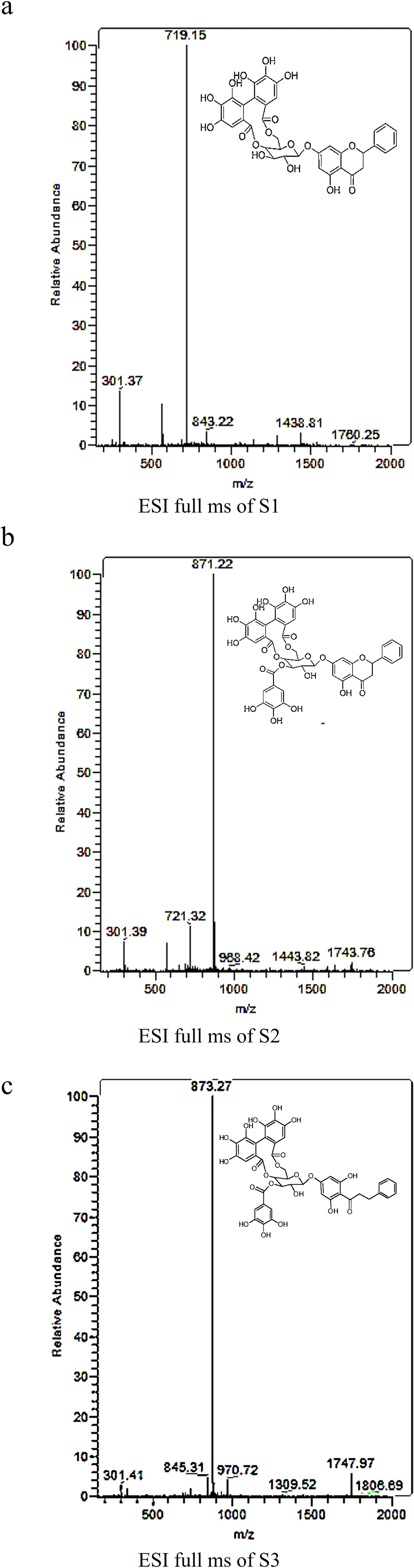
The ESI-MS analysis and structural formula of S1 (a), S2 (b), and S3 (c). ESI-MS, electrospray ionization mass spectrometry.
The 3 Pinocembrin Derivatives Are Antioxidants
Highly reactive free radicals and oxygen species are produced either by exogenous chemicals or by body metabolic process, leading to the oxidation of nucleic acids, lipids, and proteins. Oxidative stress is related to cell damages and apoptosis and results in various diseases. In this study, the antioxidation capabilities of S1, S2, and S3 were evaluated by a DPPH scavenging assay and H2O2 treatment. The DPPH assay utilizes the reaction between the antioxidants and the stable free radical, DPPH (deep violet color). The reaction generates a colorless product. The free radical scavenging capabilities of S1, S2, and S3 are shown in Figure 3(a). The percentages of radical scavenging by the 3 compounds increased linearly with the increase of concentrations. The calculated EC50s of S1, S2, and S3 were 26.75, 9.06, and 5.50 µg/mL (Figure 3(c)), respectively.
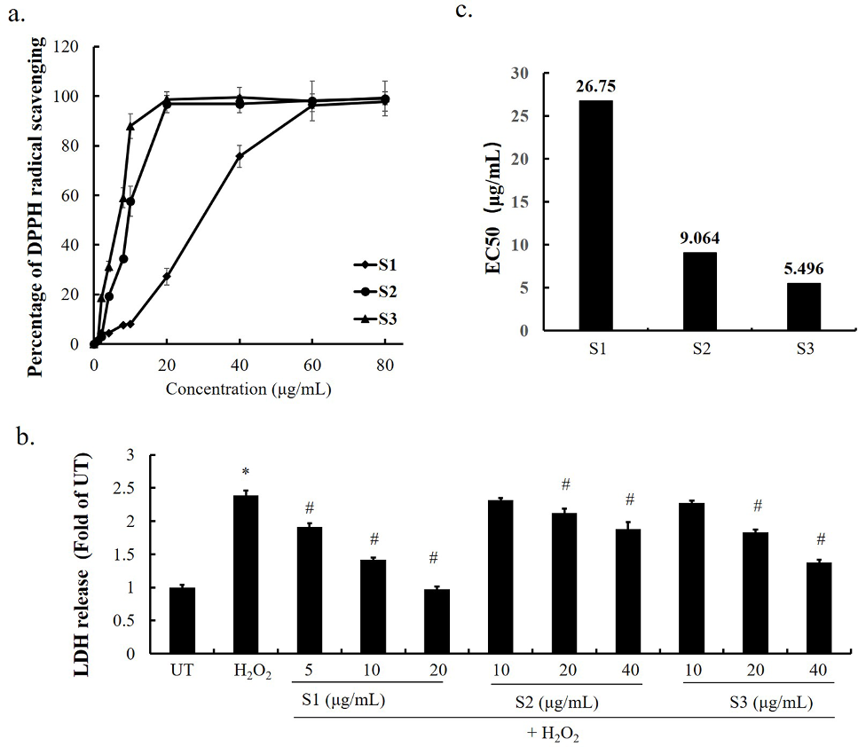
The 3 Pinocembrin derivatives are antioxidants. a) Various concentrations of the 3 Pinocembrin derivatives were used to scavenge DPPH radicals. The bars in (c) represent the EC50s (mean ±SD) of 3 independent experiments. b) AML-12 cells were treated with 0.4 mM H2O2 in the presence of various doses of the compounds for 12 hours. LDH release in the culture medium was detected as described in Materials and Methods. All the values are denoted as means ± SD from 3 or more biological repeats. *P < 0.05 vs UT (the control group); # P < 0.05 vs the H2O2 group. DPPH, 2,2-diphenyl-1-picrylhydrazyl; EC50, half-maximal effective concentration; LDH, lactate dehydrogenase.
H2O2 is an extremely active chemical compound, which is easily reduced to form hydroxyl radicals and induces tissue and cell damages. To investigate the protective effects of S1, S2, and S3 on H2O2-induced cell damages, an LDH assay was conducted to determine the cell viability. The AML-12 cell viability was significantly suppressed upon the treatment with 0.4 mM H2O2 compared with the control (Figure 3(b)). The pretreatment of S1, S2, and S3 protected cells from LDH releases, and S1 showed the best protective effect while S3 showed better protection than S2.
S1, S2, and S3 Reduced Lipid Accumulation In Vitro
OA treatment of hepatocytes is frequently used to evaluate lipid accumulation in vitro. The supplied OA in medium induces TG synthesis in cells, which can be quantified with a sensitive chromogenic reagent assay. In our study, 0.5 mM OA induced a marked TG accumulation in AML-12 cells in comparison with the control cells (Figure 4). S1, S2, and S3 reduced the TG contents in AML-12 cells in a dose-dependent manner. The lowest concentrations of S1, S2, and S3 that reduced the TG contents significantly were 5, 20, and 20 µg/mL, respectively (P < 0.05).

The 3 Penthorum chinense Pursh compounds protected AML-12 cells from OA-induced lipid accumulation. AML-12 cells were treated with 0.5 mM OA in the presence of various doses of S1, S2, and S3 for 12 hours. Glycerin trimyristate was detected as described in “Materials and Methods”. All values are denoted as means ± SD from 3 or more independent biological repeats. *P < 0.05 vs UT (the control group); # P < 0.05 vs the OA group. OA, oleic acid; TG, triglyceride.
S1, S2, and S3 Protected Cells From Alcohol-Induced Cell Damages
Next, we examined whether S1, S2, and S3 could play a hepatoprotective role in alcohol-induced cell damage in vitro. The excessive alcohol in the culture medium induces cell damages. The LDH analysis in our study showed that alcohol treatment was significantly associated with increased cell damages compared with the control cells. S1, S2, and S3 pretreatment significantly reversed the alcohol-induced cell damages in a dose-dependent manner (Figure 5). S1 and S2 showed the best and least protective effects, respectively.

S1, S2, and S3 attenuated alcohol-induced cell damages. AML-12 cells were treated with 400 mM alcohol in the presence of various doses of S1, S2, and S3 for 12 hours. LDH release in the culture medium was detected as described in “Materials and Methods”. All the values are denoted as means ± SD from 3 or more biological repeats. *P < 0.05 vs UT (the control group); # P < 0.05 vs the alcohol group. LDH, lactate dehydrogenase.
S1, S2, and S3 Showed Antibacterial Activity
The bacteriostatic activities of S1, S2, and S3 against 5 Gram-positive strains (B. subtilis, 2 S. aureus, L. rhamnosus, Enterococcus faecalis) and 1 Gram-negative strain Escherichia coli were examined. The IZDs demonstrated that S1 and S3 inhibited the growth of all the tested strains. IZDs of S1 were from 6.00 to 7.67 mm, and S3 presented larger IZDs (from 7.33 to 11 mm) against the most test strains except L. rhamnosus (5.67 mm). In comparison, S2 showed antibacterial capabilities against all the Gram-positive strains (IZDs 6.33-9 mm) but not the Escherichia coli strain. L. rhamnosus was least sensitive to S1, S2, and S3 in comparison with other tested bacterial strains. Interestingly, S. aureus ATCC 43300 were more sensitive to S2 and S3 than S. aureus ATCC 29213. The IZDs of each compound listed in Table 1.
Evaluate the Cytotoxicity of S1, S2, and S3
In vitro cytotoxicity study is a useful initial step toward determining the potential toxicity of the tested compounds. The selectivity index is an important parameter to identify drug candidates with promising biological activity and negligible cytotoxicity. At last, the cytotoxicities of S1 (Figure 6(a)), S2 (Figure 6(b)), and S3 (Figure 6(c)) on AML-12 cells were evaluated with the MTT assay. The IC50s of S1, S2, and S3 were 74.19, 85.86, and 80.43 µg/mL, respectively (Figure 6(d)).

The cytotoxicity of the Pinocembrin derivatives on AML-12 cells. AML-12 cells were treated withS1 (a), S2 (b), or S3 (c) for 24 hours. And the cell viability was evaluated with MTT. (d) showed half-maximal inhibitory concentrations of S1, S2, and S3 in bar. All the values are denoted as means ± SD from 3 or more independent repeated assays of cells. *P < 0.05 vs the control group. MTT, 3-(4, 5-dimethylthiazol-2-yl)−2, 5-diphenyltetrazolium bromide.
Discussion
The pharmacological benefits of PCP, a traditional medicinal herb and a source of phytochemicals, are widely studied in China nowadays. Some chemical studies have been done; 34 however, very little biological investigations have been done. Therefore, we compared some biological activities of 3 pinocembrin derivatives in PCP stem extracts in this study.
Pinocembrin derivatives are common constituents in herbs, 35 as well as in bee propolis. 36 These compounds have been used as nutrient supplements because of their potential antioxidative capability, which is implicated in their benefits for human health. 37,38 The results of our previous study showed that the ethyl acetate fraction of the PCP 70% ethanol extract contained many flavonoids. In the current study, we showed that 250 g PCP stem powder contained 1.01 g S1, 1.81 g S2, and 0.59 g S3, the 3 Pinocembrin derivatives. The mass spectra of the isolated S1, S2, and S3 (Figure 2) were identical to the result of Era et al. 39
Liver diseases, such as nonalcoholic fatty liver disease (NAFLD), are a significant health burden worldwide. According the statistical data reported by Asrani et al, 40 liver diseases account for approximately 2 million deaths per year worldwide. Oxidative stress is one of the key contributors in the progression of NAFLD. 41,42 Several studies have demonstrated that oxidative stress induces the damage of functional molecules, such as proteins and DNAs, in hepatocytes. Many studies revealed that Pinocembrin and its derivatives displayed high antioxidative ability and free radical scavenging activities. 37,38 In addition, the antioxidative activity of Thonningianin A (S3) from an African medicinal herb, Thonningia sanguine, was reported in 2002. 24 However, S1 and S2 were less studied. In this study, the antioxidative and free radical scavenging activities were evaluated. Our results suggest that S1 is the strongest radical scavenger in the 3 derivatives (Figure 3(b)). The lack of 1 gallic acid subunit in Pinocembrin-7-O of S1 might make the disequilibrium of the molecule’s electron clouds compared with S2 and S3. This characteristic also determined the molecular polarity. S1 was the first component eluted from the reversed-phase C18 column, followed by S2 and S3 (Figure 1). Interestingly, S2 showed markedly higher radical scavenging activity than S3, but exhibited a similar H2O2 protective efficiency as S3 (Figure 3). It is known that the free hydroxylates of gallic acid and ellagic acid (constituted by 2 gallic acid molecules) subunits can react with free radicals 43 and donate hydrogens, which are absorbed by free radicals to form inert radicals. 44,45 The H2O2 protective efficiency of an antioxidant not only results from its reactive capability with -OH released by H2O2. The cell damage induced by H2O2 treatment is a complex process. Therefore, we speculate that S3 has also other capability to protect the cells against H2O2 treatment beside the chemical mechanism.
The etiology of NAFLD is complex. Beside reactive oxygen species (ROS), the dysfunction of liver cells also arises from the imbalance between hepatic lipid synthesis and lipid clearance (free fatty acid oxidation). 46,47 In clinic, TG accumulation in the liver is a biomarker of NAFLD. One of our previous studies demonstrated that the extract of PCP stem protected cells from OA-induced lipid accumulation in hepatoma cells (accepted manuscript). In this study, we showed that S1, S2, and S3 reduced OA-induced lipid accumulation in AML-12 cells in a dose-dependent manner (Figure 4). The anti-lipid accumulation activity of S1 is the strongest among the 3 compounds.
Other causes of NAFLD include lipopolysaccharide (LPS) and alcoholic products produced by gut bacteria, 48 such as ethanol acetate or acetaldehyde, which promote the transport of endotoxins (LPS) in the gut vessels 49 and induce rapid formation of ROS. 50 It has been demonstrated that high levels of circulating LPS 49 and ethanol 51 stimulate the accumulation of hepatic fat. Our result showed that S1, S2, and S3 have protective activity on alcohol-induced cell damages (Figure 5) and bacteriostatic activities on some intestinal bacteria (Table 1). So we considered that S1, S2, and S3 can protect NAFLD through an indirect way.
Although PCP extracts have been used as a herb medicine in decades in China,34 their cytotoxicity has not been investigated well. 52,53 The toxicity of a candidate medicine should be determined before clinical studies. The toxicity of S1, S2, and S3 was determined in this study using AML-12 cells. The IC50s of S1, S2, and S3 were 74.19, 85.86, and 80.43 µg/mL (Figure 6(d)), which are markedly higher than their EC50s (26.75, 9.06, and 5.50 µg/mL) in the DPPH test. The notable protective effects against H2O2-induced cell damages and OA-induced lipid accumulation were also observed. The lowest effective concentrations of S1, S2, and S3 were 5, 20, and 20 µg/mL, respectively.
To conclude, our study has demonstrated that S1, S2, and S3 in PCP are potential active components of PCP for NAFLD treatment. S1, S2, and S3 protect cells from ROS and prevent hepatic lipid accumulation. Their inhibitory effect on enteric bacterial growth may reduce circulating LPS and alcoholic products produced by intestinal bacteria and benefit NAFLD treatment indirectly. The mechanisms of these activities will be further explored in future studies.
Inhibition Zone Diameters (Millimeters) of Different Fractions of 3 Components.

In the experiment, 70% ethanol-impregnated paper discs, the negative control, showed no effect and were not in show. “-” represented that no visible inhibition zone was observed. “Nd” means not detected.
Footnotes
Acknowledgement
We thank Dr Zhenyuan Song (Department of Kinesiology and Nutrition University of Illinois at Chicago) and Dr Songtao Li (College of Basic Medicine, Zhejiang Chinese Medical University) for providing guidance and valuable advice on our experiments.
Declaration of Conflicting Interests
The author(s) declared no potential conflicts of interest with respect to the research, authorship, and/or publication of this article.
Funding
The author(s) disclosed receipt of the following financial support for the research, authorship, and/or publication of this article: This work was supported by grants from the Natural Science Foundation of China (