
Abstract
In our effort to find materials for drugs and cosmetics from tropical natural resources, we screened 21 methanol extracts from 7 Macaranga trees species (Macaranga bancana, Macaranga gigantea, Macaranga hullettii, Macaranga pruinosa, Macaranga tanarius, Macaranga trichocarpa, and Macaranga triloba) for antioxidant and antimelanogenesis. The antioxidant and melanogenesis (tyrosinase enzyme assay and melanin inhibitor in B16 melanoma) assays were used to determine the activities. The fractionation and the isolation of active compound were done by various chomatographic methods and the structure was determined by spectroscopic analysis data. We isolated a prenylated flavonoid, named Glyasperin A, from M. pruinosa leaf. This compound showed a potency as antioxidant and inhibited melanin in B16 melanoma but not tyrosinase activity. These results showed that the methanol leaf extracts of M. pruinosa could be developed for cosmetic applications especially as a skin whitening agent.
In search of material for drugs and cosmetics, we have screened 7 species of Macaranga trees (Macaranga bancana, Macaranga gigantea, Macaranga hullettii, Macaranga pruinosa, Macaranga tanarius, Macaranga trichocarpa, and Macaranga triloba) in cancer (MCF-7, B16 melanoma, HCT116, and Hela) and normal (TIG-1 and NHDF) cells, previously. 1 Macaranga pruinosa is the tree with up to 15 m of height, diameter of breast height up to 20 cm. The bark is smooth, hooped, whitish-gray, and with red sap. The inner bark is reddish-brown and granular, the male flowers are pale green, and the fruits are green. 2 The previous biological or chemical studies on M. pruinosa have shown antioxidant, antityrosinase, anticholinesterase, nitric oxide inhibition, and antibacterial activities of leaf extracts 3,4 and chemical compounds such as flavonoids (macapruinosin B, C, D, E, F and Nymphaeol C) 5,6 and Stilbene (Macapruinosin A) were produced. 5 The young shoots’ extracts of M. gigantea, M. pruinosa, and M. triloba are used for treating fungal infections and the leaves for treating stomach aches. 7 In this study, we reported the antioxidant and antimelanogenesis (antityrosinase and antimelanin) of the extract and the isolated compound.
Results and Discussion
The reactive oxidants such as reactive oxygen species (ROS) and H2O2 are produced in melanogenesis, which create oxidative stress in melanocytes. Some ROS scavengers and inhibitors of ROS generation inhibit UV-induced melanogenesis, and antioxidants like reduced glutathione and ascorbic derivatives are applied to treat various skin problems such as hyperpigmentation. Hence, antioxidants regulate the important role in hyperpigmentation or melanogenesis. 8 In this study, we have screened those extracts for antioxidants and antimelanogenesis as seen in Table 1 in order to evaluate its potency as cosmetic ingredient. Based on Table 1 , Figure 1, and thin layer chromatography (TLC), the leaf extract of M. pruinosa was chosen for further fractionation to isolate the active compound. A total of 27 fractions were collected and their antioxidant activities were determined. Fraction 11 was the potent fraction for antioxidant and focused on the isolation of the active compound. By using some chromatographic methods and nuclear magnetic resonance (NMR) assigments (1H, 13C, distortionless enhancement by polarization transfer [DEPT], heteronuclear single quantum coherence [HSQC], and heteronuclear multiple bond correlation [HMBC]), Glyasperin A 9,10 was obtained.
Effect of Macaranga Trees Extracts on Antioxidant, Tyrosinase Enzyme Activity, and Melanin Inhibition in B16 Melanoma Cells at 100 mg/mL.
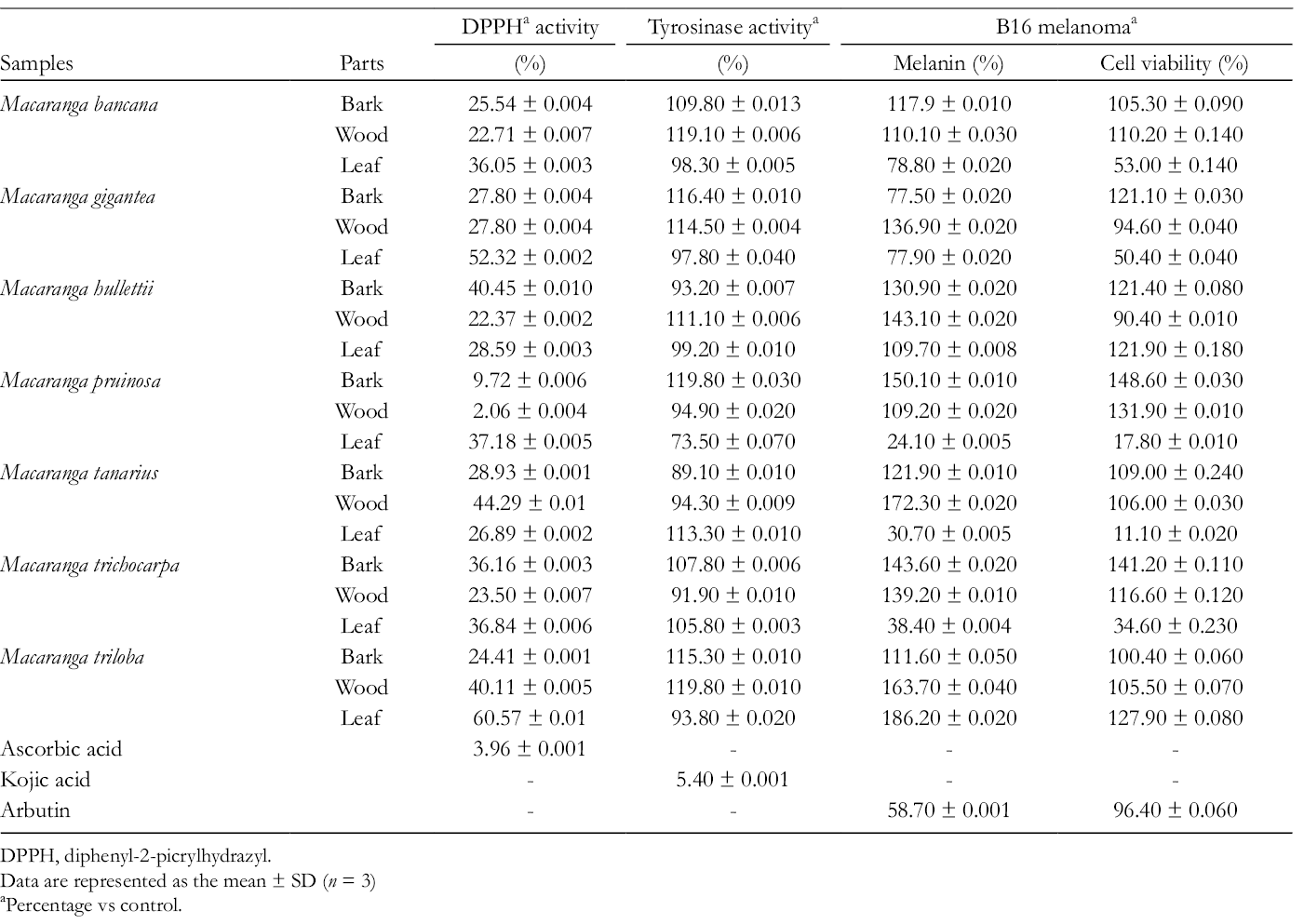
DPPH, diphenyl-2-picrylhydrazyl.
Data are represented as the mean ± SD (n = 3)
Percentage vs control.

Antimelanogenesis in B16 melanoma cells of Macaranga pruinosa leaf extracts (all values: mean ± SD; Arb 100, Arbutin 100 µg/mL). Significantly different from the control value (Student’s t test) : P < 0.05 (*) and P < 0.01 (**).
Next, we determined antioxidant and antimelanogenesis activities of Glyasperin A. Table 2 depicts the antioxidant activities such as diphenyl-2-picrylhydrazyl (DPPH), (2,2’-azino-bis-3-ethylbenzthiazoline-6-sulphonic acid) [ABTS], superoxide dismutase-like activity (SOSA), and oxygen radical absorbance capacity (ORAC). All the IC50 of Glyasperin A showed almost a half lower than the positive control (Kaempferol), excepted ORAC activity which was stronger. In Figure 2, Glyasperin A showed a potency for antimelanogenesis, especially inhibited melanin in B16 melanoma cells but less cytotoxicity to the cells. Glyasperin A inhibited melanin in B16 melanoma cells at concentration 1 to 2 μg/mL up to 40% but less cytotoxicity (<7%) which is better than arbutin as positive control. Unfortunately, Glyasperin A did not inhibit tyrosinase enzyme activity (data not shown). Based on the above results, Glyasperin A could be a candidate for cosmetic applications especially as a skin whitening material with antioxidant capacity. Further experiments need to address the mechanism of melanogenesis and its safety for human use.

Antimelanogenesis in B16 melanoma cells of Glyasperin A (all values: mean ± SD; Arb 367, Arbutin 367 µM). Significantly different from the control value (Student’s t test) : P < 0.05 (*) and P < 0.01 (**).
Antioxidant Activities of Isolated Compound (Glyasperin A) From Macaranga pruinosa Leaf Extracts (All Values: Mean ± SD).
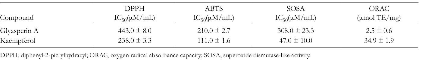
DPPH, diphenyl-2-picrylhydrazyl; ORAC, oxygen radical absorbance capacity; SOSA, superoxide dismutase-like activity.
Experimental
General
The 1H, 13C, DEPT, HSQC, and HMBC NMR were recorded on a Bruker DRX 600 NMR (Bruker Daltonics, Billerica, MA, United States). High resolution electrospray ionisation mass spectrometry (HR-ESI-MS) was determined with liquid chromatograph mass spectrometer ion trap time of flight (LC-MS-IT-TOF) (Shimadzu, Tokyo, Japan). The silica gel column chromatography was conducted using Wakogel C-200. The preparative high-performance liquid chromatography (HPLC) column, Inertsil Prep-octadecyl silica (ODS) (20 mm i.d. × 250 mm, GL-Science) was used. UV spectra were recorded on JASCO-V-530 spectrophotometer. All chemicals were commercially available, such as methanol (MeOH) (Wako, Japan), DPPH and dimethylsulfoxide (DMSO) (Wako, Japan), ethylenediaminetetraacetic acid (Dojindo, Japan), fetal bovine serum (FBS), and Eagle’s minimum essential medium (EMEM, Gibco, United States). The 3-(4,5-dimethyl-thiazol-2-yl)-2,5-diphenyl tetrazolium bromide was from Sigma (United States).
Plant Materials
All Macaranga trees were collected in November 2015 from the Forest Education of Mulawarman University, Samarinda, East Kalimantan, Indonesia. The specimen were identified by Raharjo S.Hut and deposited in the Forest Products Chemistry Laboratory, Department of Forest Product Technology, Faculty of Forestry, Mulawarman University. The plant parts (wood, leaf, and bark) were dried, powdered, and extracted (each 50 g) with 360 mL of MeOH at room temperature for 48 hours. The extract solutions were filtered and concentrated to gain the methanol extracts. The leaves of M. pruinosa were recollected in May 2016 in Samarinda, Indonesia.
Extraction, Fractionation, and Isolation of Active Compound
The leaf M. pruinosa (1 kg) was re-extracted into MeOH at 72 hours in room temperature and yielded 128.02 g. About 10 g of extracts were fractionated with silica gel column chromatography (130 g of Wakogel C-200, 4.14 × 50 cm) and eluted with n-hexane/EtOAc in the ratio of 10:0, 9:1, 8:2, 7:3, 6:4, 5:5, 4:6, 3:7, 2:8, 1:9, and 0:10 and EtOAc/MeOH in the ratio of 8:2, 6:4, 4:6, 2:8, and 0:10 to give 27 fractions (F1-F27). Based on antioxidant (DPPH), tyrosinase activity, TLC, and HPLC results, F11 (204 mg) was subjected to isolate the active compound by preparative HPLC (Inertsil Prep-ODS:20 mm i.d. × 250 mm). Isolation process was eluted with MeOH/H2O (0.1% trifluoroacetic acid, TFA), 95:5, 5 mL/min, yielded 3 fractions and fraction F11-3 (57.5) was focused for purification. The fraction F11-3 was repreparative HPLC with MeOH/H2O (0.1% TFA), 75:25, 5 mL/min to obtain Glyasperin A (8.1 mg).
Antioxidant Assays
Radical Scavenging (Diphenyl-2-Picrylhydrazyl)
The sample was dissolved in DMSO and used for the actual experiment at 30 times dilution. The assay was performed as previously described. 11 Ascorbic acid/Vitamin C and Kaempferol were used as a positive control.
Oxygen Radical Absorbance Capacity
Samples were directly dissolved in acetone/water/acetic acid (70:29.5:0.5, v/v/v) and diluted with 75 mM potassium phosphate buffer (pH 7.4) for analysis. Trolox, fluorescein sodium (FL), and (2,2’-Azobis(2-amidinopropane) dihydrochloride) [AAPH] solutions were prepared with 75 mM phosphate buffer (pH 7.4). The ORAC assay was performed as described by Ou et al 12 and Kaempferol was used as a positive control.
ABTS Radical Cation Decolorization
The working solution of ABTS was prepared with 5 mL of 7 mM ABTS solution and 88 µL of 140 mM potassium persulfate.The ABTS assay was performed as described previously. 13 The results were calculated in the same way as for the DPPH and expressed in terms of Trolox equivalent antioxidant capacity (TEAC µg/mg). Kaempferol was used as a positive control.
Superoxide Dismutase-Like Activity
Samples were added to the (2-(2-methoxy-4-nitrophenyl)-3-(4-nitrophenyl)-5-(2,4-disulfophenyl)-2H-tetrazolium) [WST] working solutions (200 µL) containing 2-(4-iodophenyl)-3-(4-nitrophenyl)-5-(2, 4-disulfophenyl)-2H-tetrazolium in 50 mM carbonate buffer (pH 10.2). This assay was performed as described previously 13 and Kaempferol was used as a positive control.
Tyrosinase Enzyme Assay
Although mushroom tyrosinase differs somewhat from the other sources, this fungal source was used for the present experiment due to its ready availability. The tyrosinase activity was determined with the method as previously described. 11 Kojic acid was used as a positive control.
Determination of Melanin Content and Cell Viability in B16 Melanoma Cells
A mouse melanoma cell line, B16, was obtained from RIKEN Cell Bank. The cells were maintained in EMEM supplemented with 10% (v/v) FBS and 0.09 mg/mL theophylline. The cells were incubated at 37°C in a humidified atmosphere of 5% CO2. These assays for determining melanin content and cell viability were determined as described by Arung et al. 11 Arbutin was used as a positive control.
Footnotes
Declaration of Conflicting Interests
The author(s) declared no potential conflicts of interest with respect to the research, authorship, and/or publication of this article.
Funding
The author(s) disclosed receipt of the following financial support for the research, authorship, and/or publication of this article: This work was financially supported by Islamic Development Bank Grant for Mulawarman University (No. 137/UN17.11/PL/2019).