
Abstract
We explored the potential application of sulforaphane against influenza A virus and elucidated the underlying cytopathic effect (CPE) and cytotoxicity. In the present study, 2 sulforaphane products were used to investigate the CPEs on influenza A virus replication in Madin-Darby canine kidney cells and to conduct a cytotoxicity test. One was the standard sample and the other was extracted from broccoli seeds. The 2 products of sulforaphane were each diluted to different concentrations. The results indicated that sulforaphane possessed antiviral activity against influenza A/WSN/33 (H1N1) virus, and the standard sulforaphane sample showed biological activity against influenza virus with low cytotoxicity at concentrations of 6.25 to 12.5 μM. The same phenomenon was observed with a broccoli seed extract concentration of sulforaphane of 6.25 μM. Both samples displayed higher cytotoxicity at 50 μM of sulforaphane, and the extract samples showed stronger cytotoxicity at sulforaphane concentrations of 12.5 to 100 μM compared with the standard, particularly at 100 μM. In conclusion, natural sulforaphane from broccoli seeds showed potential as an agent against influenza A virus infection, and the CPE after treatment with sulforaphane was concentration dependent; a suitable sulforaphane concentration of 6.25 μM is suggested and was demonstrated as effective, with high antiviral activity and low cytotoxicity.
Influenza A virus, a member of the Orthomyxoviridae family, is a major human pathogen that typically causes annual epidemics and occasional pandemics. 1 -3 Furthermore, the recent emergence of highly pathogenic avian and swine flu has become a global issue for humans. 4,5 In the past 100 years, 5 influenza epidemics and pandemics have caused increasing morbidity and mortality worldwide, including H1N1 in 1918, H2N2 in 1957, H3N2 in 1968, H5N1 in 2009, and H7N9 in 2014. Particularly, influenza A (H7N9) causes high mortality in China. 2,6,7 Influenza A virus is an RNA virus with a rough spherical shape. These viruses are negative sense, single-stranded, and segmented with 7 or 8 pieces, and each piece encodes 1 or 2 genes. Two large glycoproteins, hemagglutinin (HA) and neuraminidase (NA), have been identified in influenza A viruses of different subtypes on the surface of the viral envelope. Thus far, 18 types of HA and 11 types of NA have been identified among different influenza viruses. 8,9
HA is a glycol protein expressed on the viral surface along with NA, and these proteins are responsible for the attachment of the viral particle to the host cell through cell surface sialic acid receptors. 3,10 HA subsequently completes the fusion of the viral envelope with the host membrane to release the viral genome into the target cells, initiating infection. NA is an enzyme that cleaves the sialic acid residue tethering the progeny virus and detaches it from infected cells, accomplishing virus propagation. 10,11 Currently, 2 main strategies, using vaccination and anti-influenza drugs, are widely employed. However, effective vaccination may require sustained reproduction to match the antigenic changes of viruses, implying that it is almost impossible to produce efficient and timely vaccines to control influenza outbreaks. 12,13 Currently, 2 classes of anti-influenza drugs, amantadine and rimantadine, have been developed for the interruption of certain processes of influenza infection by targeting either the M2 channel or NA. 3,12 Additionally, oseltamivir (OSV) and zanamivir target NA protein by the inhibition of its enzymatic activity, rendering the tethered progeny virus unable to escape from the host cells. 14 However, resistant variants have continued to emerge from patients after treatment, regardless of both classes of drugs and other drugs, making it urgent to identify novel anti-influenza drugs with safe and effective activity. 3,15 Sulforaphane [1-isothiocyanato-4-(methylsulfinyl) butane] is an isothiocyanate produced by the hydrolysis of glucoraphanin-rich broccoli (Brassica oleracea var. italica). 16 -19 Epidemiological studies have shown that sulforaphane exhibits anticancer activity, cardiovascular disease prevention, hypotensive effects, and myopia prevention. 20,21 The anticancer activities of sulforaphane have been widely demonstrated and studied in many cancers, including those of the lung, 22 stomach, 19 liver, 23 colon, 24 breast, 25 prostate, 20 and bladder. 26 This mechanism of sulforaphane is attributed to its induction of phase II detoxification enzymes and prevention of the generation of phase I detoxification enzymes and mutagenesis. 27,28
Many studies have reported that sulforaphane plays a key role in sulforaphane preconditioning of the Nrf2 defense pathway, which protects cerebral vasculature against blood-brain barrier disruption, neurological deficits in stroke, and tumor growth and spread. 29 Additionally, in the past 30 years, studies have shown that sulforaphane plays an important role in regulation and as an inducer of Nrf2 signaling and efficacy as an inhibitor of carcinogenesis, as well as preventing infectious disease, cardiovascular disease, and recently new medical areas. 30 Measures of the Nrf2 pathway response and function serve as guideposts for the optimization of dose, schedule, and formulation as clinical trials with sulforaphane and broccoli-based preparations have become more commonplace and more rigorous in design and implementation. 3,12 Thus, it is necessary to investigate the response of standard sulforaphane and the broccoli extract sulforaphane against HA cells, which can aid in new drug research and support human health via the consumption of broccoli. 19,22,31 Thus far, few reports have shown whether sulforaphane could prevent influenza virus infection; therefore, it is necessary to explore this issue. In the present study, influenza A/WSN/33(H1N1) virus was selected to examine sulforaphane in cytopathic effect (CPE) reduction assays and cytotoxicity tests, which are beneficial for research on influenza prevention, the development of new drugs, and epidemiology studies.
In the present study, we selected broccoli seeds with high sulforaphane content as material for extracting sulforaphane. The sulforaphane content in broccoli seeds was 5512.63 mg/L DW, as detected by HPLC, which is a high content in Brassica. 32,33 The chromatography of sulforaphane in the standard and extracted samples showed that the system was effective and stable. The linearity was good within the range of 5.0 to 300.0 mg/L, and the linear equation was Y = 2.76 × 10⁻⁴ X −0.73 (R 2 = 0.9998), where the X-axis represents the peak area and the Y-axis represents the concentration (mg/L).
The HPLC method, with low-temperature working conditions and good stability, has been widely used for the determination of sulforaphane and glucosinolates and their hydrolysis products. 33,34 Sulforaphane, the second product derived from glucoraphanin, is not stable and easily changes into either nitrile material or other compounds at high temperatures or under different chemical conditions. 33 Thus, GC and GC-MS are not suitable for the determination of sulforaphane or glucosinolates. Most studies have successfully detected sulforaphane in cruciferous vegetables, particularly in B. oleracea, with HPLC and HPLC-MS methods. Sulforaphane is particularly rich in broccoli; the ripe seed contains the highest levels, followed by seedlings, buds, florets and stems, and leaves, and almost no sulforaphane has been detected in the roots. 33 Thus, HPLC and HPLC-MS have become typical and simple determination methods for sulforaphane. 33,35
Initially, we examined the cytotoxicity of sulforaphane in Madin-Darby canine kidney (MDCK) cells by using the CellTiter-Glo® assay. Culture medium containing 0.5% dimethyl sulfoxide (DMSO) served as a negative control. The compound showed cytotoxicity in MDCK cells at concentrations of more than 25 µM, while the compounds did not show significant cytotoxicity to MDCK cells at concentrations of less than 12.5 µM (Figure 1). As shown in Figure 1, concentrations of 6.25 and 12.5 µM of sulforaphane showed significant bioactivity against influenza A virus, and the cytotoxicity in MDCK cells was not significantly different from that of the control.

The induction of viral resistance was examined by continuous sulforaphane treatment. Plaque formation was observed at different concentrations of sulforaphane (6.25, 12.5, 25.0, 50.0, and 100 µM) by microscopy. (a) Standard, (b) the broccoli seed extract. Madin-Darby canine kidney cells were infected with influenza A/WSN/33 (MOI = 0.01) and treated with sulforaphane and dimethyl sulfoxide. At 40 hours postinfection, the supernatants were collected and used for infection in the next round of investigation. Virus yields of mock-treated cells were set at 100%.
CPE screening, an assay for measuring the damage to host cells during virus invasion, was utilized to screen and identify compounds that display a reduction in the CPE on influenza A/WSN/33 virus. 2 We found that 6.25 and 12.5 µM concentrations of standard sulforaphane showed significant bioactivity against influenza A virus (Figure 1a), and the cytotoxicity to MDCK cells was not significantly different from that of the control. The reduction in CPE was confirmed by direct microscopic observation, which detected far less CPE than in the DMSO control (Figure 1). In addition, this compound exerts a well-defined dose-dependent response against the A/WSN/33 virus based on plaque formation (Figure 1). This is an alternative assay for the evaluation of potency, with an EC50 of approximately 6.25 µM, which is almost 2-fold lower than that of OSV. 3,36 The same effect was observed at a concentration of 6.25 µM of sulforaphane seed extract (Figure 1b), consistent with that of the standard sulforaphane.
According to microscope observations, there were different reflections of MDCK cells alone and those infected with virus to different gradient concentrations of sulforaphane diluted with DMSO based on the same magnification (Figure 1). MDCK cells with virus showed a greater reduction in CPE at sulforaphane concentrations of 100 and 50 µM than that of standard with more transparent cells. The extract from broccoli seed showed higher CPE reduction at sulforaphane concentrations of 100, 50, and 25 µM, and both samples demonstrated that sulforaphane could prevent influenza A virus, especially at low concentrations of 12.5 and 6.25 µM, compared with the control without virus (DMSO).
The most characteristic feature of sulforaphane is its high chemical reactivity due to the electrophilicity of the central carbon of the isothiocyanate (-N=C=S) group, 37 which readily reacts with sulfur-, nitrogen-, and oxygen-centered nucleophiles. 38 Chemical modification of the sensor cysteine of KEAP1 by sulforaphane impairs its substrate adaptor function, leading to Nrf2 accumulation and the enhanced transcription of Nrf2-dependent genes. 38 These genes have antioxidant response elements (AREs) in their upstream regulatory regions, which are the sites of binding Nrf2 as a heterodimer with a small Maf transcription factor. 39
Nrf2-dependent genes encode multiple functionally diverse enzymes and other proteins with cytoprotective activities. 29,30 Several studies have indicated that there is an inverse relationship between the levels of Nrf2 expression and the viral entry and replication, and an attractive therapeutic intervention demonstrated that supplementation with Nrf2-activating antioxidants inhibits viral replication in human NEC. 29,40 Sulforaphane could increase the accumulation of Nrf2; therefore, additional studies are needed to explore the relation between the virus and standard sulforaphane or its extract from broccoli. 21,40
Studies have reported that glucoraphanin is also taken up from the gut to the liver where it is interconverted to its reduced glucosinolate analog, glucoerucin, while sulforaphane is converted to its corresponding reduced isothiocyanate analog, erucin [1-isothiocyanato-4-(methylthio) butane]. 41,42 Glucoerucin and glucoraphanin have poor bioavailability; thus, the bioavailability found in humans or other mammals is typically due to sulforaphane in a specific test. 43 These findings suggested that sulforaphane at concentrations of 6.25 and 12.5 µM were good for living cells and effective against influenza A virus. Higher cytotoxicity was observed at concentrations higher than 25.0 µM in both the standard and extracted samples (Figure 1). Moreover, the extract contained more toxic compounds from the extraction process than the standard, which should be investigated in future research.
We examined the inhibitory activity of the test compounds against virus replication in MDCK cells using the influenza A/WSN/33 (H1N1 subtype) virus strain. The results are shown in Figure 2, including OSV as a positive control. The compounds showed significant anti-influenza A/WSN/33 virus activity at 25, 12.5, and 6.25 µM of the standard sulforaphane, and 6.25 µM of the sulforaphane from broccoli seed extract (Figure 2) (P < 0.05).
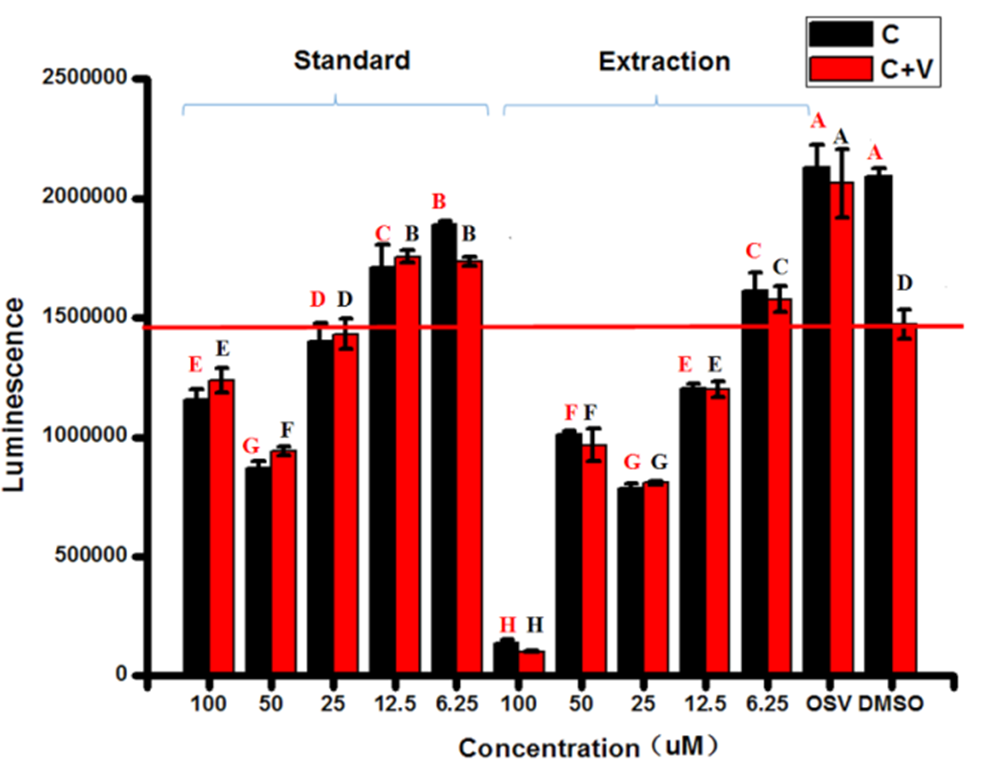
The cytotoxicity toward influenza A virus in Madin-Darby canine kidney cells treated with different concentrations of sulforaphane from the standard and extract samples. The capital letters show significant differences at the level of 0.01 based on one-way ANOVA. Capital letters shown in red and black represent parallel comparisons of black bars and red bars, respectively
The MDCK cells without the influenza A virus (control) indicated that DMSO and OSV showed no significant cytotoxic activity on MDCK cells (Figure 2), whereas 6.25 µM sulforaphane of the standard slightly reduced the activity of MDCK cells maintaining 90.2% of initial cell numbers (DMSO). Notably, 12.5 µM standard sulforaphane and 6.25 µM sulforaphane in the extract from broccoli seed showed the same level of activity, maintaining 81.7% and 77.0% of initial cell numbers, respectively. A concentration of 25.0 µM standard sulforaphane maintained 67.0% of initial cell numbers, and both 100 µM standard sulforaphane and 12.5 µM sulforaphane in the extract from broccoli seed showed the same level of activity (57.5% and 55.3%, respectively, responding to initial cell numbers). This result suggested that sulforaphane is effective against influenza (EC50 > 12.5 µM). The concentration of 50.0 µM sulforaphane in the extract of broccoli seed was better than 50.0 µM sulforaphane standard and 25.0 µM sulforaphane in the extract of broccoli seed with regard to cytotoxic activity for MDCK cells. The 100.0 µM concentration of sulforaphane in the extract from broccoli seed showed strong cytotoxic activity toward MDCK cells, maintaining only 6.5% of initial cell numbers.
The positive OSV control treatments showed that the numbers of MDCK cells infected with influenza A virus and treated with the OSV positive control (Figure 2), 6.25 µM sulforaphane standard and in the extract samples, and 12.5 µM sulforaphane standard were higher than the numbers of MDCK cells treated with DMSO (negative control), suggesting that sulforaphane at a concentration of 6.25 µM from both standard and broccoli extract showed better bioactivity against influenza A virus in MDCK cells. Standard and extract samples showed 19.2% and 17.8% increased cell numbers, respectively, compared with DMSO-treated cells, and the positive control increased cell numbers by 40.0%. The same function against virus was also found at 12.5 and 6.25 µM sulforaphane, while 25 µM sulforaphane standard had no significant effect on MDCK cells infected with influenza A virus compared with the negative control. Additionally, 100 µM of sulforaphane standard and 12.5 µM sulforaphane in the extract from broccoli seed showed the same effect, maintaining 84.0% and 81.4% of initial cells (DMSO), respectively. Moreover, 50 µM sulforaphane standard or in the extract samples maintained 65.6% and 63.9% of the initial cells (DMSO), respectively, and 25.0 µM sulforaphane standard could maintain 55.0% of the initial cells, whereas 100 µM sulforaphane in the extract only maintained 7% of the initial cells, suggesting strong cytotoxicity to MDCK cells.
During the hydrolysis of glucoraphanin, more sulforaphane product is catalyzed by myrosinase at neutral or high pH, in the presence of Zn2+ at low temperature. In contrast, at acidic pH, in the presence of Fe2+ or Cu2+, and at high temperature, nitrile will be favored. 44,45 According to the observed effects of the sulforaphane standard on MDCK cells, we concluded that sulforaphane was the main component, but there might be a small amount of nitrile or residual product(s) from broccoli seeds in the extract, which could be present in samples of a higher concentration, causing toxicity to MDCK cells (Figure 2). Moreover, many studies have demonstrated and validated that sulforaphane is readily absorbed in humans or mammals and is rapidly eliminated, and more than 70% of the administered dose of sulforaphane can be recovered as thiol conjugates in the urine with a biological half-life of only a few hours, providing further evidence of sulforaphane with no significant toxicity to animals and mammals. 46 Additionally, according to the cytotoxicity test activity of influenza A virus treated with different concentrations of sulforaphane from the standard and extract samples, except for the 100 µM concentration of sulforaphane derived from the extract with higher cytotoxicity to MDCK cells, low concentrations of sulforaphane (6.25–50 µM) from the standard and the extract showed lower cytotoxicity to MDCK cells (C and C+V). Moreover, some additional chemical compounds in the sulforaphane extract contributed to the cytotoxicity to MDCK cells.
This result suggested that the cytotoxicity test activity of influenza A virus treated with different concentrations of sulforaphane in MDCK cells demonstrated concentration dependence, and the concentrations of 6.25 to 12.5 µM based on the standard were effective for MDCK cell activity with no obvious cytotoxicity. Additionally, the purification methods, such as preparative liquid chromatography and DEAE-Sephacel, could be used for better purification of sulforaphane before application in food engineering. 44,45
HA represents an attractive target for discovering new anti-influenza agents because of its popular role in host cell attachment and fusion processes. 10 Recent reports reveal that several compounds with small molecules can interfere with the HA functions by hindering one or both functions. The major function is binding to the receptor-binding site and competing with sialic acids, such as triterpenoids, 2,12 and sialic acid mimetic peptides. 8 Recently, arbidol has been commercialized in China, 4,47 and stachyflin derivatives, CL-61917, polyphenols, BMY 27709, and some others have recently been explored. 48
The Nrf2 signaling pathway can regulate N600 genes, of which N200 encodes cytoprotective proteins that are also associated with inflammation, cancer, neurodegenerative diseases, and other major diseases. Recently, Nrf2 expression was shown to modify influenza A entry and replication in nasal epithelial cells, which provided good cross evidence for exploring the function of sulforaphane in anti-influenza A virus. 21,29 Recently, several studies have focused on extracting sulforaphane from broccoli seeds and sprouts to examine its anti-cancer activity, showing that sulforaphane and sulforaphane broccoli extract have the same or similar functions against cancers. 49
Since the recognition of the bioactivity of sulforaphane in 1992, 2,12 several studies have examined its action in cells, animals, and humans. Additionally, increasing evidence has shown that broccoli, particularly as seeds and young sprouts, is a rich source of sulforaphane, and broccoli-based preparations are now used in clinical studies probing their efficacy in health preservation and disease mitigation. 23 The transcription factor Nrf2 is a master regulator of cell survival responses to endogenous and exogenous stressors. 50 Studies have revealed that many putative cellular targets are affected by sulforaphane, although only KEAP1-Nrf2 signaling can be considered a validated target at this time. 21,29 Thus, we propose that sulforaphane plays a role in preventing HA by interfering in the Nrf2 signaling pathway, which provided the premise for the present study. In addition, sulforaphane has therapeutic effects on inflammatory diseases through the Nrf2 signaling pathway. 51,52
Currently, sappanone, an anti-influenza, antiallergic, and neuroprotective medication, is widely distributed in Southeast Asia. 53 Bixin extracted from the seeds of Bixa orellana is used to treat infectious and inflammatory diseases in Mexico and South America, 54 and both sappanone and bixin play medical roles via Nrf2-dependent mechanisms. According to the above-mentioned studies and other recent reports, sulforaphane has activities against cancer, inflammation, cardiovascular disease, and neurological diseases and improves immunity. 27,37 Thus, sulforaphane might play an important role as an anti-influenza virus by increasing the accumulation of Nrf2 factors and decreasing the replication of the virus. These plant compounds activate the Nrf2 signaling pathway mainly in the form of electrophilic materials that modify the cysteine residues of KEAP1, leading to free nuclear Nrf2 binding with the ARE, resulting in Nrf2 accumulation and the activation of the transcription of the corresponding genes. 2,55,56
Experimental
Cells and Materials
MDCK cells were grown in Dulbecco’s modified Eagle medium (DMEM) (Gibco BRL, Inc., Gaithersburg, MD, USA) supplemented with 10% fetal bovine serum (FBS) (PAA Laboratories, Linz, Austria) at 37°C under 5% CO2. Influenza A/WSN/33 (H1N1) virus, provided by HAKE Genetics Co., Ltd., was used in the present study. “WSN” is the acronym for the influenza A/Wilson Smith/1933 (H1N1) neurotropic variant, which was deliberately selected by repeatedly passaging its parent virus, influenza A/Wilson Smith/1933 (H1N1) virus (WS), in mouse brain. The WS virus was isolated in 1933 by Wilson Smith and colleagues from human influenza by inoculating ferrets. 2 Broccoli “B61” seeds were cultured at the Institute of Vegetables and Flowers, Chinese Academy of Agricultural Sciences, and subsequently collected for extracting the bioactive compound sulforaphane.
Chemicals
Sulforaphane standard was purchased from LKT Labs (LKT Laboratories, Inc., St Paul, MN, USA), and the purity was more than 98% (HPLC grade). Methanol, ethyl acetate, and DMSO were obtained from Sigma (Sigma Chemical Company, St Louis, MO, USA). Phosphates were purchased from Beijing Chemical Company (Beijing, China). The standard samples were dissolved in 10 mL of DMSO (Sigma Chemical Company) with a concentration of 1.0 g/L and then serially diluted to concentrations of 6.25, 12.5, 25, 50, and 100 µM for CPE assays and cytotoxicity tests; another concentration gradient was used to determine the linearity of sulforaphane.
HPLC Conditions
The Shimadzu LC-20A HPLC system was equipped with an SPD-20 UV detector and a reverse-phase C18 column (250 × 4.6 mm, 5 µm, Shiseido, Japan). The gradient mobile phase consisted of 5% tetrahydrofuran for pump A and 100% methanol for pump B. The solvent for pump B was initially set at 40%, then linearly changed to 60% by the 10th minute, and subsequently returned to full methanol (100%) after an additional 10 minutes, maintained at 100% for 15 minutes at a flow rate of 0.80 mL/min, and finally returned to the initial condition. The absorbance value was 254 nm, and the column oven temperature 32°C. A total of 10 mg of the sulforaphane standard was dissolved in 10 mL of methanol to generate a dilution series: 5.0, 50.0, 100.0, 200.0, and 300.0 mg/L. The precision of the system was measured by standard peak areas (n = 6, 100 mg/L), and the recovery was defined by adding standard samples (100 mg/L) at known concentrations (5.51, 12.55, 20.43, 45.51, 60.27, 80.69 mg/L) (n = 6). The determination method was performed as previously described, 57 and this method is popularly used for the determination of sulforaphane in plants. 32
Extraction of Sulforaphane
The extraction method was performed according to Liang, 44 with some modifications, as detailed in previous studies. 32,33 A total of 0.5 g of seed powder was homogenized in 15 mL of neutral phosphate buffer (0.1 M and pH 7.0). The homogenate was then transferred to a beaker with stirring for 2 hours, after which 30 mL ethyl acetate was injected. Thirty minutes later, the mixture was centrifuged for 10 minutes at 6000 × g (Thermo, MA, USA). The supernatant in the tubes was collected, and the remaining mixture was transferred to a fresh beaker. Then, 30 mL of ethyl acetate was added to the mixture and treated by the initial process. The same operation was repeated again. Finally, the 3 supernatants were evaporated in a rotavapor (RII, BÜCHI TM, Switzerland) at 35°C. The residue was then dissolved in 10 mL of methanol and filtered through a 0.22 µm (D 13 mm) nylon filter paper (Agela, China). The solution was stored at -20°C until HPLC analysis.
CPE Reduction Assay
This assay was performed as previously described, 2 with some modifications. MDCK cells were seeded onto 96-well plates, incubated overnight, and infected with influenza virus (MOI = 0.1) suspended in DMEM supplemented with 1% FBS, containing test compound and 2 mg/L TPCK-treated trypsin, with a final DMSO concentration of 1% in each well. After 40 hours of incubation, CellTiter-Glo reagent (Promega Corp., Madison, WI, USA) was added, and the plates were read using a plate reader (Tecan Infinite M2000 PRO; Tecan Group Ltd, Mannedorf, Switzerland). The CPE in virus-infected cells was observed through microscopy. MDCK cells were infected with influenza A/WSN/33 (MOI = 0.01) and treated with different concentrations of sulforaphane from the standard and extract samples diluted with DMSO to 6.25, 12.5, 25, 50, and 100 µM. Each treatment was designed in triplicate (n = 3). The postinfection supernatants were collected and used for infection in the next round of investigation. 3
Cytotoxicity Test
Cells grown on 96-well plates overnight were cultured in 1% FBS with increasing amounts of the test compounds for 40 hours, with OSV as a positive control. Cytotoxicity was assessed with the CellTiter-Glo assay as described above. The cells were treated with different concentrations of sulforaphane from the standard and extract samples after 40 hours. The cytotoxicity was measured by the neutral red uptake assay (Tecan Infinite M2000 PRO; Tecan Group Ltd). The cytotoxicity was computed by comparisons to normal cells of wells containing compounds with wells containing DMSO.
Data Analysis
All statistical analyses were performed by using SPSS 12.0. The results are expressed as the means ± standard deviation (SD) from experiments performed in triplicate. The statistical significance between 2 groups was analyzed by Student’s t test, and one-way ANOVA with Duncan multiple comparisons was used in the present study. A P value of <0.05 was regarded as statistically significant.
Footnotes
Declaration of Conflicting Interests
The author(s) declared no potential conflicts of interest with respect to the research, authorship, and/or publication of this article.
Funding
The author(s) disclosed receipt of the following financial support for the research, authorship, and/or publication of this article: The present study was funded by grants from the National Key Research and Development Program of China (2017YFD0101805), the National Nature Science Foundation (31501761), the China Agriculture Research System (CARS-23-A8), and the Science and Technology Innovation Program of the Chinese Academy of Agricultural Sciences (CAAS-ASTIP-IVFCAAS).