Two new megastigmane glycosides, phoebenoside A (1) and phoebenoside B (2) and 4 known compounds, dendranthemoside A (3), lyoniresinol (4), (+)-3-O-L-rhamnopyranoside-5-methoxyisolariciresinol (5), and (+)-lyoniresinol 3α-O-β-D-glucopyranoside (6) were isolated from the leaves of Phoebe tavoyana (Meisn.) Hook. Their chemical structures were elucidated by analyses of their spectroscopic data, and comparison with those reported in the literature.
Phoebe genus (Lauraceae family), evergreen trees and shrubs, is distributed in tropical and subtropical areas.1 There are about 12 Phoebe species, often used to treat skin injuries and headaches, in Vietnam.2 Phytochemistry of Phoebe genus indicated the presence of alkaloids and flavonoids.1 There are few reports about chemical and biological studies related to Phoebe tavoyana.3 Herein, we report the isolation, structural elucidation of 2 new and 4 known compounds from the leaves of P. tavoyana.
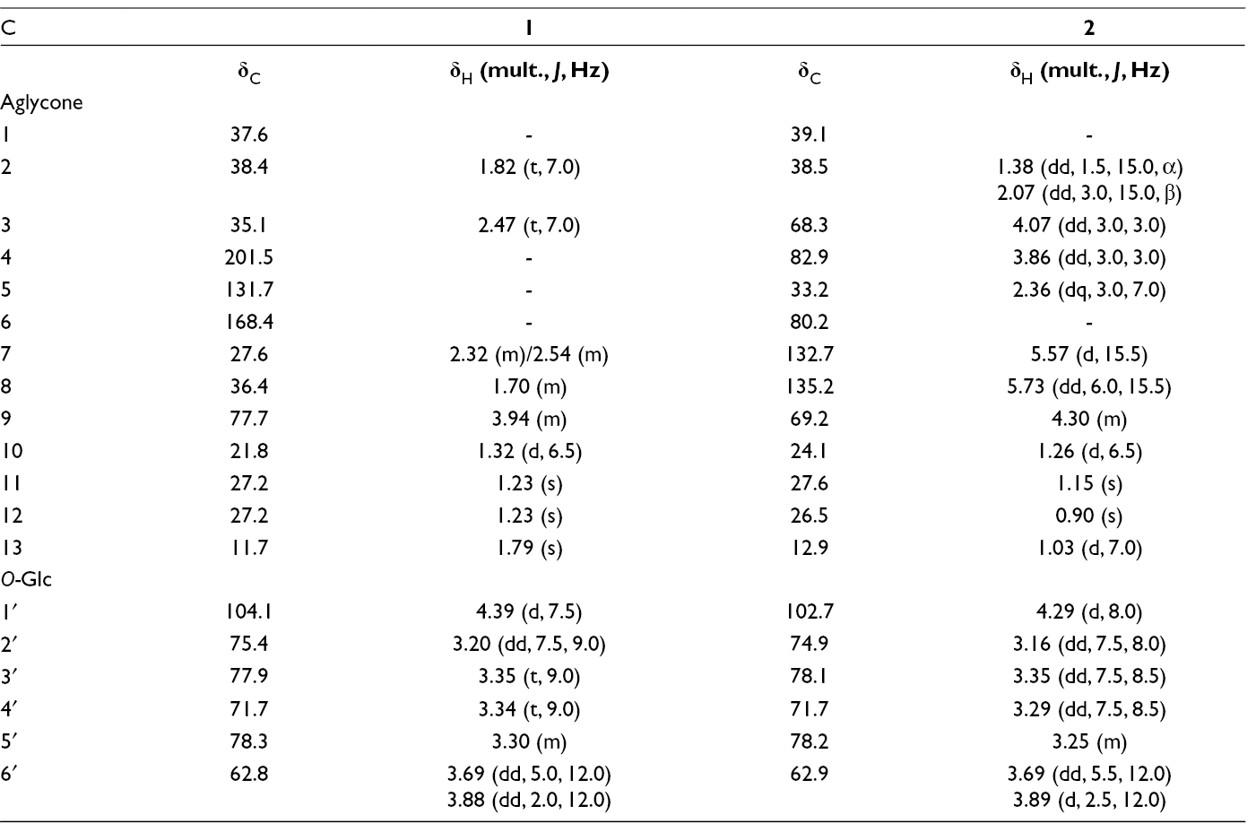
The molecular formula of 1 was determined to be C19H32O7 by high resolution-electrospray ionization-mass spectrometry (HR-ESI-MS) ion at m/z 407.1843 [M+Cl]− (calcd. for C19H32O7Cl, 407.1842). The 1H-nuclear magnetic resonance (NMR) spectrum of 1 showed the signals of 3 tertiary methyl groups at δH 1.23 × 2 and 1.79, 1 secondary methyl group at δH 1.32 (d, J = 6.5 Hz) assigning to a megastigmane aglycone; an anomeric proton at δH 4.39 (d, J = 7.5 Hz) suggesting the presence of 1 sugar unit (Table 1). The 13C-NMR and heteronuclear single quantum correlation (HSQC) spectra of 1 revealed the signals of 19 carbons, including 1 carbonyl, 3 quaternary carbons, 6 methines, 5 methylenes, and 4 methyl carbons. In the 1H-1H correlation spectroscopy (COSY), cross peaks between protons H-2 (δH 1.82) and H-3 (δH 2.47), H-8 (δH 1.70) and H-7 (δH 2.32/2.54)/ H-9 (δH 3.94), and between H-9 and H-10 (δH 1.32) were observed. The heteronuclear multiple bond correlation (HMBC) from H-13 (δH 1.79) to C-4 (δC 201.5)/C-5 (δC 131.7)/C-6 (δC 168.4); from H-11/H-12 (δH 1.23) to C-1 (δC 37.6)/C-2 (δC 38.4)/C-6 (δC 168.4) suggested the positions of an oxo group at C-4 and the double bond at C-5/C-6. The HMBC correlations between H-10 (δH 1.32) and C-8 (δC 36.4)/C-9 (δC 77.7) suggested the oxygenated group at C-9 (Figure 1). Furthermore, acid hydrolysis of 1 revealed D-glucose. The multiplicity of glc H-1′ [δH 4.39 (d, J = 7.5 Hz)] showed the presence of β-d-glucopyranosyl. The HMBC between H-1′ (δH 4.39) and C-9 (δC 77.7) suggested the location of β-d-glucopyranosyl at C-9. The chemical shifts of C-9 (δC 77.7), C-10 (δC 21.8), and C-1′ (δC 104.1) confirmed the 9S configuration by comparing with the corresponding data of (3R,9S)-megastigman-5-en-3,9-diol 9-O-β-d-glucopyranoside [C-9 (δC 77.9), C-10 (δC 21.8), and C-1′ (δC 103.9)] and (3R,9R)-megastigman-5-en-3,9-diol 9-O-β-d-glucopyranoside [C-9 (δC 76.1), C-10 (δC 19.8), and glc C-1′ (δC 102.2)] in the same solvent CD3OD.4 Consequently, the new structure of 1 was determined to be (9S)-hydroxymegastigman-5-en-4-one 9-O-β-d-glucopyranoside and named as phoebenoside A (Figure 1).
The 1H- and 13C-NMR Data for Compounds 1 and 2 in CD3OD.
C
1
2
δC
δH (mult., J, Hz)
δC
δH (mult., J, Hz)
Aglycone
1
37.6
-
39.1
-
2
38.4
1.82 (t, 7.0)
38.5
1.38 (dd, 1.5, 15.0, α)2.07 (dd, 3.0, 15.0, β)
3
35.1
2.47 (t, 7.0)
68.3
4.07 (dd, 3.0, 3.0)
4
201.5
-
82.9
3.86 (dd, 3.0, 3.0)
5
131.7
-
33.2
2.36 (dq, 3.0, 7.0)
6
168.4
-
80.2
-
7
27.6
2.32 (m)/2.54 (m)
132.7
5.57 (d, 15.5)
8
36.4
1.70 (m)
135.2
5.73 (dd, 6.0, 15.5)
9
77.7
3.94 (m)
69.2
4.30 (m)
10
21.8
1.32 (d, 6.5)
24.1
1.26 (d, 6.5)
11
27.2
1.23 (s)
27.6
1.15 (s)
12
27.2
1.23 (s)
26.5
0.90 (s)
13
11.7
1.79 (s)
12.9
1.03 (d, 7.0)
O-Glc
1′
104.1
4.39 (d, 7.5)
102.7
4.29 (d, 8.0)
2′
75.4
3.20 (dd, 7.5, 9.0)
74.9
3.16 (dd, 7.5, 8.0)
3′
77.9
3.35 (t, 9.0)
78.1
3.35 (dd, 7.5, 8.5)
4′
71.7
3.34 (t, 9.0)
71.7
3.29 (dd, 7.5, 8.5)
5′
78.3
3.30 (m)
78.2
3.25 (m)
6′
62.8
3.69 (dd, 5.0, 12.0)3.88 (dd, 2.0, 12.0)
62.9
3.69 (dd, 5.5, 12.0)3.89 (d, 2.5, 12.0)
Chemical structures, key heteronuclear multiple bond correlation (HMBC), correlation spectroscopy (COSY), and nuclear Overhauser effect spectroscopy (NOESY) correlations of compounds 1 and 2.
The molecular formula of 2 was established as C19H34O9 based on its ion at m/z 441.1900 [M+Cl]− (calcd. for [C19H34O9Cl]−, 441.1897) in the HR-ESI-MS spectrum. The 1H-NMR spectrum of 2 exhibited the signals of 4 methyl groups at δH 0.90 (s), 1.03 (d, J = 7.0 Hz), 1.15 (s), and 1.26 (d, J = 6.5 Hz), assigning to a megastigmane and an anomeric proton at δH 4.29 (d, J = 8.0 Hz) suggesting the presence of 1 sugar moiety. The 13C-NMR and HSQC spectra (Table 1) showed the signals of 19 carbons, including, 2 non-protonated carbons, 11 methines, 2 methylenes, and 4 methyl carbons. The HMBC correlations between H-13 (δH 1.03) and C-4 (δC 82.9)/C-5 (δC 33.2)/C-6 (δC 80.2); between H-11 (δH 1.15)/H-12 (δH 0.90) and C-1 (δC 39.1)/C-2 (δC 38.5)/C-6 (δC 80.2); between H-4 (δH 3.86) and C-3 (δC 69.2)/C-6 (δC 80.2)/C-13 (δC 12.9) as well as 1H-1H COSY cross peaks of H-2/H-3/H-4/H-5/H-13 suggested the positions of hydroxyl groups at C-3 and C-6; oxygenated group at C-4 (Figure 1). Acid hydrolysis of 2 revealed D-glucose (see Supporting information). In addition, the location of β-d-glucopyranosyl at C-4 was confirmed by HMBC correlations between H-1′ (δH 4.29) and C-4 (δC 82.9). Moreover, the HMBC correlations from H-10 (δH 1.26) to C-8 (δC 135.2)/C-9 (δC 69.2); from H-7 (δH 5.57) to C-6 (δC 80.2)/C-8 (δC 135.2)/C-9 (δC 69.2), and coupling constant of H-7 and H-8, J = 15.5 Hz) suggested the position of double bond (E geometry) at C-7/C-8 and a hydroxyl group at C-9. The relative configuration of 2 was deduced from analysis of coupling constants and the NOESY spectrum (Figure 1). The small coupling constants of H-2 and H-3 (J = 3.0 Hz); H-3 and H-4 (J = 3.0 Hz) and H-4 and H-5 (J = 3.0 Hz), suggested configurations of both hydroxyl groups at C-3 and C-4 to be axial orientation. The NOESY correlation between H-5 and H-11 indicated that H-5 and C-11 were axial (β-configuration). The hydroxyl group at C-6 was determined to be α-oriented based on the strong NOESY correlations between H-7 and H-5/H-11. Enzymatic hydrolysis of 2 gave aglycone. The aglycone of 2 was found to be identical to (3S,4S,5R,6R,9R)-3,4,6,9-tetrahydroxymegastigman-5-ene (2a), a compound reported from Cinnamomum wilsonii and confirmed by single-crystal X-ray5 by comparing their spectroscopic data and optical rotation. Thus, structure of 2 was elucidated as (3S,4S,5R,6R,9R)-3,4,6,9-tetrahydroxymegastigman-5-ene 4-O-β-d-glucopyranoside and named as phoebenoside B (Figure 1).
The known compounds were elucidated as dendranthemoside A (3),6 lyoniresinol (4),7 (+)-3-O-l-rhamnopyranoside-5-methoxyisolariciresinol (5),8 (+)-lyoniresinol 3α-O-β-d-glucopyranoside (6)9 by analyses of spectroscopic data and comparison with those reported in the literature.
All the isolated compounds were evaluated against 3 human cancer cell lines, HT-29 (human colon cancer), A-549 (human lung cancer), and A-2058 (human melanoma cancer cell) using the MTT assay. However, they did not show any cytotoxic activity (IC50 > 50 µM).
The leaves of P. tavoyana (Meisn.) Hook. were collected at Me Linh, Vinh Phuc province, Vietnam in May, 2016 and identified by Dr Nguyen The Cuong, Institute of Ecology and Biological Resources, VAST. A voucher specimen (NCCT-P59) was deposited at the Institute of Marine Biochemistry.
Extraction and Isolation
The dried leaves of P. tavoyana (3.2 kg) were extracted with hot MeOH (3 × 5 L) using sonicator to yield 200 g of extract. The MeOH extract was suspended in water and successively partitioned with CH2Cl2, ethyl acetate (EtOAc) to obtain CH2Cl2 (PT1), EtOAc (PT2), and water (PT3) fractions. PT3 was chromatographed on a Diaion HP-20 column eluting with water to remove sugars, then increasing concentration of methanol in water (25%, 50%, 75%, and 100%, each 1 L) to obtain 4 fractions, PT3A-PT3D, respectively. PT3A was subjected on a RP-18 column chromatography (CC) using MeOH/water (1/3, v/v) to give 5 fractions, PT3A1-PT3A5. PT3A2 was chromatographed on a Sephadex LH-20 column using MeOH/water (1/1, v/v) to give 3 fractions, PT3A2A-PT3A2C. PT3A2B was chromatographed on a silica gel CC (CH2Cl2/acetone/water, 1/4/0.3, v/v/v) to yield 5 (10.0 mg) and 6 (8.0 mg). PT3A4 was chromatographed on a silica gel CC eluting with CH2Cl2/MeOH/water (2.5/1/0.1, v/v/v) to give 2 fractions, PT3A4A-PT3A4B. PT3A4A was chromatographed on a silica gel CC eluting with EtOAc/MeOH/water (7/1/0.05, v/v/v) to yield 1 (8.8 mg). Compound 2 (20.0 mg) was obtained from PT3A4B on a silica gel CC eluting with CH2Cl2/acetone/water (1/4/0.3, v/v/v). PT3A5 was loaded on a silica gel CC eluting with CH2Cl2/MeOH/water (2.5/1/0.1, v/v/v) to give 3 fractions, PT3A5A-C. Compound 3 (10.0 mg) was obtained from PT3A5A using a RP-18 CC eluting with MeOH/water (1/3, v/v). PT3C was chromatographed on silica gel CC eluting with CH2Cl2/MeOH (8/1, v/v) to give 4 (50.0 mg).
The author(s) declared no potential conflicts of interest with respect to the research, authorship, and/or publication of this article.
Funding
The author(s) disclosed receipt of the following financial support for the research, authorship, and/or publication of this article: This research is funded by Vietnam National Foundation for Science and Technology Development (NAFOSTED) under grant number 104.01-2015.22.
Supplemental Material
References
1.
Kumar SemwalD.Badoni SemwalR.Deepak KumarS.Ruchi BadoniS. Ethnobotany, pharmacology and phytochemistry of the genus Phoebe (Lauraceae). Mini-Rev Org Chem. 2013;10(1):12-26.doi:10.2174/1570193X11310010002
2.
ChiVV. The dictionary of Vietnamese medicinal plants. Hanoi: Hanoi Medicine; 1999.
3.
OmarHB. Chemical constituents of Phoebe grandis (Nees) Merr, Phoebe tavoyana (Meissn.) HK. F., and Actinodaphne sesquipedalis Hook. F. var. glabra and their biological activities. Kuala Lumpur: University of Malaya; 2015.
4.
YamanoY.ShimizuY.ItoM. Stereoselective synthesis of optically active 3-hydroxy-7,8-dihydro-β-ionol-glucosides. Chem Pharm Bull. 2003;51(7):878-882.doi:10.1248/cpb.51.878
5.
ShuP.WeiX.XueYet al. Wilsonols A-L, megastigmane sesquiterpenoids from the leaves of Cinnamomum wilsonii. J Nat Prod. 2013;76(7):1303-1312.doi:10.1021/np4002493
6.
OtsukaH.KamadaK.OgimiC.HirataE.TakushiA.TakedaY. Alangionosides A and B, ionol glycosides from leaves of Alangium premnifolium. Phytochemistry. 1994;35(5):1331-1334.doi:10.1016/S0031-9422(00)94848-9
7.
OhashiK.WatanabeH.OkumuraY.UJIT.KitagawaI. Indonesian medicinal plants. XII. four isomeric Lignan-Glucosides from the bark of Aegle marmelos (Rutaceae. Chem Pharm Bull. 1994;42(9):1924-1926.doi:10.1248/cpb.42.1924
8.
Y-HH.DouD-Q.TereshimaK. Two lignan glycosides from Vitis thunbergii. Heterocycles. 2004;63:871-877.
9.
NhiemNX.KiemPV.MinhCVet al. Diarylheptanoids and flavonoids from Viscum album inhibit LPS-stimulated production of pro-inflammatory cytokines in bone marrow-derived dendritic cells. J Nat Prod. 2013;76(4):495-502.doi:10.1021/np300490v
Supplementary Material
Please find the following supplemental material available below.
For Open Access articles published under a Creative Commons License, all supplemental material carries the same license as the article it is associated with.
For non-Open Access articles published, all supplemental material carries a non-exclusive license, and permission requests for re-use of supplemental material or any part of supplemental material shall be sent directly to the copyright owner as specified in the copyright notice associated with the article.