Five quassinoids and 2 alkaloids were isolated from the roots of Eurycoma longifolia. The structures of 3 new quassinoids, eurycomalide F, G, and H (1-3), were elucidated using a variety of spectroscopic methods. The known compounds were identified as eurycomalide A (4), laurycolactone B (5), 5-methoxycanthin-6-one (6), and canthin-6-one (7). Among the isolated compounds, canthin-6-one (7) exhibited the strongest inhibitory effect on nitric oxide production (IC50 = 16.9 μM). 5-Methoxycanthin-6-one (6) and eurycomalide F (1) exhibited weak inhibition with IC50 values of 23.4 and 32.7 μM, respectively.
Eurycoma longifolia Jack (Simaroubaceae) has been used in Southeast Asia to treat sexual dysfunction, constipation, exercise fatigue, fever, loss of libido, aging, stress, malaria, osteoporosis, diabetes, high blood pressure, cancer, leukemia, and glandular swelling.1 Quassinoids, canthin-6-one alkaloids, β-carboline alkaloids, biphenyl neolignans, and triterpenes have been identified as major phytochemical compounds in E. longifolia.2,3 This article describes the isolation and identification of 3 new quassinoids (1-3), together with eurycomalide A (4),4 laurycolactone B (5),5 5-methoxycanthin-6-one (6),6 and canthin-6-one (7)7 from a methanol extract of E. longifolia roots.
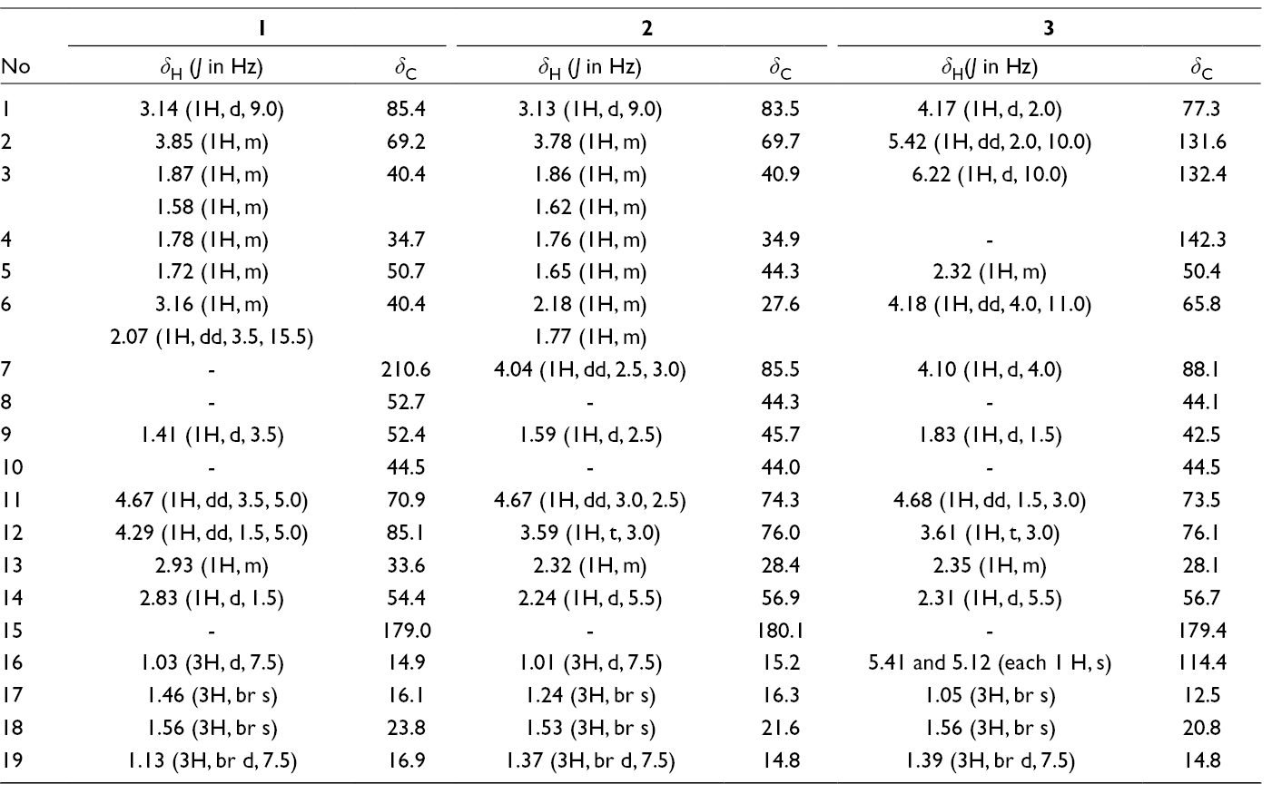
Eurycomalide F (1) was obtained as colorless solid. The molecular formula C19H28O6 for 1 was established from an ion peak at m/z 353.1968 [M + H]+ in its high resolution electrospray ionization mass spectrometry (HR-ESI-MS) spectrum. The 1H nuclear magnetic resonance (NMR) spectrum showed 2 secondary methyl doublets at δH 1.03 (3H, br d, J = 7.5 Hz, CH3-16) and 1.13 (3H, br d, J = 7.5 Hz, CH3-19) and 2 tertiary methyl singlets at δH 1.56 (3H, br s, CH3-18) and 1.46 (3H, br s, CH3-17). The 13C NMR and heteronuclear single quantum correlation (HSQC) spectra indicated the presence of 4 methyl, 2 methylene, 9 methine, and 4 quaternary carbon atoms, including 1 carboxylic group and 1 carbonyl group (Table 1). These data were similar to those of eurycomalide A4 with differences attributed to the presence of a methine group (δC 44.3, C-5) and a methylene group (δC 27.6, C-6) instead of double bonds.4 Thus, compound 1 was considered to be a hydrogenated derivative of 4, which was confirmed by heteronuclear multiple bond correlation (HMBC) spectral analyses (Figure 1). The relative configurations of this compound were confirmed through nuclear Overhauser effect spectroscopy (NOESY) experiments. The nuclear Overhauser effect (NOE) cross peaks between H-1 (δH 3.14) and H-5 (δH 1.72), and between H-9 (δH 1.41) and H-12 (δH 4.29), indicated a β-orientation of hydroxyl groups at C-1 and C-12. An α-orientation at 2-OH was supported by a NOESY correlation between H-2 (δH 3.85) and CH3-17 (δH 1.56) and a large coupling constant between H-1 and H-2 (J = 9.0 Hz). The couplings between CH3-16 (δH 1.03) and CH3-17, and H-13 (δH 2.93) and H-18 (δH 1.56), respectively, confirmed that CH3-16 was β-oriented and CH3-19 was α-oriented. Together, these data confirm the structure of eurycomalide F (1).
NMR Spectroscopic Data of Compounds 1 to 3.
No
1
2
3
δH (J in Hz)
δC
δH (J in Hz)
δC
δH(J in Hz)
δC
1
3.14 (1H, d, 9.0)
85.4
3.13 (1H, d, 9.0)
83.5
4.17 (1H, d, 2.0)
77.3
2
3.85 (1H, m)
69.2
3.78 (1H, m)
69.7
5.42 (1H, dd, 2.0, 10.0)
131.6
3
1.87 (1H, m)
40.4
1.86 (1H, m)
40.9
6.22 (1H, d, 10.0)
132.4
1.58 (1H, m)
1.62 (1H, m)
4
1.78 (1H, m)
34.7
1.76 (1H, m)
34.9
-
142.3
5
1.72 (1H, m)
50.7
1.65 (1H, m)
44.3
2.32 (1H, m)
50.4
6
3.16 (1H, m)
40.4
2.18 (1H, m)
27.6
4.18 (1H, dd, 4.0, 11.0)
65.8
2.07 (1H, dd, 3.5, 15.5)
1.77 (1H, m)
7
-
210.6
4.04 (1H, dd, 2.5, 3.0)
85.5
4.10 (1H, d, 4.0)
88.1
8
-
52.7
-
44.3
-
44.1
9
1.41 (1H, d, 3.5)
52.4
1.59 (1H, d, 2.5)
45.7
1.83 (1H, d, 1.5)
42.5
10
-
44.5
-
44.0
-
44.5
11
4.67 (1H, dd, 3.5, 5.0)
70.9
4.67 (1H, dd, 3.0, 2.5)
74.3
4.68 (1H, dd, 1.5, 3.0)
73.5
12
4.29 (1H, dd, 1.5, 5.0)
85.1
3.59 (1H, t, 3.0)
76.0
3.61 (1H, t, 3.0)
76.1
13
2.93 (1H, m)
33.6
2.32 (1H, m)
28.4
2.35 (1H, m)
28.1
14
2.83 (1H, d, 1.5)
54.4
2.24 (1H, d, 5.5)
56.9
2.31 (1H, d, 5.5)
56.7
15
-
179.0
-
180.1
-
179.4
16
1.03 (3H, d, 7.5)
14.9
1.01 (3H, d, 7.5)
15.2
5.41 and 5.12 (each 1 H, s)
114.4
17
1.46 (3H, br s)
16.1
1.24 (3H, br s)
16.3
1.05 (3H, br s)
12.5
18
1.56 (3H, br s)
23.8
1.53 (3H, br s)
21.6
1.56 (3H, br s)
20.8
19
1.13 (3H, br d, 7.5)
16.9
1.37 (3H, br d, 7.5)
14.8
1.39 (3H, br d, 7.5)
14.8
Structures, and key HMBC (→) and NOESY (↔) correlations of 1 to 3.
Compound 2 was obtained as a colorless solid with the molecular formula C19H30O6, as deduced from a molecular ion peak at m/z 355.2129 [M + H]+ in its HR-ESI-MS spectrum. The NMR data of 2 closely resembled those of 1 with 19 carbon signals, including 2 secondary methyl and 2 tertiary methyl groups, which indicate the same quassinoid skeleton. The replacement of the carbonyl signal in 1 by an oxymethine signal (δC 85.5) and a 2 Da difference in molecular weight suggest hydroxylation of the ketone group at C-7. Heteronuclear multiple bond correlation correlations between H-6, H-9, and CH3-18 to C-7 confirm the location of hydroxylation (Figure 1). However, a larger coupling constant of H-14 (5.5 Hz) compared to that of 1 (1.5 Hz), and an HMBC correlation between H-7 (δH 4.04) and C-15 (δC 180.1), suggest lactone cyclization at C-15 and C-7.8 Similarities in the NOESY correlation patterns of 2 and 1 were used to determine the relative configurations of 2. Furthermore, NOE coupling between H-7, H-14 (δH 2.24), and CH3-18 (δH 1.53) suggest β-orientations for H-7 and H-14. Therefore, the structure of 2 was determined as shown and the compound was named eurycomalide G.
Eurycomalide H (3) was obtained as a white solid. The HR-ESI-MS spectrum of 3 revealed a molecular ion peak at m/z 351.1813 [M + H]+, corresponding to the molecular formula C19H27O6. The 1H NMR spectrum showed 1 secondary methyl doublet at δH 1.39 (3H, br d, J = 7.5 Hz, CH3-19), 2 tertiary methyl singlets at δH 1.56 (3H, br s, CH3-18) and 1.05 (3H, br s, CH3-17), and 4 olefinic proton signals at δH 6.22 (1H, d, J = 10.0 Hz, H-3), 5.42 (1H, dd, J = 2.0, 10.0 Hz, H-2), 5.41, and 5.12 (each 1 H, s, H-16a and 16b). 13C NMR and HSQC spectra showed 19 signals including 1 carboxylic group (δC 179.4), 1 exomethylene (δC 142.4/114.4), 1 double bond (δC 131.6/132.4), 5 oxygenated methines at δC 77.3 (C-1), 65.8 (C-6), 88.1 (C-7), 73.5 (C-11), and 76.1 (C-12), 4 sp3 methines, and 2 sp3 quaternary carbon atoms. By comparing these NMR data with those reported for E. longifolia, the structure of 3 resembled that of the Δ4(18)-isomer of longilactone for rings B, C, and D.8,9 They differed in the presence of a double bond instead of a C-2 oxygenated methine and C-3 methylene, which was confirmed by HMBC correlations between H-2 (δH 5.42) and C-3 (δC 132.4), C-4 (δC 142.4), and C-10 (δC 44.5), as well as H-3 (δH 6.22) and C-1 (δC 77.3), C-4, C-5 (δC 50.4), and C-18 (δC 114.4) (Figure 1). The relative configurations of 3 was deduced from NOESY correlations shown in Figure 1. Accordingly, OH-1, H-7, CH3-17, CH3-18, OH-11, H-13, and H-14 were β-oriented, while H-5, OH-6, H-9, and CH3-19 were α-oriented.
All of the isolated compounds were evaluated for their inhibition of nitric oxide (NO) production. The most active compound was canthin-6-one (7), followed by 5-methoxycanthin-6-one (6), with the IC50 values of 16.9 and 23.4 µM, respectively. Among the isolated quassinoids, only eurycomalide F (1) exhibited a weak effect (IC50 = 32.7 µM).
Experimental
General
Optical rotation, JASCO P-2000 digital polarimeter; IR, Tensor 37 FT-IR spectrometer. NMR, Bruker AM500 FT-NMR Spectrometer. HR-ESI-MS, API Q-STAR PULSAR I of Applied Biosystem. Absorbance of bioassay solutions, xMark microplate spectrophotometer (Molecular Devices, CA). Thin-layer chromatography was performed using precoated Kiesel gel 60 F254 (Merck) and visualized by UV light 254 nm and 10% H2SO4 reagent with heat.
Plant Material
The roots of E. longifolia were collected at Quang Ninh province, Vietnam in March, 2017. The sample was identified by Dr. Nguyen The Cuong, Institute of Ecology and Biological Resources, VAST. The voucher specimens (BB-01) were deposited at the Advanced Center for Bio-organic Chemistry.
Extraction and Isolation
The air-dried and powdered roots of E. longifolia (2 kg) were extracted with methanol (5 L × 3 times) at room temperature for 24 hours. The combined extracts were evaporated under reduced pressure to give the crude extract (82.5 g), which was then resuspended in water (2 L) and successively with n-hexane and ethyl acetate (each 1 L × 3 times) to obtain n-hexane (17.5 g) and ethyl acetate (47.4 g) extracts, respectively. The later was chromatographed on a silica gel column using mobile phase of a gradient of 0% to 100% methanol in dichloromethane to obtain 6 fractions EL1-6. Fraction EL2 was subjected to a silica gel column eluted by dichloromethane-methanol 30:1 (v/v) to obtain 6 (2.6 mg) and 7 (4.4 mg). Compound 5 (7.8 mg) was purified from EL3 by a Sephadex LH20 column eluted by dichloromethane-methanol 2:1 (v/v). Fraction EL4 was passed through a silica gel column using dichloromethane-acetone 4:1 (v/v) as eluent to obtain 1 (13.3 mg) and 4 (4.0 mg). Repeated silica gel column (eluted with dichloromethane-methanol 5:1) and C-18 column (eluted with methanol-water 1:1) applied for EL5 to give 3 (4.1 mg). Compounds 2 (5.5 mg) was obtained from EL6 by a C-18 column eluted by methanol-water 1:1 (v/v).
The effects of compounds on the NO production in LPS-stimulated RAW264.7 cells were evaluated as previously described.10 Cardamonin was used as a positive control (IC50 = 2.80 µM). The MTT assay showed that all compounds had no significant toxicity at their effective doses for the NO inhibition (data not shown).
Footnotes
Declaration of Conflicting Interests
The author(s) declared no potential conflicts of interest with respect to the research, authorship, and/or publication of this article.
Funding
The author(s) disclosed receipt of the following financial support for the research, authorship, and/or publication of this article: This research is funded by Vietnam Academy of Science and Technology under grant numbers VAST04.03/17-18 and VAST.TD.TP.02/16-18.
Supplemental Material
References
1.
BhatR.KarimAA. Tongkat Ali (Eurycoma longifolia Jack): a review on its ethnobotany and pharmacological importance. Fitoterapia. 2010;81(7):669-679.doi:10.1016/j.fitote.2010.04.006
2.
RehmanSU.ChoeK.YooHH. Review on a traditional herbal medicine, Eurycoma longifolia Jack (Tongkat Ali): its traditional uses, chemistry, evidence-based pharmacology and toxicology. Molecules. 2016;21(3):331.doi:10.3390/molecules21030331
3.
BräuerP.AnielskiP.SchwaigerSet al. In vitro metabolism of selected bioactive compounds of Eurycoma longifolia root extract to identify suitable markers in doping control. Drug Test Anal. 2019;11(1):86-94.doi:10.1002/dta.2449
4.
KuoPC.SuR.DamuAG.WuTS. Eurycomalin A, a new dimeric dihydrobenzofuran from Eurycoma longifolia. Heterocycles. 2004;63(9):2123-2129.
5.
SuongNN.BhatnagarS.PolonskyJ.VuilhorgneM.PrangéT.PascardC. Structure of laurycolactone A and B, new C18 - quassinoids from Eurycoma longifolia and revised structured of Eurycomalactone (X - ray analysis). Tetrahedron Lett. 1982;23(49):5159-5162.doi:10.1016/S0040-4039(00)85785-8
6.
ReadelKE.SeiglerDS.YoungDA. 5-methoxycanthin-6-one from Leitneria floridana (Simaroubaceae). Biochem Syst Ecol. 2003;31(2):167-170.doi:10.1016/S0305-1978(02)00074-1
7.
KoikeK.OhmotoT. Carbon-13 nuclear magnetic resonance study of canthin-6-one alkaloids. Chem Pharm Bull. 1985;33(12):5239-5244.doi:10.1248/cpb.33.5239
8.
ItokawaH.QinX-R.MoritaH.TakeyaK.IItakaY. Novel quassinoids from Eurycoma longifolia. Chem Pharm Bull. 1993;41(2):403-405.doi:10.1248/cpb.41.403
9.
MoritaH.KishiE.TakeyaK.ItokawaH.TanakaO. New quassinoids from the roots of Eurycoma longifolia. Chem Lett. 1990;19(5):749-752.doi:10.1246/cl.1990.749
10.
HoiTM.ThaiTV.HaCT.AnhHT.MinhPX.DatNT. Flavonoids from Anoectochilus annamensis and their anti-inflammatory activity. Nat Prod Commun. 2015;11:613-614.
Supplementary Material
Please find the following supplemental material available below.
For Open Access articles published under a Creative Commons License, all supplemental material carries the same license as the article it is associated with.
For non-Open Access articles published, all supplemental material carries a non-exclusive license, and permission requests for re-use of supplemental material or any part of supplemental material shall be sent directly to the copyright owner as specified in the copyright notice associated with the article.