
Abstract
Fluid biphasic systems are one of the most interesting dynamic systems in chemistry and biochemistry. In nuclear magnetic resonance (NMR) spectroscopy, the study of the solute dynamics across fluid biphasic systems requires the introduction of dedicated NMR methods, due to their intrinsic heterogeneity. Diffusion and spatially resolved NMR techniques represent a useful approach for dealing with the study of solutes in biphasic systems and have been applied lately with success. Nevertheless, other potential applications of NMR spectroscopy for biphasic systems remain to be explored. In this proof-of-concept communication, we specifically aimed to investigate whether solute exchange between two immiscible phases can be followed by NMR experiments involving transfer of magnetization. To that aim, we have used spatially resolved saturation transfer difference NMR (SR-STD NMR) experiments to analyze solute exchange by transfer of saturation from one phase to the other in a biphasic system and have explored which are the underlying mechanisms leading to the transfer of magnetization between phases and the limits of the approach. We hereby demonstrate that SR-STD NMR is feasible and that it might be implemented in pharmacological screening for binders of biological receptors or in the study of chemical and biochemical reactions occurring at interfaces.
The study of reactions at interfaces along with solute diffusion or transport through fluid biphasic systems is of major interest both in chemistry and biochemistry. In chemistry, interface reactions have opened the way to novel methodologies of synthesis of important products impossible to obtain via monophasic approaches. 1 In biochemistry, interface reactions are at the heart of very important mechanisms for life itself, spanning from enzymatic reactions (eg, lipases), shuttle systems, and transport to diffusion of solutes across the membranes.
Some latest nuclear magnetic resonance (NMR) applications have successfully explored phenomena related to Brownian molecular diffusion or to distribution of solutes between two phases. In fact, DOSY 2 and spatially resolved NMR techniques 3 are all well suited for the study of solutes diffusing through fluid interfaces. Lately, a hybrid methodology, known as chemical exchange saturation transfer (CEST), has been applied with success in the field of magnetic resonance imaging (MRI) to enhance the contrast between phases. 4,5 In the CEST approach, either exchangeable protons or molecules are selectively saturated. The transfer of this saturation is detected indirectly by monitoring the bulk water signal, which decreases as a consequence of the exchange with saturated protons. The difference between the saturated and not saturated spectra plotted as a function of the saturation frequency is called the CEST spectrum. 4 For the CEST experiment to be informative, the system under investigation should involve nuclei under slow-to-intermediate chemical exchange and the molecular species in analysis should have separated resonances. Typically, in a slow-to intermediate exchange regime the following is true:

where k ex is the chemical exchange rate constant and Δω is the frequency difference between the exchanging nuclei (in rad*s−1). 6 Only if such condition is satisfied the saturation will be transferred by chemical exchange. We have recently demonstrated that a CEST-like NMR approach (spatially resolved saturation transfer difference NMR [SR-STD NMR]) can be successfully applied to characterize internal rotations in small molecules. 7
Translating the principles underlying the CEST experiment to the transfer of magnetization between two immiscible phases, we first probed the feasibility of the approach on a simple biphasic system. Hence, we carried out the study of the immiscible fluid biphasic system constituted by CDCl3 as organic phase and 10% D2O in H2O as aqueous phase (CDCl3/H2O:10% D2O). The system was prepared in an NMR tube as described in the Experimental section and shown in Figure 1a. From the 1H NMR spectrum of the biphasic system (Figure 2), we can easily identify the H2O peak at 4.7 ppm in the water phase and the HDO/H2O peak at 1.53 ppm in the CDCl3 phase. The exchange between HDO and H2O in the water phase and the following transfer through the interface (transport) to the CDCl3 phase can be described by the model in Figure 1b.

(a) Scheme of the biphasic CDCl3/H2O system in a 5 mm nuclear magnetic resonance sample tube. The origin of z-coordinates is considered at the center of the 18 mm active coil window. (b) Proposed mechanism for the transfer of saturation from the H2O species in water phase to the HDO/H2O species found in CDCl3 phase. The water species in the aqueous (top) phase are directly saturated by the saturating on-resonance train of selective pulses, and saturation is transferred by diffusion and transport to the organic phase, where it accumulates and is detected.

1H NMR spectra of the biphasic system CDCl3/H2O:10% D2O. The H2O peak in the aqueous phase at 4.7 ppm and the H2O/HDO peak in CDCl3 at 1.53 ppm are easily identifiable. The shape of the peaks is not due to poor homogeneity correction on the spectrometer (shimming) but to the intrinsic inhomogeneity in magnetic susceptibility of the biphasic system.
In this model, the hydrogen isotope exchange (H-D) between H2O and HDO species occurs in the H2O phase and is to be considered in fast exchange regime (only one peak is observed in H2O:D2O solutions). 8 In contrast, the interface transfer (transport) of the HDO/H2O to the CDCl3 phase is slower and can be considered the rate-limiting step of the entire system (Figure 1b). In this case, the CEST requirement (k ex ≤ Δω) is fulfilled by the interface transfer rate of HDO/H2O to the CDCl3 phase. We have to emphasize that in this system the chemical species in exchange with each other (HDO/H2O in water phase and HDO/H2O in CDCl3 phase) are not only in two different chemical environments but are found in two different immiscible and physically separated phases (Figure 1a,b).
To experimentally demonstrate the transfer of magnetization from one phase to the other following the CEST approach, we decided to resort to STD NMR experiments, which, to the best of our knowledge, have not been previously applied to this type of system. 9
As biphasic systems are intrinsically inhomogeneous, a new SR-STD NMR pulse sequence was specifically written (Figure 3).
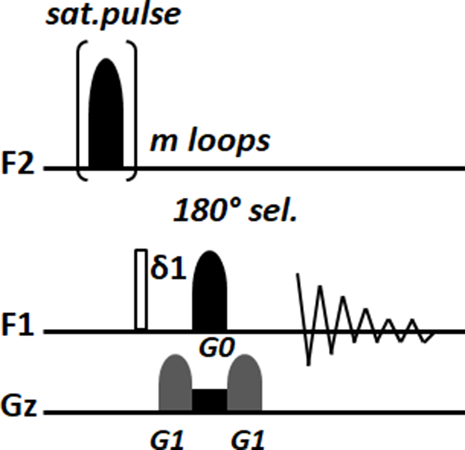
Spatially resolved saturation transfer difference NMR pulse sequence. The saturation pulse was a Gaus1.1000 shaped pulse while the 180° selective pulse was a Reburp.1000. G1 gradient shape was SMSQ10.100 while the rectangular gradient G0 is applied for the whole duration of the selective 180° pulse; for this reason, care should be taken. All the pulses and gradients have to be calculated accordingly as usual for the NMR spectrometer in use.
The pulse sequence resembles the original STD NMR experiment, in which a 180° selective pulse flanked by two shaped gradients and one rectangular gradient (simultaneous to the 180° pulse) are added. With this modification, the intrinsic inhomogeneities of the biphasic systems are avoided and it is possible to read the output of the experiment in the desired phase. From the 180° selective pulse bandwidth (Δω p) and its carrier frequency (Ω) it is possible to calculate the thickness and position of the reading slice in the desired phase using the following equations 10,11 :


The experiments were acquired by positioning the on-resonance saturation pulse at 4.7 ppm (H2O resonance in the aqueous phase) and the off-resonance pulse at 40 ppm. The saturation time was increased from 1.5 to 18 seconds. Both, reference and saturated spectra were acquired reading in a single slice at −5.5 mm from the origin by positioning the 180° selective pulse carrier frequency in the CDCl3 phase. As a convention, the origin of the z-axis is considered the center of the length (L) of the active volume coil (L = 18 mm, Figure 1a).
The feasibility of the proposed approach was demonstrated by the observation of saturation transfer from the H2O phase to the CDCl3 phase, detected as a decrease of the HDO/H2O peak intensity at 1.53 ppm. Furthermore, a buildup of saturation as a function of the saturation time was obtained, indicating efficient accumulation of saturated water molecules in the organic phase. Figure 4a shows the intensity of the saturation transferred to the CDCl3 phase as a function of the saturation time. The saturation transfer to the chloroform phase increases as the saturation time increases, reaching a plateau around 12% after 6 seconds.

(a) Saturation transfer from the aqueous to the organic phase detected by monitoring the HDO/H2O peak intensity (calculated as I 0-I sat/I 0) in the CDCl3 phase. (b) Spatially resolved saturation transfer difference nuclear magnetic resonance experiments reading at different distances from the interface, with a constant saturation time of 9 seconds. The transferred saturation decreases moving away from the interface in agreement with the general mechanism depicted in Figure 1b. The theoretical lower point at −9 mm and its symmetric counterpart at +9 mm are impractical points for the measurement purposes due to radiofrequency inhomogeneity.
We further investigated the system performing the SR-STD NMR experiments with a fixed saturation time (9 seconds) while moving the position of the carrier frequency of the 180° selective pulse on the CDCl3 phase, to read at different distances (−4.1, −5.5, and −6.8 mm) from the interface (Figure 4b). In agreement with our model (Figure 1b), the level of transferred saturation increased in the organic phase from the bottom toward the interface. Indeed, the saturation at the interface can be considered as 100% due to the fact that the HDO and H2O resonance frequencies are indistinguishable in this region. Moving toward the bottom of the active coil the saturation decreases. This decrease of the transferred saturation is dependent on the mechanisms of interface transfer (k tr), Brownian diffusion (D), and self-relaxation (R 1) of the HDO protons. Taken together, these results show unequivocally that observation of interface transport via transfer of saturation between the two phases of this model of an aqueous/organic biphasic system is possible.
Although the above study was on a simple model, it is a solid proof of principle supporting that our approach could be applied and transferred to other systems. To this aim, we explored the use of a similar experimental setup to study the case of a ligand-receptor system. Saturation transfer difference NMR experiments have been successfully used in the past to study protein-ligand systems, providing structural information on biomolecular complexes. 12-17 In that sense, our novel experimental setup could be of interest when a protein-ligand interaction cannot be studied due to either a strong inhomogeneity of the sample, or to a low aqueous solubility of the ligand. In this latter case, the ligand would accumulate efficiently in an organic phase, while the protein would be present in the aqueous one.
The ligand-receptor system selected for this experiment was naproxen and bovine serum albumin (BSA) (Figure 5). For protein-ligand systems we need to consider that ligand molecules are bigger than the water molecules, hence diffusing through the phases with a slower interface transport, slower Brownian diffusion in both phases, and more efficient self-relaxation. To avoid these pitfalls, we resorted to (1) increase the interface surface by incorporating a 2% agarose gel in the water phase and (2) move the position of the reading slice nearer to the interface between the agarose/aqueous phase and the other one (from 1.1 to 2.5 mm from the interface). In this case, the phase to which the saturation is transferred to is positioned on the upper half of the active coil volume, while the 2% agarose gel containing the BSA protein is positioned for practical convenience in the bottom half of the active coil volume (Figure 5). Two different setups, involving two different types of upper phase in contact with the 2% agarose gel phase, were considered: first, an aqueous D2O phase (Figure 5a) and, second, an organic CDCl3 phase (Figure 5b). This choice was made to evaluate the impact of the interface transport on the transfer of saturation.

Schematic of the proposed mechanism for the saturation transfer between phases in the naproxen:BSA:D2O:agarose system. In (a) the upper phase is D2O while in (b) the upper phase is CDCl3. (For the sake of clarity on the right of each proposed mechanism are shown cartoon representations of the nuclear magnetic resonance tube with the biphasic systems.)
The SR-STD NMR experiments were carried out following the routine protocol for STD NMR used for ligand-receptor interactions. 18 In the first case, the upper phase (D2O) containing naproxen (20 mM) was added to the top of the 2% agarose gel in D2O containing BSA (60 µM) in a 5 mm diameter NMR sample tube. Then, naproxen was allowed to diffuse in the agarose gel phase. The on-resonance saturation pulse was positioned at 0.7 ppm (aliphatic residues of BSA), the off-resonance saturation pulse was at 40 ppm, and the reading 180° selective pulse was positioned at 1.1 mm from the origin (on the upper part of the active coil).
The feasibility of the proposed approach for protein-ligand interactions was demonstrated by the observation of interface magnetization transfer by the detection of a decrease of the naproxen aromatic peaks in the region from 7.0 to 8.0 ppm. In this case, the interface transport step is considered absent between the agarose:D2O phase (bottom) and the D2O phase (top) (Figure 5a) as D2O is the solvent in both phases. Thus, the transfer of saturation from the agarose:D2O phase to the D2O phase depends on the dissociation constant (K D ) of the BSA/naproxen complex, the diffusion constant (D) of naproxen and its protons self-relaxation rate (R 1). Accordingly, in the model of the saturation transfer between the two phases in Figure 5a the interface transfer step is absent due to free diffusion of naproxen in both phases.
The outcome of the experiment is shown in Figure 6a. As in the case of the biphasic CDCl3/H2O:10%D2O system, for the naproxen:BSA:D2O:agarose system the results indicated an efficient ligand accumulation in the second phase, after saturation transfer from its interaction with the receptor protein. Figure 6a shows that the transfer of saturation increases with the increase of saturation time, reaching the plateau after about 4 seconds. In this case, the level of saturation observed is higher (50%) than in the model system studied before, as the reading slice was positioned closer (at 1.1 mm and not at −5.5 mm) from the interface.

(a) Saturation transfer detected monitoring the aromatic peaks of naproxen for the 2% agarose:D2O:BSA as lower phase and D2O as upper phase. (b) Example of the spectra obtained using the SR-STD NMR pulse sequence confirming the transfer of saturation to the naproxen found in the upper phase (see general mechanism in Figure 5a). The measurement was performed at 1.1 mm from the interface.
The last series of experiments on the naproxen:BSA system was then performed using an organic solvent (CDCl3) as the upper phase. The model for the transfer of saturation on this system is shown in Figure 5b. In contrast to the previous system with D2O as the top phase, in this system an interface transfer step has to be considered. This is usually the rate-limiting step of the system and might impact negatively the whole saturation transfer efficiency, preventing the detection of the naproxen-BSA interaction via saturation observation on the organic phase.
The output of the experiment is shown in Figure 7a. Again, but now in an aqueous:organic biphasic system, the feasibility of this approach for protein-ligand interactions was demonstrated by the observation of interface saturation transfer via the detection of saturated signals of naproxen in the organic phase. As in the previous cases, the ligand saturation was also efficiently accumulated on the second phase (CDCl3), as it increased with saturation time, reaching the plateau after 4 seconds (Figure 7a). In this case, the level of saturation transfer was lower, as expected, due to the interface transport step constituting a bottleneck of the transfer process, compared to the experiment with D2O as the upper phase. Still, surprisingly, this reduction amounted only to 10%. The overall small reduction is due to the increase of the interface surface area using the gel that might compensate for a lower velocity of interface transfer of naproxen. In fact, data acquired without the presence of the agarose gel in the lower aqueous phase showed no transfer of saturation between the phases (data not shown).

(a) Saturation transfer detected monitoring the aromatic peaks of naproxen for the 2% agarose:D2O:BSA as lower phase and CDCl3 as upper phase. (b) Example of the spectra obtained using the SR-STD NMR pulse sequence confirming the transfer of saturation to the naproxen found in the upper phase (see general mechanism in Figure 5b). The measurement was performed at 1.1 mm from the interface.
Finally, to confirm that the saturation transfer from one phase to the other has its origin in the transfer of saturation from BSA to naproxen, control experiments without BSA were analyzed under identical conditions (Figure S1 Supplementary Data). None of them showed saturation transfer between the phases, confirming that the saturation generated on the protein is transferred to naproxen during the binding process, and it is further transported to the top phase of the sample.
In conclusion, using model systems, we have shown promising and conclusive results supporting the applicability of the SR-STD NMR approach to the analysis of solute transport on fluid biphasic systems. We foresee that this novel approach can become a powerful tool for the in vitro studies of complex chemical or biochemical systems that involve transport between hydrophilic and lipophilic phases. Several applications may be developed using a similar setup either for screening or mechanistic studies. Most importantly, protein-ligand interactions in which the small binder shows moderate solubility in water may also be studied through the detection of the saturation in an immiscible organic phase, where the solubility is higher and the signal accumulates more efficiently.
Experimental
Naproxen, D2O, CDCl3, and BSA were purchased from Sigma-Aldrich.
CDCl3/ H2O:10% D2O Biphasic System
The biphasic system was obtained by initially adding the CDCl3 phase directly in the 5 mm NMR tube reaching exactly the center of the active coil volume. Shortly after that the 10% D2O water solution was added slowly on the top. The sample was then transferred to a 500 MHz Avance I NMR Bruker machine for the SR-STD measurements.
2% Agarose:D2O:BSA/D2O:Naproxen Biphasic System
The 2% agarose gel in D2O was obtained by heating the sample up to 80°C in order to obtain a clear solution. After this the solution was cooled to 38°C and maintained in a water bath for the time needed to add the BSA and mixed gently. Quickly after that, the “sol” phase was transferred in the 5 mm NMR tube covering exactly the half of the active coil volume (see scheme in Figure 5a inset) and was let to make its transition into the “gel” phase. At the top of the gel a solution of 20 mM of naproxen was then added which was allowed to slowly diffuse into the gel phase.
2% Agarose:D2O:BSA/CDCl3:Naproxen Biphasic System
For this system the D2O phase of the previously described 2% agarose:D2O:BSA/D2O:naproxen system was discarded and fresh naproxen (7.0 mM) in CDCl3 was added on the top of the gel phase.
NMR Parameters and SR-STD Pulse Sequence
The SR-STD NMR experiment derives from the original STD experiment and maintains the same experimental setup. For the CDCl3/H2O:10% D2O system the on-resonance saturation pulse was positioned on the water peak resonance at 4.7 ppm while the off-resonance saturation pulse was positioned at 40 ppm. The spectra were obtained by positioning the selective 180° pulse at the CDCl3 phase (at −5.5 mm from the interface) calculating the distance using equation (2) while the saturation time was increased from 1.5 to 18 seconds with a D1 of 18 seconds.
For the 2% agarose:D2O:BSA/D2O:naproxen and 2% agarose:D2O:BSA/CDCl3:naproxen the same experimental setup as before was followed. The on-resonance saturation pulse was positioned on at 0.7 ppm (aliphatic residue side chains of BSA) while the off-resonance saturation pulse was positioned at 40 ppm. The saturation time was increased from 0.5 to 12 seconds with a D1 of 12 seconds. Further, in this case, the selective 180° pulse was positioned on the upper part of the active coil at 1.1 and at 2.5 mm from the interface. Sixty-four scans were acquired for all the SR-STD NMR experiments while the temperature of the samples was maintained at 298 K.
Footnotes
Acknowledgments
We are grateful for the use of the University of East Anglia (UEA) Faculty of Science NMR facility.
Declaration of Conflicting Interests
The author(s) declared no potential conflicts of interest with respect to the research, authorship, and/or publication of this article.
Funding
The author(s) disclosed receipt of the following financial support for the research, authorship, and/or publication of this article: J.A. supported by the Biotechnology and Biological Sciences Research Council (BBSRC) through a New Investigator grant (BB/P010660/1). R. N. and S. M. supported by BBSRC through that research grant and a UEA School of Pharmacy studentship, respectively. Y.K., J.A., and J.C.M.G. supported by the Engineering and Physical Sciences Research Council (EPSRC, EP/N033337/1).
Supplemental Material
References
Supplementary Material
Please find the following supplemental material available below.
For Open Access articles published under a Creative Commons License, all supplemental material carries the same license as the article it is associated with.
For non-Open Access articles published, all supplemental material carries a non-exclusive license, and permission requests for re-use of supplemental material or any part of supplemental material shall be sent directly to the copyright owner as specified in the copyright notice associated with the article.