
Abstract
Background:
One of the most common consequences in individuals with diabetes is the diabetic foot, which can cause foot ulcers and even lead to limb amputation. Since an increase of the temperature in the plantar region is directly correlated with an increased risk of ulceration, infrared thermography (IRT) has been used in multiple studies as an automatic tool for detecting problems in diabetic foot. Artificial intelligence-based computer-aided diagnosis systems are being more frequently used to improve decision-making and minimize errors. These technologies are designed to increase examination accuracy, consistency in image interpretation, prognosis evaluation support, and examination accuracy. They also have the ability to offer insightful information and help medical professionals to manage diabetic foot issues successfully.
Methods:
In this work, 37 papers that used thermography and artificial intelligence (AI) to identify diabetic foot complications and/or predict the risk of developing diabetic foot are analyzed.
Results:
The results demonstrate the potential of IRT imaging implementation with AI for the identification and prediction of diabetic foot complications.
Conclusions:
The combination of IRT and AI shows significant potential for diabetic foot assessment; however, the great majority of these studies show that the research is confined to classification of foot thermograms using pre-prepared data sets. In particular, there is limited research on segmentation methods and constraints in the use of deep learning due to the lack of large and diverse datasets.
Introduction
Diabetes mellitus (DM), also referred to as diabetes, is a chronic condition characterized by the body’s inability to use its main energy source, glucose (sugar), properly, resulting in increased blood glucose levels (hyperglycemia). Diabetes is a chronic disease that can have catastrophic implications if not managed properly. 1 Individuals with diabetes can be classified according to the type of diabetes they have. The two most common types of diabetes are “insulin-dependent” or type 1 diabetes and “non-insulin-dependent” or type 2 diabetes.
The diabetic foot, which can be characterized as infection, ulceration, and/or destruction of deep tissues, is one of the most common problems observed in individuals with diabetes. 2 Between 12% and 25% of individuals with diabetes are at risk of developing foot ulcers during their lifetime, which are mostly linked to peripheral neuropathy and frequently to peripheral vascular disease. 3
Diabetic foot ulcers (DFUs) can be prevented with early identification and treatment. As a result, conducting regular evaluations is critical. However, there are a variety of constraints that may be connected with diabetes-related health problems or any social challenges, making self-examination difficult. 4 An increase in skin temperature can also indicate tissue damage or inflammation caused by trauma or excessive pressure on the foot. As the risk of ulceration is directly related to increased temperature in the plantar region, infrared thermography (IRT) is one of the non-invasive methods that can be used to predict risk, as temperature differences in the foot can indicate problems associated with diabetic foot. 5
Artificial intelligence (AI) refers to the utilization of technology and computers to emulate intelligent behavior and critical thought. Different approaches deal with various and expanding volumes of health data, enabling greater patient autonomy and individualized care. The diagnosis and prognosis of DM and its consequences, such as diabetic foot, have been studied.6,7 Due to the simplicity of high-volume data collecting and sophisticated computational processing, the automation of health care administration has caused a change both in the industry and the development of AI-based solutions. It made possible to predict delayed diagnoses and locate preventive therapies. These prediction models can be used in clinical practice to better identify the individuals with diabetes at high risk, who need closer monitoring and more intense care. 8
In this article, a review is presented, with the aim to emphasize the potential of combining AI with IRT imaging for the diagnosis and prediction of diabetic foot problems, while also recognizing its current limitations.
In the work of Gosak et al, 8 articles that address the prediction of the risk of developing DFU using AI techniques are reviewed, but limited to the studies that use thermography with models just to predict the risk of developing DFU or to classify thermograms. In this review, studies that can be used to identify foot complications, including segmentation and classification techniques, are also included.
This review is structured as follows: The “Materials and Methods” section describes the search strategy, inclusion/exclusion criteria, and data extraction process. The “Results” section presents the findings, beginning with an overview of the target groups studied, followed by a description of how the foot is divided into anatomical regions. It, then, categorizes the analyzed literature based on the techniques applied, including studies focused exclusively on segmentation, those addressing only classification, and those combining segmentation and classification. The “Discussion” section interprets the main findings and analyzes limitations in current research. Finally, the “Conclusions” section summarizes the main ideas and suggests directions for future work.
Materials and Methods
For this literature review, the “Web of Science” and “Scopus” databases were searched, as they provide broad coverage of peer-reviewed scientific literature in medical imaging, AI, and biomedical engineering. Only articles written in the English were considered, with no restrictions on the publication year. The search was performed using the query ((“
Studies were included if they (1) used IRT images, (2) focused on diabetic foot assessment or early detection, (3) applied machine-learning (ML), deep-learning (DL), or neural-network-based classification or segmentation techniques, and (4) included a predictive modeling component.
Figure 1 presents a PRISMA (Preferred Reporting Items for Systematic Reviews and Meta-Analyses) flow diagram 9 summarizing the identification, screening, eligibility, and the inclusion/exclusion stages of the literature search. A total of 106 records resulted from the initial search, with duplicates removed in the first phase, leaving a total of 68 articles for screening. During the screening stage, review articles and records for which the full text was not available were excluded. During the eligibility (full-text assessment) stage, studies were excluded if they did not involve IRT images, did not have a clear focus on early detection, did not implement classification or segmentation techniques via ML, DL, or neural networks, or did not include a predictive modeling component. As a result, a total of 37 articles were included in the final review.
PRISMA flow diagram.
Results
The following subsections describe the cataloging that was performed; a synthesis of the main topics based on the chosen characteristics is presented.
The articles were categorized into three main groups, accordingly to the used techniques: only segmentation techniques, only techniques of classification and both techniques combined.
Target Group
Depending on their specific research objectives, the included studies were categorized based on the population under analysis: some focused exclusively on individuals with diabetes, while others focused on individuals without diabetes or healthy individuals, while several included both groups:
Studies that aim to investigate specific aspects related to diabetic foot conditions, such as temperature variation with different types of complications in individuals with diabetes, 10 focus exclusively on this population.
Some studies included individuals without diabetes or healthy individuals as a comparison group to enable the analysis of thermographic differences, particularly in foot temperature distribution, between populations with and without diabetes. In such cases, the primary objective was to identify specific thermal patterns or anomalies associated with diabetic conditions by contrasting them with those observed in healthy individuals, especially in the context of foot complications.
Foot Division Into Regions
Several studies have conducted thermal analysis based on the observation of specific points or specific foot regions. Considering the temperature of the entire foot allows a comprehensive analysis of its overall state. However, since the foot does not have a uniform temperature, it is important to consider regional divisions of the foot. 11 For example, in studies like 12 temperatures were recorded in some regions of interest (ROIs) on both feet (focusing on areas with a higher likelihood of ulceration) prior to analysis. However, as one of the main causes of diabetic foot ulceration is the decrease in blood supply, the most commonly used and discussed division, divides the feet into four regions based on the concept of angiosomes, which are tissue regions supplied by a single artery. 13
Techniques
An automatic system can easily be connected and processed using various ML and image processing algorithms, such as image segmentation and classification. 14 These systems involve multiple stages of processing, either in a serial or parallel sequence, for extracting and classifying features from thermal images, enabling health care professionals to access more precise diagnoses.
Segmentation
Image segmentation involves dividing an image into several parts/segments with similar characteristics or attributes. This technique forms the basis for image analysis and is an essential step for feature extraction and recognition. Image segmentation techniques can play a crucial role in segmenting and extracting the hottest or coldest regions from clinical infrared images. 15 For example, the shape, size, and boundaries of the hottest regions in thermal images can help determine the features used to detect abnormalities. So far, various techniques have been applied to extract different regions, such as contouring, background isolation, even the hottest regions in thermograms, and convolutional neural networks (CNNs). By observing the hottest or coldest regions, potentially suspicious regions in real-time thermal images can be of interest to physicians.15,16 Another crucial aspect of segmentation is the distinction between a plantar foot and its bottom. It can accurately detect the edges of the foot in thermal images with varying contrast efficiently, laying the foundations for further exploration of the detection or classification of diabetic foot status. 17 Table 1 summarizes the studies that utilize segmentation techniques.
Basic Characteristics of the Included Studies that Use Segmentation Techniques.
Classification
In this context the term “classification” is used to describe the procedure of grouping patients’ plantar thermograms according to their state of health. The majority of articles specifically seek to categorize the thermograms as belonging to both healthy individuals and individuals with diabetes.
To perform this categorization, both ML and DL models have been applied to plantar thermogram analysis. Support vector machines (SVMs) classifiers have been widely employed for the binary classification of healthy and diabetic feet,12,19 whereas K-nearest neighbors (KNNs) and artificial neural networks (ANNs) classifiers have also been employed for the classification of diabetic feet based on thermal patterns.20,21 Ensemble methods like AdaBoost and Random Forest classifiers are another approach investigated for enhancing the robustness and accuracy of diabetic foot screening systems.22,23
More recent studies have been using DL methods, especially CNNs, because of their success in the classification of thermographic images. Some studies have utilized transfer learning using established CNN models, including AlexNet, 24 VGG networks, 25 ResNet models, 26 DenseNet, 27 and MobileNet models. 28 Moreover, more advanced models like DarkNet-based networks 29 and vision transformers (ViT) have recently been explored for the classification of plantar thermograms, and they have shown promising results on small data sets. 30 During the training process, the classification model extracts distinctive thermal features from the labeled plantar thermograms, which are, then, utilized for distinguishing between the healthy and diabetic classes or between the stages of diabetic foot complications. After the training process, the model can be used for predicting the health status of patients from unseen thermograms. Table 2 summarizes the studies that utilize classification techniques.
Basic Characteristics of the Included Studies That Use Classification Techniques.
Segmentation and classification
Some studies integrate both segmentation techniques and classifications to present more complete systems. In these methods, segmentation is employed as a pre-processing step to detect ROIs, as explained in the “Segmentation” section, and classification is, then, carried out using the information obtained from these regions.
Unlike the methods explained in the “Classification” section, where classification is carried out directly on the whole thermogram or on pre-defined ROIs for the plantar area, combined segmentation and classification methods are used when there is a need to detect localized thermal irregularities before making any decisions. In these scenarios, segmentation helps in the classification process by demarcating ROIs that are of anatomical or thermal significance, while classification offers subject-level or region-level diagnostic results.
In diabetic foot analysis, segmentation is particularly beneficial when the aim is to examine localized patterns, such as asymmetric angiosomes, ulcerative prone areas, or localized areas of temperature increase. On the contrary, classification only methods are adequate for global screening applications, such as distinguishing normal subjects from ones with diabetes without the need for spatial localization.
Some methods combine both segmentation and classification by first segmenting to define ROIs, followed by classification using ML or DL models, thereby offering both spatial localization of irregular regions and good classification accuracy.14,46-49 Table 3 summarizes the studies that utilize segmentation and classification techniques in combination.
Basic Characteristics of the Included Studies That Use Segmentation and Classification Techniques.
Discussion
Individuals with diabetes who develop DFUs face significant costs and disabilities. It is crucial for all patients with diabetes to receive comprehensive education about foot care and preventive measures. However, the conventional method of diagnosing DFUs by clinicians and DFU experts is costly and time-consuming. Deep learning in medical imaging offers the potential for automatic DFU diagnosis. Given the complexity of DFUs, AI approaches are well-suited to address issues, such as prompt screening to identify the likelihood of foot ulcers or amputation using appropriate sensor technology.
In this review, articles are categorized as: articles presenting segmentation techniques, articles presenting classification techniques, and articles presenting the combination of both techniques. From Figure 2, it can be observed that the majority of the studies primarily focus on the classification process, with a lack of studies presenting methods for segmenting thermograms with relevant information, such as extracting the foot from the background or isolating the specific foot area required for analysis. The majority of the existing studies focus primarily on classification approaches, while significantly fewer explore segmentation, a fundamental step that can greatly enhance the precision and interpretability of computer-aided diagnostic systems.
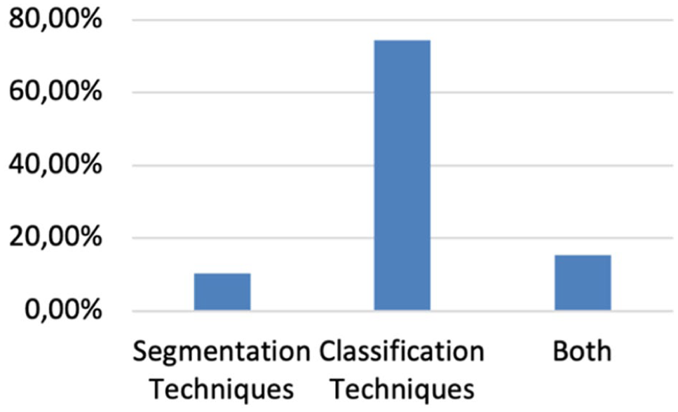
Distribution of the articles according to the type of techniques used.
In the four studies presented in Table 1, foot images were segmented and analyzed to identify anomalies or areas of interest. Accurate segmentation and localization of foot sole areas were achieved using a variety of techniques, including clustering, Mask R-CNN, U-Net, Skin approaches. Future work for these studies includes further analysis, technique validation, and optimization, with a focus on improving segmentation algorithms, exploring new imaging modalities or approaches, and conducting larger-scale studies to confirm the efficacy and generalizability of the detection techniques.
The studies summarized in Table 2 demonstrate the effectiveness of various ML algorithms, including SVM, CNNs, such as VGGNet, DenseNet, ResNet, and MobileNet, as well as conventional ML methods like AdaBoost classifier, KNN, and logistic regression. These algorithms accurately classify and identify diabetic foot complications based on thermal images of individuals with and without diabetes.
Several studies utilized the public INAOE data set, 11 which consists of thermal pictures from 167 individuals, including both individuals with and without diabetes. The proposed approaches and algorithms were developed and evaluated using this data set, yielding high accuracies, sensitivities, and specificities ranging from 90% to 100% accuracy in categorizing and stratifying the severity of diabetic foot problems.
However, the studies also acknowledge certain limitations and areas for further investigation. These include the need for larger and more evenly balanced data sets, as well as consideration of under-represented classes to increase sensitivity. The robustness and generalizability of the suggested methods need to be further explored by evaluating them using different infrared cameras, acquisition protocols, and independent data sets. Future research can also explore the integration of other modalities, such as color and texture cues, to improve classification performance and provide a more comprehensive analysis of diabetic foot problems.
Studies that utilize both segmentation and classification techniques in combination highlight the essential role of segmentation in identifying and isolating areas of interest in thermal images. When comparing the performance of various algorithms, it is evident that DL frameworks, such as AlexNet, GoogleLeNet, and the proposed DFTNet, generally outperform more conventional ML techniques, such as SVM and MLP, in terms of classification accuracy. These DL models often eliminate the need for explicit ROI definition and feature extraction, making the classification process more efficient and faster.
However, the use of DL models in diabetic foot diagnosis is still limited by the lack of available data, the cost of data acquisition and annotation, and the need for intensive computational resources.25,30,43 These factors partly explain why the use of DL models is less common in this area and the need for continued efforts in data sharing, standardization of data acquisition, and large-scale validation.27,30
Moreover, the conditions of image acquisition, such as camera placement, distance, and environment, may cause inconsistencies in thermal distribution.19,26 From a clinical standpoint, the implementation of AI-assisted diagnostic systems in a busy clinical setting also poses challenges with regard to workflow compatibility and understanding of the diagnostic output.35,42
Application-Oriented Use Cases of AI Models in Diabetic Foot Thermography
Aside from performance aspects, it can be seen from the reviewed studies that various AI models are appropriate for different application scenarios in the case of diabetic foot thermography.
Lightweight architectures like MobileNet and ShuffleNet are appropriate for resource-limited settings, such as portable devices, smartphone-based screening, and telemedicine applications, because of their low computational complexity and efficient inference.28,34,42 These architectures are highly relevant in the context of large-scale screening and telemedicine.
Conversely, more complex architectures of CNNs, such as VGGNet, ResNet, and DenseNet, are more often used in clinical workstation or hospital environments where computational power is less of a constraint.23-27,31 These architectures are usually employed in decision support systems for diagnosis.
When the application involves spatial localization and interpretability, such as marking areas that are prone to ulcers or abnormal plantar areas to help with clinical decision-making, it is particularly useful to have frameworks that integrate segmentation and classification.14,46-49 In such instances, segmentation offers complementary information to classification.
More contemporary architectures, such as ViT and ConvNeXt-based models, have until now been largely investigated in research environments,30,45 and their adoption in clinical practice awaits more data and studies.
For health care professionals, the classification results can serve as a valuable resource for identifying diabetes patients who may be at risk of developing foot issues. Early identification of potential problems enables the implementation of effective preventive measures and interventions to reduce risks and enhance patient outcomes.
It is crucial to remember that the quality and representativeness of the training data, the selection of the ML algorithm, and the choice of classification features all play a role in the effectiveness of the classification process. To ensure the accuracy and generalizability of the model, it should undergo routine validation and continuous improvement.
Conclusions
Infrared thermography imaging and AI have demonstrated great potential in detecting diabetic foot problems and estimating the potential of developing diabetic foot. The reviewed studies show that AI-based computer-aided diagnosis systems using thermography can help with quick screening and diagnosis of DFUs, giving health care professionals helpful data.
Although the classification process is the main focus of the majority of the papers evaluated, there is a noteworthy dearth of studies on segmentation methods for obtaining pertinent data from thermograms. To create precise computer-aided diagnostic systems, segmentation is a crucial component, and further study is required to enhance this area.
The DL methods are less frequently used in classification research because of the lack of data and the difficulties in acquiring it. The ML techniques like SVM and KNN were frequently used. To effectively use DL approaches, future research should concentrate on gathering larger and more varied data sets.
Health care professionals can use the categorization outcomes from these AI systems as important resources to find individuals with diabetes who are at risk of developing foot issues. Early detection enables the implementation of therapies and preventive measures, improving patient outcomes and lowering the dangers connected with DFUs.
Footnotes
Acknowledgements
The authors acknowledge the use of ChatGPT (OpenAI) for assistance in language editing and clarity improvements during manuscript preparation. All content was reviewed and finalized by the authors. The authors also gratefully acknowledge the support of University of Trás-os-Montes e Alto Douro.
Abbreviations
AUC, area under the curve; AI, artificial intelligence; ANN, artificial neural network; CAD, computer-aided diagnosis; CNN, convolutional neural network; DFU, diabetic foot ulcer; DL, deep learning; DM, diabetes mellitus; DT, decision tree; IRT, infrared thermography; K-NN, K-nearest neighbor; MLP, multilayer perceptron; ML, machine learning; PAD, peripheral arterial disease; ROIs, regions of interest; SPD, skin + depth; SVM, support vector machine; TCI, thermal change index; UPD, U-Net + Depth; ViT, vision transformer.
Funding
The authors disclosed receipt of the following financial support for the research, authorship, and/or publication of this article: The research of the Ana Paula Teixeira author was partially financed by Portuguese Funds through FCT (Fundação para a Ciência e a Tecnologia) within the Project UID/00013/2025 (![]() ).
).
Declaration of Conflicting Interests
The authors declared no potential conflicts of interest with respect to the research, authorship, and/or publication of this article.