
Abstract
Objective:
The objective was to assess the relationship of skin advanced glycation endproducts (AGEs) between first-degree relatives estimated from skin fluorescence (SF) after adjustment for skin pigmentation.
Study design:
SF was excited by LEDs centered at 375, 405, and 420 nm from children with type 1 diabetes and their mothers. Data were adjusted to generate measures of skin intrinsic fluorescence (SIF) at the various excitation wavelengths, using 2 different pairs of correction coefficients for excitation (kx) and emission (km): kx = 0.5, km = 0.5 (not associated with skin pigmentation) and kx = 1.0, km = 0.0 (strongly associated with skin pigmentation). Pearson correlation analysis was performed, as well as a multiple variable analysis with maternal SIF adjusted for the effects of maternal age and race.
Results:
There were 50 matched pairs of children and their mothers. Children were 13.3 ± 3.7 years of age and there were 19 boys/31 girls and 15 black/35 white. Mothers were 41.8 ± 6.8 years of age. The age of mother and child was highly correlated, r = .64, P < .0001. In Pearson correlation analysis, child’s SIF (kx = 1.0, km = 0.0) the had strongest association with maternal SIF, while with SIF (kx = 0.5, km = 0.5) there was a trend for association. In the multiple variable model child SIF was associated with maternal SIF for all corrections and wavelengths but was stronger for kx = 1.0, km = 0.0.
Conclusion:
Even after adjustment for skin pigmentation and race, correlation of SIF between family members persists, suggesting that other genetic and/or environmental factors shared by parent and child may influence estimated skin AGEs.
Advanced glycation endproducts (AGEs) are long lived complexes formed endogenously by nonenzymatic glycation and oxidative modification of proteins, lipids, and nucleic acids.1,2 Formation and tissue accumulation of AGEs are increased in patients with diabetes.2,3 Tissue AGE levels determined from biopsy have been shown to increase with chronologic age, duration of diabetes, and HbA1c levels. 4 AGE levels may also be influenced by environmental factors.5,6 Assessment of AGEs may be clinically relevant in diabetes care as tissue AGE levels have been associated with the development and progression of diabetes complications. 7 Previously, study of tissue AGEs had been limited as their measurement required direct chemical assay from tissue biopsy samples.7,8 An innovative, noninvasive method to estimate skin AGEs was developed based on measuring induced skin fluorescence (SF).9,10 This technology enables rapid, painless estimation of skin AGEs, which facilitates clinical studies of tissue AGEs especially in children. 11
Barat et al reported that SF levels were correlated in siblings, suggesting that hereditary or environmental factors may also influence SF. 12 The AGE-Reader used to measure SF in the Barat study has been reported to be sensitive to the degree of skin pigmentation, and patients with dark complexion are usually excluded from analysis. 13 To mitigate the influence of race on SF, we previously have found it important to use intrinsic corrections of the data to eliminate potential interference due to patient variation in skin pigmentation. 11
To further examine the phenomenon of correlation of estimated AGEs between family members, we measured SF at different wavelengths and intrinsic corrections in children with diabetes in comparison with their mothers.
Research Methods and Design
Subjects
This study was part of a larger study of skin AGEs in youth performed at the Children’s Hospital of New Orleans. Children with established type 1 diabetes (T1D) were recruited for the study from the Diabetes Clinic at the Children’s Hospital of New Orleans (LA, USA). Patients and their biological mother were studied on the same day. Individuals with a history of smoking 6 and parents known to have diabetes were excluded from the study.
Estimation of Skin AGEs
SF was noninvasively measured from the volar surface of the left forearm from each patient and parent on the same day using a Scout DS instrument (VeraLight, Inc, Albuquerque, NM, USA).11,14 Testing was not performed in relationship to timing of subject’s meals. The device sequentially excited the skin surface using different light-emitting diodes (LEDs) that had peak excitation wavelengths of 375, 405, and 420 nm. SF generated from each LED was detected over a 435-655 nm emission window. Skin reflectance was measured for each excitation LED, and a white light LED was used to measure skin reflectance over the emission region. SF induced in this manner is further adjusted mathematically to calculate skin intrinsic fluorescence (SIF) using this formula:11,14
Statistical Analysis
SAS software (SAS Institute, Cary, NC) was used to perform standard Pearson correlation analysis as well as multiple variable analysis of child SIF as a function of maternal SIF. Maternal SIF was statistically adjusted for the effects of maternal age, and race by covariate analysis in a general linear model. Results of Type III sums of squares analysis, which adjusts the influence of covariates for the presence of the others in the model without regard to order of position, are reported. Statistical significance was considered to be at P < .05.
Results
The characteristics for the patient and mothers are shown in Table 1. The ages of mother and child were correlated at r = .64, P < .0001. A simple correlation analysis showed significant association in most SIFs between the child and mother (Figure 1 and Table 2).
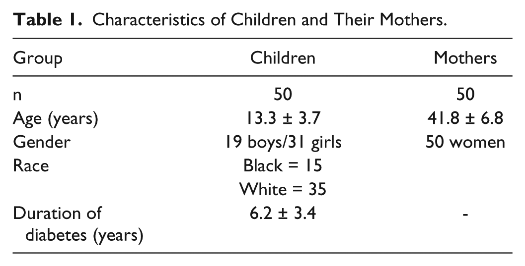
Characteristics of Children and Their Mothers.
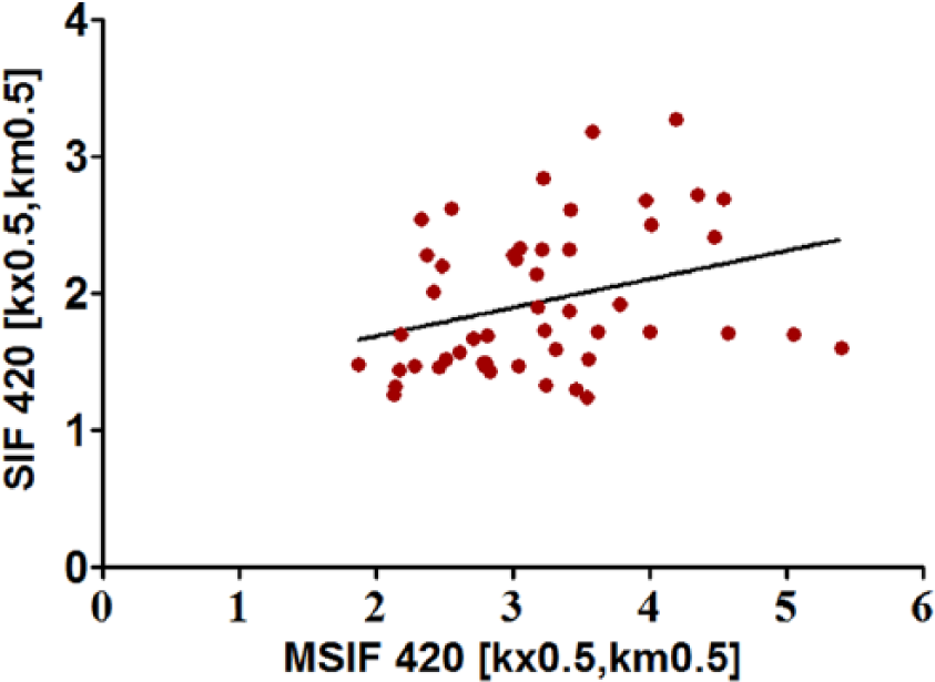
Pearson correlation of child’s versus maternal skin intrinsic fluorescence (SIF) for 420 (kx = 0.5, km = 0.5). The x-axis represents maternal SIF (MSIF). The y-axis represents child SIF.
Child’s Versus Maternal Skin Intrinsic Fluorescence (SIF) Simple Correlation and Multiple Variable Analysis With Maternal SIF Adjusted for Race and Maternal Age (n = 50).
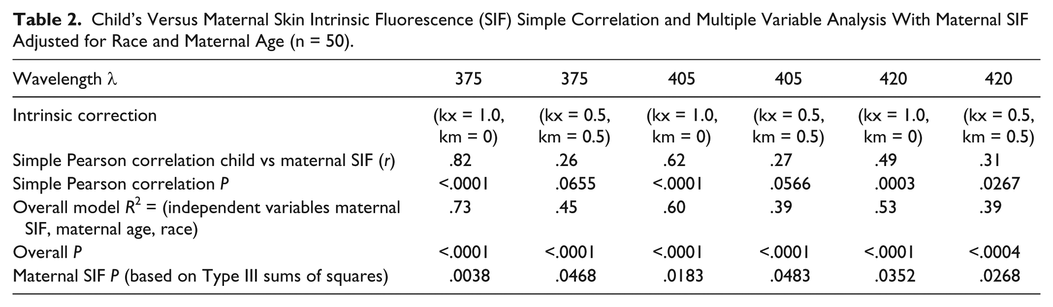
After adjustment for maternal SIF, maternal age, and race, all statistical models showed that child’s SIFs were significantly associated with maternal SIF at the same excitation wavelength and kx and km used for intrinsic correction. The R2 values of the models for different SIFs ranged from .39 to .73 (Table 2). In general, kx and km intrinsic correction of 1.0 and 0.0, respectively, yielded higher correlations between mother and child than 0.5 and 0.5 at the same excitation wavelength.
Discussion
AGEs fluoresce when exposed to near UV and blue light. This fluorescent property has been exploited for assay of AGEs from biopsied tissue3,4 and more recently has been adapted for noninvasive estimation of AGEs in the skin.9,10 Intrinsic correction of SF data from the Scout DS is useful to mitigate the impact of skin pigmentation, hemoglobin content and light scattering on data interpretation.11,15 We have previously reported that application of different intrinsic corrections can also modify the influences of chronologic age and race (due to differences in skin pigmentation) on SF data at different wavelengths. 11 In this study, 2 intrinsic corrections were examined with coefficient of excitation (kx), and coefficient of emission (km) pairs (kx = 1.0, km = 0.0) and (kx = 0.5, km = 0.5). SIFs for 405 and 420 nm corrected with (kx = 1.0, km = 0.0) were previously reported by our group to be influenced by chronologic age, HbA1c, and race, while at 375 nm there was an influence of race, but not of chronologic age. 11 SIFs at 405 and 420 nm corrected with (kx = 0.5, km = 0.5) showed no statistical influence of race, but were associated with increasing chronologic age, duration of diabetes, and HbA1c. 11
Furthermore, the Scout DS machine can generate a statistic representative of the diffuse reflectance of the skin measured over the wavelength range the emitted fluorescence measured (MREFsum). This is correlated with the degree of skin pigmentation among subjects in a population. In an internal study, using SF data from 309 pediatric patients, we found that at 375, 405, and 420 nm excitation, with intrinsic correction kx = 1 and km = 0, that these SIFs were strongly associated with MREFsum even after statistical adjustment for race. However, at the same wavelengths with intrinsic correction kx = 0.5 and km = 0.5, there was no longer an influence of MREFsum or race on SIF. Thus choice of specific intrinsic corrections with kx = 0.5 and km = 0.5 facilitates comparison between and within racial/ethnic groups with different degrees of skin pigmentation.
This is the first study to examine the relationship of SIFs between parents and their diabetic child. Even after statistical adjustment for maternal age and race, SIFs of child and mother were statistically associated at all wavelengths and kx, km pairs used for intrinsic correction. We found that correction with the kx, km coefficient pair of (kx = 1.0, km = 0.0) generally yielded a stronger mother–child association than the (kx = 0.5, km = 0.5) pair. As (kx = 1.0, km = 0.0) is sensitive to dichotomous black versus white racial difference, as well as, individual variation in skin pigmentation within racial groups, it is highly likely that mother–child similarity of skin pigmentation is a contributing component of these specific SIF associations. For reasons discussed above, mother–child SIF at 405 and 420 nm with (kx = 0.5, km = 0.5) association is unlikely to be due to similarity of skin pigmentation, and suggests a potential influence of other underlying environmental and/or genetic factors.
Using an AGE-Reader, which employs a technology similar to the Scout DS, Barat et al found a correlation of SF (r = .43, P < .01) among 27 sibling pairs, 1 sibling with and the other without diabetes. 12 AGE-Reader SF data are influenced by skin pigmentation, and Koetsier et al have proposed a method for adjustment. 16 The AGE-Reader SF data may most closely resemble SIF375 (kx = 1.0, km = 0.0) obtained with the Scout DS. 17 If this is so and SF data in the sibling study were not adjusted by the method of Koetsier et al, then potentially there was a large influence of skin pigmentation in the sibling to sibling correlation.
Barat et al noted that similarity in skin pigmentation among other genetic and environmental factors may account for the high correlation they observed between siblings in their data. 12
Genetic factors have previously been reported to influence AGE levels. Leslie et al reported a high correlation of serum levels of the AGE carboxymethyllysine among twin pairs. 18 The apparent substantial genetic effect in twins was confirmed by quantitative modeling techniques. 18 More recently, genomewide association studies of SF were conducted in 2 populations measured by the AGE-Reader and the Scout DS. 17 This study linked SF levels with the NAT2 acetylator in both cohorts. 17 Thus there is independent evidence linking serum AGE levels and SF status with genetic factors besides skin pigmentation that could account for correlation of SF among family members.
In addition to genetics, environmental factors potentially may also contribute to SF status. AGEs are contained in commonly ingested foods. 5 SF measured by AGE-Reader has been noted to be acutely influenced by meal intake. 19 As family members likely share a similar nutritional environment with prolonged exposure to similar AGE containing foods, this may contribute to sibling–sibling and mother–child correlation of estimated skin AGEs. Other possible shared environmental factors, such as air pollutants, smoking, 6 and potentially secondary smoke, might also account for the SF associations of individuals sharing the same household. The potential impact of these other factors on SF will need to be evaluated in future studies.
Conclusion
The present study indicates that the use of specific intrinsic corrections mitigates the influence of skin pigmentation on SF data. The use of these corrections is a valuable component of SF interpretation when comparing data between individuals independent of race. Failing to use a correction that mitigates skin pigmentation may prove more difficult to interpret interindividual SF relationships related to genetics, environment, and most definitely race. Even after adjustment for skin pigmentation/race on SF, an association between mother and child is present in this study. This association suggests a potential influence of genetic or environmental factors besides skin pigmentation on estimated AGEs in first-degree family members.
Footnotes
Abbreviations
AGEs, advanced glycation endproducts; AU, autofluorescence units; LEDs, light-emitting diodes; SF, skin fluorescence; SIF, skin intrinsic fluorescence; T1D, type 1 diabetes.
Declaration of Conflicting Interests
The author(s) declared no potential conflicts of interest with respect to the research, authorship, and/or publication of this article.
Funding
The author(s) received no financial support for the research, authorship, and/or publication of this article.