
Abstract
Importance
Bilateral vocal cord paralysis is a challenging problem to manage in adults, and a known complication of thyroid surgery which is more common in resource-limited settings. Posterior graft laryngotracheal reconstruction is a management option that has not been studied in this population or setting.
Objective
To report the surgical outcomes of posterior graft laryngotracheal reconstruction for bilateral vocal cord paralysis in a resource-limited setting, and to evaluate the efficacy of a hybrid system for teaching this technique in Kenya.
Design
Retrospective cohort study.
Setting
Tertiary public referral hospital, Eldoret, Kenya
Participants
Adults >18 years with tracheostomy dependence secondary to bilateral vocal cord paralysis after thyroid surgery or other iatrogenesis. The hybrid training program included Kenyan surgeons and surgical trainees.
Intervention or Exposures
Posterior graft laryngotracheal reconstruction and hybrid training system included didactic lectures, simulation-based training, case discussion and planning, and live cases.
Main Outcomes Measures
One-year airway outcomes measured by achievement of tracheostomy decannulation, and self-reported surgeon knowledge and skill acquisition.
Results
Ten patients met criteria and were included in the analysis, with mean age 42 years (range 30-62 years) and had been tracheostomy dependent for an average of 6 years (range 1-12 years). Seven (70%) had open reconstruction, and 3 (30%) had endoscopic reconstruction. All were decannulated and remained tracheostomy-free at 1 year. Three surgeons and 7 trainees participated in the hybrid teaching methods. All reported increased comfort in laryngotracheal reconstruction, with all 3 surgeons reporting comfort performing the surgeries independently.
Conclusion
Posterior graft laryngotracheal reconstruction shows promise as a potential method of treating bilateral vocal cord paralysis and achieving decannulation in a resource-limited setting.
Relevance
The methods reported in this study lend themselves to replication and expansion to other similar settings. The authors plan to replicate this work in other centers in East Africa.

Keywords
Background
One of the most common causes of bilateral vocal cord paralysis (BVCP) in adults is iatrogenic injury to the recurrent laryngeal nerves during thyroid surgery. 1 This is particularly common in resource-limited settings (RLS) and low-middle income countries (LMICs) due to the prevalence of large thyroid goiters, a relative lack of dedicated high-volume thyroid surgeons, and a varied level of training of surgeons performing these cases. 2 The mainstay of management of BVCP, at least in the short term, has traditionally been tracheostomy. 3 However, managing a tracheostomy is particularly challenging in RLS due to limited access to home tracheostomy supplies, suctioning, high burden of bacterial colonization, and social stigma. 4 Thus, LMICs have a particularly-prudent need for strategies for managing BLVCP without tracheostomy, in addition to strategies to avoid injury.
There are several strategies for improving airway patency in BVCP to obviate the need for a tracheostomy or to achieve tracheostomy decannulation. In adults, these include endoscopic arytenoidectomy and/or cordotomy, 5 vocal cord lateralization, and laryngeal reinnervation procedures.6,7 Costal cartilage graft laryngotracheal reconstruction of the posterior glottis has been studied extensively in pediatrics since the 1980s8,9; however, its use in adults for the purpose of BVCP is much more limited. More recently, authors have reported success with adapting this technique to adults, although ossification of laryngeal and costal cartilage can be a limiting factor.10-12
Posterior graft laryngotracheal reconstruction (PGLTR), which involves division of the posterior cricoid lamina and placement of a costal cartilage graft in the interaytenoid space, is a viable treatment option for BVCP and PGS. It has the advantages of lending itself to either a single-staged or double-staged approach, with permanent resolution of airway symptoms and high rates of achieving long-term decannulation. 13 It also preserves the vocal cords should they regain movement. Moreover, PGLTR provides a valuable first step in adapting and teaching LTR techniques in a RLS, where these skills can be valuable for many other surgical applications. 11 The majority of airway reconstructive surgery is performed in high-income countries where procedures are resource-intensive and rely on complex technology and equipment. 14 However, the majority of patients requiring these procedures live in LMICs, where the poorest one third of the world’s population receives less than 5% of global surgical care. 15 This disparity in global surgery has been highlighted by many, including the Lancet Commission on Global Surgery which has set out measurable and achievable targets for global surgery by 2030.16,17 Nevertheless, creating long-term scalable and sustainable programs in airway reconstruction remains a challenge due to the extensive training and expertise required, reliance on advanced technology, and need for intensive postoperative care, all of which can drive up cost and resource expenditure. 18 Many challenges remain with respect to the role of Otolaryngology—Head & Neck Surgery in global surgery, including how to integrate international outreach and training into the longitudinal health infrastructure in a resource-limited setting.19-23
One group has published on their experience developing airway surgical programs in a RLS, which were mostly in South America.24,25 This work provides a useful framework for building airway reconstruction capacity and evaluating the effectiveness of such programs. In addition to these short-term intermittent surgical outreach programs, the field of global otolaryngology lends itself well to innovative approaches for knowledge transfer such as simulation-based teaching and virtual surgical instruction. Several groups have developed simulation programs for teaching laryngotracheal surgery using animal, 26 cadaveric, and synthetic 3D-printed models.27-32 These types of innovations have high-impact potential in RLS where it can be difficult to establish longitudinal teaching relationships based on short-term missions. Some authors have also explored the use of virtual and augmented reality with the use of smart glasses as a surgical training tool for RLS. 33 These techniques allow surgeons to participate virtually in surgical procedures and provide live instruction and mentorship during cases. These innovations have significant potential to enhance longitudinal knowledge translation in airway reconstruction.
The objectives of this study were to report the clinical and surgical outcomes of PGLTR for managing BVCP in a RLS, as well as to evaluate the efficacy of a hybrid system for teaching LTR in a RLS. The ultimate goal is to develop a viable and replicable model for creating LTR surgical programs in RLS based on our experience in Eldoret, Kenya.
Methods
This was a retrospective review of patient outcomes and self-reported surgeon skill/knowledge acquisition at a tertiary public referral center in Eldoret, Kenya, from 2020 to 2023. The patient population included adults >18 years old with tracheostomy dependence secondary to BVCP after thyroid surgery or other iatrogenic injury. Participants were excluded if they had multilevel airway stenosis, previous open laryngotracheal surgery other than tracheostomy, were ventilator-dependent, medically unfit for surgery, or declined surgical intervention.
Airway Team and Structure
The personnel consisted of a visiting airway team led by a visiting airway surgeon (VAS) from the University of Alberta, Canada (A.I.). This team included another VAS who participated virtually (E.P.), a pediatric anesthesiologist (P.H.), pediatric and adult intensive care physicians (N.A., P.H.), and a teaching coordinator (A.C.). The local airway team consisted of 4 local airway surgeons (LAS) and multiple local trainee surgeons (LTS), as well as local anesthesia providers, intensive care unit (ICU), and nursing providers.
Teaching and Knowledge-Skills Transfer
Knowledge and skills transfer was a major emphasis of this project. Prior to patient evaluation, the VAS team delivered a series of didactic lectures on open and endoscopic airway surgical techniques for LAS and LTS teams tailored to their patient population and resources. This was followed by simulation-based training on airway models provided by AweSIM Medical (Toronto, Canada). LAS and LTS teams were instructed about and able to practice in costal cartilage graft carving, anterior and posterior cricoid split, cricoid dissection, graft placement, and graft suturing (Figure 1). Trainees wore head-mounted cameras so that the VAS team could provide direct and ongoing feedback on dissections in real-time either in person or virtually (Figure 2). Simulation-based training was followed by live case demonstration with a graduated level of independence, eventually transitioning to the LAS performing the surgeries from start to finish with little or no intervention from the VAS (Figure 3). This was accomplished during 3 separate 2 week surgical camps spanning 2 years. Knowledge and skills transfer was assessed using a previously-validated self-assessment questionnaire.

Laryngotracheal reconstruction simulation course with surgeons and surgical trainees.
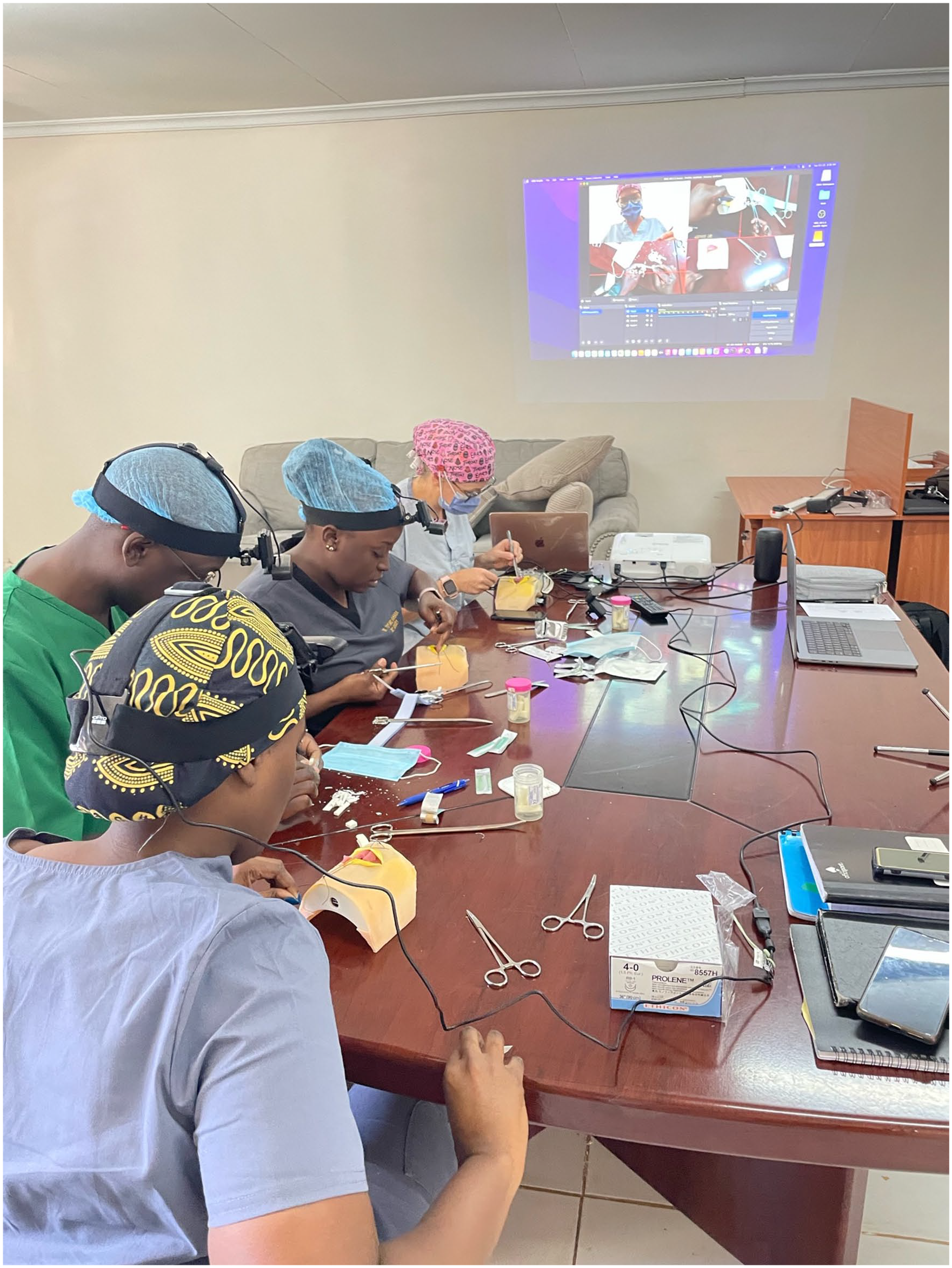
Video-monitoring of laryngotracheal reconstruction course to facilitate direct live feedback.

Local airway surgeon performing a posterior graft laryngotracheal reconstruction.
Preoperative Evaluation
All patients underwent preoperative assessment by a VAS in conjunction with a LAS. Vocal cord immobility was confirmed using in-office flexible fiberoptic laryngoscopy. Lack of multilevel airway stenosis was confirmed by in-office tracheoscopy after topicalization. Medical comorbidities were optimized by the local anesthesiologists and/or medicine team to determine fitness for surgery. Due to the extremely-high rates of bacterial colonization, tracheostomy tube aspirates were sent for culture and sensitivity, and all patients had a minimum of 48 hours of culture-directed preoperative antibiotic coverage, which was continued for 7 days postoperatively. Ciprofloxacin-dexamethasone drops were instilled into the tracheostomy twice daily starting 48 hours before surgery. Evaluation of swallowing was beyond the scope of this study.
Surgical Details and Decision-Making
Patients were consented for surgery if they met the above criteria (BLVC paralysis, lack of multilevel airway stenosis, and medically fit for surgery). All patients underwent suspension laryngoscopy and rigid bronchoscopy under spontaneous ventilation at the start of the case. This was used to determine the most appropriate surgical approach, confirm the status of the cricoarytenoid joints and subglottis, and rule out other airway lesions. Cases were performed as a team with a minimum of 1 VAS and 1 LAS with at least 1 local surgical trainee (LST). Surgery consisted of right costal cartilage graft harvest, and either open or endoscopic posterior cricoid split via cold steel microlaryngeal instruments, with insertion of a cartilaginous graft spanning the posterior glottis, infraglottis, and interarytenoid area to optimize airway patency (Figure 4).

Posterior graft position showing splaying of the vocal cords and arytenoids.
Staging and Stenting
All surgeries were performed in a double-stage approach, leaving the original, matured tracheostoma intact. An appropriately-sized Montgomery suprastomal stent was used in each case, and secured with at least one 2-0 prolene suture. The stents were removed 4 to 6 weeks later.
Postoperative Care
Patients were kept in hospital postoperatively either in an ICU or on a surgical ward with nurses experienced in caring for tracheostomies. The decision for postoperative ICU care was based on the duration of the case, medical comorbidities, patient stability under anesthetic, and anticipated complications. Patients managed in an ICU were permitted to emerge from sedation and were transferred to the surgical ward on postoperative day 1 if they remained without complications. Patients were kept in hospital for a minimum of 48 hours postoperatively to ensure stent tolerance. Oral feeding tolerance and safety was assessed via functional endoscopic evaluation of swallowing prior to discharge.
Stent Removal and Follow-Up Bronchoscopy
One week prior to stent removal, patients were re-started on antibiotic coverage and instillation of ciprofloxacin-dexamethasone drops down the tracheostomy was begun. Stents were removed under bronchoscopic guidance by an LAS. Bronchoscopy was performed 1 week later to confirm airway patency, and obstructive granulation tissue was removed. Bronchoscopy videos were reviewed by a VAS to confirm readiness for decannulation.
Decannulation
Decannulation was attempted between 3 days and 6 weeks after stent removal, depending on a number of factors including airway status, and the extent of granulation. Tracheostomies were capped for a minimum of 48 hours prior to decannulation. An occlusive dressing was placed over the stoma after decannulation, and the stoma allowed to close by secondary intention. Patients were monitored in hospital for a minimum of 48 hours prior to discharge.
Follow-Up
Patients were followed up for a minimum of 6 months postoperatively. Clinical assessment of airway, swallowing, and voice was made at each follow-up. Further bronchoscopy and/or targeted endoscopic interventions were performed when required.
Variables and Outcomes Collected
Charts were reviewed for demographic data, comorbidities, duration and type of tracheostomy, previous airway procedures, and other airway diagnoses. Surgical details including the type of surgery performed, and intraoperative and postoperative complications were collected. Short-term and long-term complications rates were analyzed and classified as major (mortality, graft failure, need for revision surgery, airway obstruction) and minor (infection, pneumothorax, granulation, non-airway related morbidity). The postoperative course, and 1, 6, and 12 month decannulation rates were assessed.
Results
Fifteen patients underwent LTR. Two were excluded as they were under 18 years of age, and 3 had multilevel airway stenosis and/or anterior graft reconstruction. Ten patients met inclusion/exclusion criteria and were included in the final analysis. Patient demographics and comorbidities are presented in Table 1. There were 7 females and 3 males, with a mean age of 42 years (range 30-62 years). Prior to surgery, patients lived with their tracheotomy for a median of 5.5 years (range 1-12 years). All patients had previous attempts at airway expansion and/or decannulation, including balloon dilation, cordotomy, and arytenoid lateralization.
Patient Demographics, Medical, and Surgical Outcomes for Patient Cohort Who Underwent Laryngotracheal Reconstruction.

Abbreviations: BLVCP, bilateral vocal cord paralysis; PTLR, posterior graft laryngotracheal reconstruction; ICU, instensive care unit.
Surgical and postoperative details are also displayed in Table 1. Seven patients underwent open PGLTR and 3 had endoscopic PGLTR. Six patients were observed postoperatively on the surgical ward and 4 were observed in the ICU postoperatively. The median postoperative length of hospital stay was 4 days (range 2-7 days). There were 2 in-hospital complications: 1 case of excessive bleeding from the costal cartilage harvest site, which was managed conservatively with pressure dressings; and 1 case of subcutaneous emphysema of the neck, which resolved spontaneously while a penrose drain was left in situ. Patients underwent stent removal a median of 6 weeks postoperatively. The median time to decannulation was 8 weeks (range 5-12 weeks). Following decannulation, 2 patients had granulation or restenosis requiring endoscopic treatments. One year postoperatively, decannulation and survival rates were 100%.
Knowledge and Skills Transfer
Three LAS and 7 LST participated in the didactic, simulation-based, and surgical training sessions. The final 3 PGLTR surgeries (1 endoscopic, 2 open) were completed without any intervention from the VAS team. Results of the self-reported knowledge and skill questionnaires are summarized in Table 2. LAS and LST indicated a significant need for training in LTR prior to initiation of the course, and an increase in comfort and knowledge of airway reconstruction following completion. Participants rated the value of training highly, with all LAS reporting feeling confident performing the procedure alone.
Results of Pre- and Post-course Surveys Among Surgeons and Trainees.


Red color represents pre-course; green color represents post-course. x: surgical trainee; o: surgeon.
Discussion
This is the first series to report on outcomes of PGLTR for BLVCP in a RLS. Our results demonstrate this technique to be highly effective for managing BLVCP and achieving tracheostomy decannulation even in patients who have been tracheostomy-dependent for many years. There are concerns with using costal cartilage graft expansion techniques in the adult population, namely, that the cricoid and costal cartilage are more ossified and the stiffness of the thyroid cartilage in adults can work against posterior expansion of the cricoid plate. Considerations for potential complications such as thyroid cartilage fracture, graft dislocation, and poor epithelialization need to be considered. Despite this however, our results show a high rate of success with a single procedure, with limited need for further airway procedures to maintain airway patency. This has important implications in settings where follow-up can be difficult. This stands in contrast to other endoscopic approaches such as cordotomy which requires a high rate of revision. 34
Moreover, PGLTR does not require the use of advanced endoscopic airway equipment such as lasers which are scarce in a RLS. The approach can easily be adapted for double-staged or single-staged procedures, and has a low complication rate and relatively-simple postoperative care when performed as a double-stage procedure.
The adaptation of airway reconstructive techniques to RLS is of critical importance. The challenges in managing tracheostomies and patients with airway stenosis in RLS have been well-documented.4,35 These issues make advanced airway reconstruction a critical skill for practitioners managing patients in RLS. Traditionally, these procedures have been resource-intensive and have required advanced equipment to perform. Despite this, there has been a movement toward adapting these and other surgical techniques to RLS.24,25 The current study adds to the growing literature on adapting airway surgery to RLS. Advances in simulation-based surgical training and enhanced virtual collaboration have made this more accessible than ever before. Our team has been able to create and establish a sustainable and longitudinal teaching relationship, which has allowed the LAS team to increase its skill, confidence, and breadth of airway expertise. Although cost analysis was beyond the scope of the current study, the relatively-short hospital stay of the patients treated, in comparison with the cost of long-term tracheostomy care, is another reason why these collaborations are important.
Traditionally, LTR has been felt to be risky and/or inadequate in adults due to concerns about graft loss and poor blood supply, resulting in a higher reliance on resection techniques. 36
In addition to the concerns for performing single-stage procedures in RLS, there is the added issue of being unable to address stenosis at the level of the vocal cords with resection techniques. Despite previous concerns, the results here add to the evidence that graft techniques are reliable and good results are achievable in adults undergoing LTR.
Lessons Learned
ICU and postoperative sedation
During the airway collaboration we have presented, there were several lessons learned and pitfalls encountered that other groups can learn from. Namely, we found that postoperative sedation and ICU care can be challenging in centers not experienced with airway reconstruction. After double-staged LTR procedures, centers use varying degrees of postoperative sedation or lack thereof depending on the comfort and volume of the hospital. Although the team initially thought that postoperative sedation would be more prudent and safer in a RLS where tracheostomy care on the ward is more sparse, we have found that achieving optimal sedation while ensuring patient safety in terms of ventilation and tracheostomy care was extremely difficult. As such, we soon realized that minimal to no sedation, as well as minimal to no ICU stay, was safer both for the patient and for the integrity of the airway reconstruction, even when there may be very high nursing ratios and limited tracheostomy care on the surgical ward. We aim to help address the lack of ICU airway experience and comfort in future collaborations.
Suctioning
Postoperative suctioning of tracheostomies was of critical importance but often of limited availability in surgical wards. This made the use of portable battery-powered suction machines of particular importance. This gave the added benefit of not losing suction power during power outages.
Bacterial contamination and infection
Bacterial colonization of tracheostomy tubes was a frequent and problematic issue, as most patients used the same tracheostomy tube for years due to supply constraints. As such, we found culture-directed antibiotics be highly important, as well as use of a new tracheostomy tube for the 6 week stenting period to limit bacterial infection of the cartilage graft.
Preoperative airway assessment and patient selection
We used communication and conferencing software such as Zoom and WhatsApp to keep open lines of communication between the LAS and VAS teams. The ability to share preoperative and postoperative bronchoscopic videos, airway assessments, and surgical outcomes greatly aids in the ability to deliver collaborative care, as well as to make maximal use of short-term surgical trips by preselecting patients for surgical collaboration.
Minimum materials, personnel, and resources
One of the main goals of this program is to allow other groups to replicate the successes and avoid the failures our team has experienced. In this vein, the authors recommend a certain number of minimum requirements that would be deemed essential for replicating such a program. These include a single LAS “champion,” who is the main point of contact and helps to coordinate preoperative and postoperative care as well as follow-up and patient selection. A VAS “champion,” who is the main point of contact for LAS team questions and serves as the primary instructor for surgical didactics, is required. A minimum of 2 VAS surgeons is recommended. An appropriately-equipped and skilled local operating room is essential, including reliable anesthesia equipment and providers, basic surgical equipment, and supplies. The ability of the OR to perform endoscopic airway procedures and bronchoscopy is essential prior to initiating any LTR teaching or training. In terms of hospital resources, a critical care unit is essential in case of unexpected airway complications that may arise. The postoperative monitoring environment must have readily-available suction ideally not dependent on a power source. In terms of minimum materials and equipment, basic bronchoscopy equipment including rigid and flexible telescopes, ventilating bronchoscopes, and basic microlaryngeal instruments are essential, as well as a method of repairing, sharpening, and sterilizing this equipment. For disposables, a variety of tracheostomy and endotracheal tubes must be available, as well as the appropriately-sized suprastomal stents. The LAS team must be capable of performing bronchoscopy, tracheostomy, and basic endoscopic procedures prior to the initiation of any LTR program. This is essential for ensuring safe postoperative care and the ability to manage complications. We found that with the use of virtual collaboration, it was not necessary for the VAS team to be present on-site for more than a few days following LTR procedures as long as there were methods available for rapid communication, and the LAS team was able to handle complications including the need for emergency tracheostomy.
Limitations of this study were that it involved a single-center experience with a small number of patients, both of which can limit generalizability to other RLS. There was no comparison or control group, which limits the ability to compare PGLTR with other techniques of glottic dilation or laryngeal reinnervation. Long-term decannulation beyond 1 year was not assessed. We also did not study functional outcomes including voice, respiratory, and swallowing outcomes, which can be a source of morbidity for patients. The period of laryngeal stenting can have quality-of-life impacts, which were also not studied. The teaching methods were not externally validated, and the LAS team may have been biased to report improved skill and comfort after the completion of teaching. A case series without a comparison or control group also cannot be used to demonstrate the superiority of one technique over another.
Conclusion
PGLTR shows promise as a potential method of treating BLVC paralysis and achieving decannulation in a RLS. Further data are required in order to establish the long-term safety and reliability of this technique for more widespread use. The collaboration and teaching methods reported in this study lend themselves to replication and expansion to other RLS. Further long-term studies with a higher number of patients are needed, as well as addressing functional outcomes of speech and swallowing after airway reconstruction in RLS.
Footnotes
Author Contributions
A.I. is the lead surgeon, helped conceive the study, participated in study design, helped analyze the data, wrote the first draft of the manuscript, and revised and approved the final version. O.M., G.R., and H.N. participated in the surgeries, helped design the study, participated in data collection, and revised and approved the final manuscript. H.N. coordinated the study design, data collection, and ethics approval. A.C. participated in data collection and data analysis, and revised and approved the final manuscript. D.M.I. helped with study design, data analysis, proofreading, and revised and approved the final manuscript. J.W. advised on data collection and analysis, and revised and approved the final manuscript. P.H. and N.A. participated in data collection and data analysis, and revised and approved the final manuscript. E.P. assisted with study design and execution, data analysis, and revised and approved the final manuscript.
Availability of Data and Material
Data available from the corresponding author upon request.
Declaration of Conflicting Interests
The author(s) declared no potential conflicts of interest with respect to the research, authorship, and/or publication of this article.
Funding
The author(s) disclosed receipt of the following financial support for the research, authorship, and/or publication of this article: The collaboration between University of Alberta and Moi Teaching and Referral Hospital is partly funded by a Royal College International Development Aid and Collaboration grant.
Ethics Approval and Consent to Participate
The study obtained ethics approval from Moi Teaching and Referral Hospital/Moi University Institutional Research and Ethics Committee IREC/693/2023 (approval number: 0004664), and consent was waived.