
Abstract
Bone fragility is the susceptibility to fracture due to poor bone strength. This condition is usually associated with aging, comorbidities, disability, poor quality of life, and increased mortality. International guidelines for the management of patients with bone fragility include a nutritional approach, mainly aiming at optimal protein, calcium, and vitamin D intakes. Several biomechanical features of the skeleton, such as bone mineral density (BMD), trabecular and cortical microarchitecture, seem to be positively influenced by micro- and macronutrient intake. Patients with major fragility fractures are usually poor consumers of dairy products, fruit, and vegetables as well as of nutrients modulating gut microbiota. The COVID-19 pandemic has further aggravated the health status of patients with skeletal fragility, also in terms of unhealthy dietary patterns that might adversely affect bone health. In this narrative review, we discuss the role of macro- and micronutrients in patients with bone fragility during the COVID-19 pandemic.
Background
Good nutrition is a major determinant of health and can help prevent or control most chronic diseases.
Nutrition is associated with bone health at multiple levels, being involved in bone metabolism, structural (e.g. bone geometry and matrix mineralization) and density (i.e. bone mineral density, BMD) changes as well as in functional issues (i.e. fall risk). Therefore, inadequate nutrition patterns might contribute to an increased risk of fragility fractures.1–3 Besides vitamin D and calcium, other micronutrients seem to bring benefits to bone health, including fluorine, magnesium, potassium, vitamin B6, vitamin C, vitamin K, and zinc.4,5
The COVID-19 pandemic significantly impacted the mental and physical well-being of the worldwide population. Psychological discomforts led people to assume wrong habits, including low levels of physical activity and an unhealthy diet, as evidenced by the increase in the consumption of alcohol and foods rich in sugars, leading to an altered ratio between caloric intake and energy expenditure.6,7 Moreover, it is expected that the impact of the sedentary lifestyle adopted during the pandemic also due to the prolonged and repeated lockdown, might have affected bone health, both in young and elderly people, increasing the risk for fragility fractures. 8
COVID-19 seems to have a detrimental effect on the musculoskeletal system also at the biological level. Indeed, a key role regards the association between severe acute respiratory syndrome coronavirus 2 (SARS-CoV-2) and angiotensin-converting enzyme 2 (ACE2). This enzyme is widely expressed in a variety of human tissues in addition to the lungs, where acts as a host cell receptor. 9 For example, low levels of ACE2 were found on human bone marrow-derived stem/progenitor cells (BMSPCs), where monocytes/macrophages, the osteoclast precursors, synthesize also other RAS (renin-angiotensin system) components as Mas receptor (MasR), a class of G-protein-coupled receptor. Moreover, osteoblasts and osteoclasts synthesise ACE2 and MasR. 10 In this context, ACE2 promotes AngII degradation and synthetizes Ang-(1–7), which acts via MasR promoting skeletal repair through the ACE2/Ang-(1–7)/Mas axis. More specifically, the activation of this pathway increases osteocalcin and collagen 1A mRNA levels promoting osteoblast activity and, at the same time, it reduces RANK and IL-1β mRNA levels decreasing osteoclast differentiation. 11
Therefore, the downregulation of ACE2 induced by SARS-CoV-2 infection may affect bone homeostasis, causing bone fragility.
Among the other potential factors that may influence bone metabolism in COVID-19 patients, it was observed that the ‘cytokine storm’ and other inflammatory factors, as well as depletion of B and T lymphocytes, contribute to the receptor activator of nuclear factor-kappa B ligand (RANKL) upregulation, increasing bone resorption. It seems that Ang-(1–7) can decrease the expression of pro-inflammatory cytokines, such as interleukin (IL)-1β, IL-6, and tumor necrosis factor (TNF), that are directly involved in bone resorption in a wide range of inflammatory conditions;11–13 therefore, the depletion of ACE2 and the high levels of cytokine caused by SARS-CoV-2 infection might act synergistically inducing osteoclastogenesis in COVID-19 patients.
Evidence also suggested a putative role of oxidative stress in bone remodeling in SARS-CoV-2 infected patients, through the amplification and perpetuation of biological events, such as cytokine storm, coagulopathy, and cellular hypoxia, and the association with excessive levels of reactive oxygen species (ROS). 14 The ROS are involved in the regulation of RANKL-dependent osteoclast differentiation and seems to influence the process of bone resorption with a dual role. At physiological levels, ROS production induced by osteoclasts regulates RANKL signaling pathways, essential for osteoclast differentiation through the recruitment of TNF receptor-associated factor (TRAF) 6, Rac1, and NADPH (nicotinamide adenine dinucleotide phosphate) oxidase (Nox) 1. 15 On the other hand, at higher concentrations, as those observed in aging or inflammatory states like in COVID-19, ROS lead to cell death and bone loss.16,17 In this scenario, adequate intake of nutrients is essential for an optimal immune response able to prevent infections, and to counteract inflammatory state and oxidative stress, strengthening a relationship between nutrition and individual response to disease burden.18,19 Consistent with this concept, a recent cross-sectional study showed the importance of adequate levels of vitamin D and zinc for their immunomodulatory and direct antiviral effects during SARS-CoV-2 infection. Similarly, these nutrients are noteworthily required for normal skeletal growth and bone homeostasis,20,21 further supporting their adequate intake during COVID-19 to prevent bone loss.
Finally, hypoxemia and subsequent acidosis observed in the severe COVID-19 inhibits osteoprotective factors like osteoprotegerin (OPG) through hypoxia-inducible factor (HIF)-1α and further upregulated RANKL and nuclear factor of activated T cells, cytoplasmic 1 (NFATc1) disrupting bone homeostasis. 22
Considering all the above-mentioned potential risks for bone health during the pandemic, we provide an overview to address how inadequate micro- and macronutrient intakes could contribute to bone fragility during the COVID-19 pandemic.
Bone fragility and nutrition: what does it matter?
Bone fragility is an umbrella term that includes quantitative (e.g. density) and qualitative (e.g. geometry, microarchitecture, material composition) changes that modify the internal stress state of bone tissue predisposing to fragility fractures. 23 All structural and material changes characterizing bone fragility are potentially related to nutritional factors, including water, macronutrients, micronutrients, and trace elements. 24
Macronutrients and micronutrients
Correct nutrition consists of the ingestion of macronutrients, as proteins, lipids, and carbohydrates, 25 as well as micronutrients, such as vitamins and minerals contained in food and in water (minerals). Water can be considered an essential food, with several roles in the human body, from building material to solvent and carrier for nutrients. 26 Bone has a complex structure and consists of water for 10–20% of total volume, available as free within pores and matrix bounded, the latter as an essential component for the integration of hydroxyapatite and collagen to modulate mechanical behavior of bone tissue in terms of better ductility. 27 Collagen conformation change (i.e. from triple helix to partially unfolded fragments) expected occurring in aging leads to poor water adsorption mode, strength, and density on hydroxyapatite with consequent bone fragility. 28 It should be underlined that calcium bioavailability of mineral water rich in calcium is comparable with that of dairy products, 29 and that bicarbonate-rich alkaline water decreases serum parathyroid hormone (PTH) and C-telopeptides (CTX). 30
Proteins are among the main components of bone, accounting for about 50% of bone volume. 31 These nutrients influence bone health particularly because of their positive effects on insulin-like growth factor-1 (IGF-1) production.32–34 Low dietary protein intake reduces intestinal calcium absorption 35 and negatively affects the activation of calcium-sensing receptor 36 leading to increased serum PTH. 37 On the other side, an excess of dietary protein intake increases intestinal calcium absorption with increased urinary excretion of calcium. 38 In a healthy pediatric population, high daily dietary protein intake (1.5–2 g/kg body weight) usually observed for growth requirements, is associated with greater periosteal circumference, cortical area, bone mineral content, and strength-strain index. 39 Interestingly, those consuming high sulfur-containing aminoacids (i.e. methionine and cysteine) did not report a significant association between protein intake and bone fragility components, suggesting the relevance of the quality of proteins for bone health. It has been suggested that alanine, lysine, arginine, leucine, and glutamine support osteoblast growth and differentiation by stimulating insulin secretion,40–42 and lysine and arginine seem to contribute to type I collagen synthesis. 43 Furthermore, a finite element analysis (FEA) demonstrated a positive association between bone strength and animal protein intake, while no significant finding was reported in people consuming vegetable-derived proteins.44,45
Several meta-analyses investigated the association between daily dietary protein intake and BMD, reporting that 0.8–1.2 g/kg body weight of protein consumption accounted for 2–4% of BMD variance.46–49 On the other side, no randomized controlled trials (RCTs) evaluated the efficacy of dietary protein on fracture risk, 24 and observational data suggest that intake over the recommended dietary allowance (RDA) might be able to reduce hip fracture risk up to –16% compared with lower intake, 48 without adverse effect on bone quality. 46
It is key to note that the benefits of proteins on bone health seem to be significantly affected by their interaction with calcium supplementation. Indeed, significantly higher fracture risk (+51%) was reported in patients receiving high dietary proteins but low calcium intake (<400 mg/1000 kcal). 50
Lipids are essential macronutrients accounting for 20–35% of daily energy intake and contribute to the absorption of fat-soluble vitamins, including vitamin D. 51 High-fat diet (HFD), especially saturated fatty acids (SFAs), might reduce BMD, impair bone microarchitecture52–54 reduce marker of bone formation (serum osteocalcin, procollagen type 1 amino-terminal propeptide, P1NP, carboxy-terminal propeptide of type 1 procollagen, P1CP), increase bone resorption markers (cross-linked N-telopeptides of bone type I collagen, NTx, urine pyridinoline, Pyr, and deoxypyridinoline, Dpyr), 55 and increase fracture risk. 52 In animal models, trabecular bone volume fraction, mineral content, trabecular number, and bone strength significantly decreased after HFD, without recovery after weight loss, despite reduced marrow adipose tissue accumulation. 54 Moreover, increased bone marrow adiposity in response to HFD has deleterious effect on the skeleton, and the progenitor cells exhaustion due to continuous recruitment to adipogenesis reduced recruitment to osteoblastic cells, and decreased bone formation. 56
Several mechanisms have been proposed for the negative effects of lipids on bone health. High dietary fat intake induces hyperinsulinemia that led to high urinary calcium and magnesium excretion, along with poor calcium absorption and increased retinol intake that stimulates bone resorption. 57 Furthermore, high lipid intake enhances sclerostin expression and damages the osteocyte network. 58
In clinical studies, a high intake of SFAs is associated with reduced femoral neck BMD and increased hip fracture risk (+31%).59,60
Carbohydrates are macronutrients widely available in Mediterranean and Western diets as mono- and disaccharides, oligosaccharides, polysaccharides, and soluble dietary fibers. 61 Diets with a high intake of carbohydrates, particularly monosaccharides (i.e. glucose) and disaccharides (i.e. sucrose), seem to reduce BMD. 62 Basic research suggests that high concentrations of glucose impair osteoblast proliferation and differentiation, 63 while sucrose-fed animal models reported lower bone strength in long bones. 64 High fructose intake reduces calcitriol-dependent intestinal and renal calcium transport, 65 and the insulin spike triggered by high glucose ingestion is proportional to urinary calcium. 66 The increased glucose intake induces lactic acid formation in osteoclasts resulting in the dissolution of calcium and magnesium from the bone surfaces. 66 The consumption of sugar-sweetened beverages, a major source of carbohydrates, is associated with poor calcium intake, 67 increased urinary calcium excretion, presumably due to kidney damage (i.e. glomerular congestion and intertubular bleeding), low BMD, 68 and poor bone quality. 69 On the other hand, increased intestinal calcium absorption (up to 58%) 70 and BMD have been reported after the consumption of fruits and vegetables rich in water-soluble fibers containing inulin.71,72
Micronutrients and trace elements
Micronutrients and trace elements with effects on bone fragility are calcium, fluoride, magnesium, potassium, phosphorus, vitamin B group, vitamin C, vitamin D, vitamin K, boron, copper, manganese, selenium, silicon (Si), strontium, and zinc.
Calcium contributes to bone strength by being stored for more than 99% in the skeleton to form hydroxyapatite crystals.34,73 Calcium is largely contained in milk, yogurt, cheeses, legumes, nuts, and sardines.74,75 The bioavailability of this element is superior when taken through dairy products and mineral waters. 24 A decreased calcium intake might adversely affect bone health by promoting bone remodeling through secondary hyperparathyroidism. 24 Different studies investigated the effects of calcium intake through nutrition or supplementation on BMD improvements, reporting similar benefits in trials of increased intake of calcium through diet or calcium supplements. Considering the mild and nonprogressive BMD change, the authors concluded that increased calcium intake is unlikely to lead to a reduction in fracture risk. 76 However, in a study conducted on an Italian outpatient population, higher fracture risk was correlated with a lower calcium intake, particularly in individuals with vertebral fractures. 77 To prevent fragility fractures, it has been recommended 1 g/day of calcium in postmenopausal women. 78
Fluoride is available particularly in tea, seafood, and fruits and vegetables. 79 The daily supplementation of approximately 5 mg of fluoride might be effective in enlarging osteoblasts volume, bone formation, and BMD. 80 However, literature is not univocal on this topic as reported in an RCT showing no beneficial effect of fluoride on cortical BMD and fracture risk.81,82 The improvement of bone metabolism by fluoride administration was reported in a double-blinded and placebo-controlled trial in which 180 postmenopausal women with osteopenia received 2.5, 5, or 10 mg of fluoride. Compared with placebo, the groups treated with 5 and 10 mg of active intervention reported a significant increase of the P1NP (p < 0.04 and p < 0.005, respectively), suggesting a modulation of bone formation. 83
Magnesium (Mg) contributes to PTH secretion and potassium homeostasis. 84 Foods containing an adequate amount of Mg are nuts, leafy green vegetables, and dairy products. 24 About 50% of Mg present in our body is accumulated in the bone where Mg modulates the size and formation of hydroxyapatite crystals. Mg deficiency may inhibit osteoblasts and activate osteoclasts,85,86 and promotes vitamin D production. 87 A lower Mg intake seems associated with a lower hip BMD. 88 On the other hand, a higher Mg intake than the recommended dose is associated with a higher risk for wrist fracture. 88 Otherwise, more recently, it has been reported significant association between high Mg intake and low fracture incidence both in women (–62%) and men (–53%). 89
Potassium dietary intake has a protective action on age-related bone loss.90,91 Foods with a high content of potassium are potatoes, milk, cereals, and coffee 92 Potassium salts seem to have a positive effect on bone by lowering renal extraction of calcium and acids and significantly reduce NTX. 93 A prospective cohort study on 266 older women reported that those consuming food rich in potassium (3676 mg daily) had significantly higher BMD at different sites compared with women in the lowest quartile of potassium intake. 94 It was also noted that an adequate potassium intake was related to an increase of BMD in postmenopausal women but not in men over 50 years. 95 In a large observational study conducted in Korea, a decreased risk for low BMD at the lumbar spine (–32%) in postmenopausal women, but not in men, was reported in people with an adequate potassium intake. 96 The most accredited hypothesis for potential benefits of dietary potassium on bone health is its effect on modulation of acid–base equilibrium. Western diet rich in meats and cereal grains but poor of fruits and vegetables predisposes to metabolic acidosis that worsens with age due to renal function decline. 97 Buffering of this acidic serum pH by the alkaline calcium salts in the skeleton may enhance bone resorption. Thus, alkaline potassium salts intake from fruits and vegetables or potassium supplements may prevent bone resorption.
Another important ion is phosphorus, which is present in dairy products, meats, beans, nuts, lentils, and cereals. 98 Phosphorus is a structural component of bones and teeth,99,100 and it is important for the optimal mineralization of cartilage and for osteoblasts activity 101 with the recommended daily administration of 700 mg for adults and 1250 mg for adolescents. 98 An adequate level of phosphorus is required to ensure apoptosis of mature chondrocytes to trigger the invasion of blood vessels and generation of new bone, thus preventing rickets and delayed growth. 102 A high intake of phosphorus increases bone mineral content and BMD. 103
B vitamins (i.e. folate, B6, and B12) seem to improve bone health by regulating plasma homocysteine concentrations (tHcy). Serum levels of vitamin B9 (folic acid) and B12 are inversely correlated with serum tHcy. 104 Meta-analyses, cross-sectional, and cohort studies suggest a role of vitamin B12 on fracture risk reduction but not on BMD changes. 105 In a 1-year double-blind placebo-controlled trial B-vitamins administration did not affect bone turnover nor BMD in patients with osteoporosis, except in those with hyperhomocysteinemia, in which B vitamins supplementation increased lumbar spine BMD.106,107
Vitamin C is an important nutrient for bone health by contributing to collagen production in the bone matrix and counteracting the production of ROS. 108 Foods that provide the highest level of vitamin C are tomatoes, potatoes, and citrus fruits, such as limes, oranges, and lemons. 109 This vitamin appears to modulate osteoclastogenesis and osteoblastogenesis 110 and stimulate the production of type 1 collagen by osteoblasts. 111 Increased vitamin C intake has been associated with a reduced risk of hip fragility fractures as well as an increase in BMD at the femoral neck and lumbar spine.111,112
Vitamin D is a fat-soluble vitamin involved in calcium metabolism and bone health. 113 Vitamin D status mainly depends on sun exposure, although it can also be influenced by diet by consuming fatty fish, mushrooms, and eggs and, also, vitamin D-fortified foods, such as low-fat cheeses and biofortified eggs, seem to be useful for increasing intake.75,114 It has been reported that the combined administration of calcium and vitamin D is more effective than the administration of the two elements alone, as confirmed by the efficacy of this intervention in reducing the risk of hip fractures and non-vertebral fractures in osteoporotic patients compared with placebo.115,116 Moreover, vitamin D deficiency needs to be corrected to guarantee the effectiveness of anti-osteoporotic drugs. 117
Vitamin K has been attributed benefits in preventing vascular calcification and cancer as well as in improving insulin sensitization and bone formation. 118 There are two different kinds of vitamin K produced by plants (K1) and bacteria (K2). Vitamin K1 is the most present in food while vitamin K2 is contained only in some cheeses. 119 Vitamin K favors the gamma-carboxylation of osteocalcin thus promoting bone mineralization. 120 However, clinical studies not reported low BMD in patients with low vitamin K intake.121,122 A RCT including 244 post-menopausal women showed that vitamin K supplementation for 3 years significantly improves BMD compared with those receiving placebo, 123 whereas a meta-analysis showed that vitamin K supplementation has little effect on BMD changes with favorable effect for clinical fragility fractures of the spine. 119
Several studies evidence the relationship between single vitamins in monotherapy and the percentage of fracture, but only a few studies have investigated the role of multivitamins on bone health. In a study conducted in an Australian care setting multivitamin supplementation significantly increased serum 25(OH)D, folate, and vitamin B12 along with higher BMD and reduced fall risk. 124 Moreover, in a meta-analysis, multivitamins intake has been associated with a decreased probability of hip fracture [odds ratio (OR) = 0.49, 95% confidence interval (CI): 0.32–0.77]. 125
Boron contributes to bone health by reducing calcium excretion and influencing the metabolism of steroid hormones, including vitamin D. 126 Boron protects from rickets regulating bone remodeling and improving bone stiffness. 127 In animal studies, an optimal intake of boron seems to have a positive effect on bone trabecular microarchitecture and cortical bone strength. 128
Copper functions as an enzymatic cofactor and removes bone free radicals imputable to osteoclasts activation. 129 In in vitro study, copper appears to block osteoclastic resorption, 130 while in experimental animal models, a lower intake of copper leads to reduced bone strength. 131 Moreover, a significant correlation between low serum copper and the occurrence of hip fractures in elderly subjects has been reported. 132
Manganese is an osteotropic element. It favors bone matrix formation and stimulates calcification. In a study conducted on 40 post-menopausal women, it has been found a positive relationship between serum manganese and BMD, and a negative association between serum manganese and the number of fragility fractures. 133
Selenium seems to bring benefits in terms of BMD improvement and fracture risk reduction,134,135 likely by a mechanism linked to high osteoclasts activation in low antioxidative status.
Interestingly, studies in rodents show that selenium deficiency increases bone resorption and damages bone microarchitecture, 136 and low levels of selenium lead to osteopenia in young animals. 137
Si is highly concentrated as bound to glycosaminoglycans in connective tissues, particularly in bone, playing a key role in the crosslinks between collagen and proteoglycans.138,139 Si concentration progressively reduces from osteoid tissue to mature bone mineral, suggesting a putative role in bone mineralization, 140 as also suggested by poor growth, cortical thinning, and skeletal fragility in animal models with low dietary Si intake. 141 Most bioavailable sources of Si are mineral water and beer, where it is present as orthosilicic acid, and vegetables.142,143 It has been estimated that the adequate daily intake of this element for bone health is about 25 mg. 144 Si seems to contribute to bone mineralization, type 1 collagen synthesis, osteoblast differentiation, and osteoclasts inhibition presumably by reducing the RANKL/OPG ratio and antagonizing the activation of nuclear factor kappa B (NF-κB),138,145–148 In animal studies, dietary Si intake is associated with increased bone content of calcium and phosphorus, enhanced activity of alkaline phosphatase, structural rigidity, and quantity of force absorbed before breaking at femur compared with lower Si intake.149,150 Moreover, low serum Si seems to inhibit growth plate closure resulting in higher longitudinal growth. 151
In human studies, high dietary Si intake resulted in increased femoral BMD, not spine BMD, in pre-menopausal women and adult men, whereas no significant BMD change was reported in post-menopausal women.152,153
Strontium is chemically related to calcium and is almost completely stored in bone and teeth after ingestion. 154 The administration of this element as the salt of ranelic acid has been widely studied for osteoporosis treatment. Strontium is adsorbed onto the mineral surface of the new bone, particularly the trabecular component, and increases structural properties of the skeleton, such as bone volume and trabecular thickness, without affecting hydroxyapatite crystal features (i.e. mineralization).155,156
Zinc is required for normal skeletal growth and bone homeostasis as well as for promoting bone healing. 157 However, the cellular and molecular pathways through which zinc promotes these effects are poorly understood. Zinc can positively affect osteoblast functions while inhibiting osteoclast activity, consistent with a beneficial role in bone homeostasis and regeneration. 157 In a murine model, zinc deficiency increased serum PTH through a reduction of serum calcium resulting in bone fragility, 158 but human studies to understand if zinc deficiency predisposes to osteoporosis are still lacking.
Table 1 synthetizes evidence about the role of nutrients on bone health.
Benefits of nutrients on bone fragility in adults.

BMC: bone mineral content; BMD, bone mineral density; CTX, C-telopeptides; IGF, growth factor-1; N/A, not available; OPG, osteoprotegerin; PTH, parathyroid hormone; RANKL, receptor activator of nuclear factor-kappa B ligand; MUFA, monounsaturated fatty acid.
Dietary reference intakes according to the National Institutes of Health. Office of Dietary Supplements. 159
Risks of the overintake of micronutrients
The evidence seems to be clear about the damages related to an insufficiency of micronutrient intake and the benefits of supplementation when needed. On the other side, pending questions remain about the potential harms of overintake of these substances, particularly on bone. It is key to note that few studies have investigated this issue. Considering vitamin D, severe hypercalcemia and confusion, abdominal pain, vomiting, and dehydration, occur in patients with vitamin D toxicity (VDT). 160 However, this condition is rare and is usually caused by the intake of extremely high doses of pharmacological preparations of vitamin D (exogenous VDT) as well as other diseases that produce the calcitriol (i.e. granulomatous disorders) (endogenous VDT).
Poor evidence reported the effects on bone of an overintake of calcium. However, overuse of calcium supplementation could lead to cardiovascular diseases and malignancy. Some studies, including a meta-analysis, estimated that calcium supplements have up to 30% increased risk for myocardial infarction and mortality, particularly in men.161,162
An overconsumption of proteins seems to have indirect effects on bone. 163 Indeed, an excess of protein intake increases glomerular filtration rate and urinary calcium. Negative calcium imbalance could negatively affect bone turnover, as demonstrated by increased urinary N-telopeptide excretion. 164
Similarly, sugar consumption impacts negatively bone tissue and is associated with low BMD. A meta-analysis found that sugar-sweetened beverages significantly reduce bone density, increasing the risk of fracture. 165 This condition seems to be related to several mechanisms, such as the increased renal excretion of calcium, but also the reduction of intestinal calcium absorption, unbalancing osteoblast, and osteoclast activity promoting bone resorption. 166
Also, excess vitamin A intake seems to have negative effects on bone health. An observational study found that increased dietary retinol intake promotes a reduction of BMD at the femoral neck, Ward’s triangle, trochanter region of the proximal femur, lumbar spine, and total body with an increased hip fracture risk. 167 Recently, an animal study observed that a massive dose of vitamin A suppresses the loading-induced gain of bone mass decreasing cortical bone area by 12%, marrow area by 19%, endocortical perimeter by 10%, and periosteal perimeter by 8%. This effect seems to be related to the suppression of osteoblastic genes Sp7, Alpl, and Col1a1 caused by vitamin A. 168 High concentration of vitamin A and its active form, all-trans retinoic acid (ATRA), inhibits both bone differentiation and mineralization. 169
Even the overconsumption of B vitamins seems to be associated with poor bone health. Particularly, it has been demonstrated that higher dosages of niacin (vitamin B3) reduce bone strength with an unclear mechanism. 170 Moreover, a clinical study suggests a putative role in increasing the incidence of hip fracture in men with higher intake of niacin. 171
These data raise further questions considering the abuse of multivitamin supplementation in clinical practice not carefully taking into account the potential negative consequences. During theCOVID-19 pandemic, the intake of these substances has been sometimes excessive, adding an additional risk factor for the occurrence of fragility fracture.
Bone damage due to the COVID-19 pandemic
Bone damage due to SARS-CoV-2 infection
Severe COVID-19 patients may be affected by pulmonary and extrapulmonary manifestations, including neurological, gastrointestinal, and musculoskeletal complications, in both acute and long-term care (i.e. long COVID).172–174 Long-COVID-19 is ‘the persistence of signs and symptoms that develop following an infection consistent with COVID-19 which continue for more than 12 weeks and are not explained by an alternative diagnosis’. 175 In this syndrome, malnutrition, dysphagia, appetite loss, taste/smell alterations, gut microbiota changes, and sarcopenia have been reported, requiring an adequate nutritional approach.176–178 On the other side, inadequate nutrition might be associated with long-COVID-19. 179
More recently, new insights are emerging about the role of SARS-CoV-2 infection as a contributor to bone fragility. Clinical data showed that COVID-19 patients that received intensive care reported lower BMD compared with those treated in other settings, 180 and that spine BMD seems to be a predictor of mortality in this population. 181 Follow-ups for patients recovering from COVID-19 are usually focused on cardiorespiratory and neurological alterations rather than skeletal disorders, despite these clinical issues typically occurring in the long term. On the other side, severe COVID-19 commonly affects older people and patients with comorbidities, on corticosteroid and immunosuppressive therapy, therefore, the identification of COVID-19-related bone damage in these patients is challenging. 182 Metabolic bone disorder can be observed in chronic inflammatory diseases, such as chronic obstructive pulmonary disease (COPD), 183 and the degree of inflammatory response is associated with bone loss that persists even if the inflammatory disease is well treated. 184 In COVID-19, it has been demonstrated that the inflammatory patterns are closely correlated with the clinical manifestations, so that, severe diseases are characterized by higher serum pro-inflammatory cytokines than mild COVID-19 (‘cytokine storm’).185,186 It is well known that inflammation negatively affects bone metabolism by enhancing bone resorption and persisting inflammation in bone marrow has been reported in COVID-19 survivors after recovery.187,188 A recent study characterized the effects of SARS-CoV-2 infection on bone metabolism in the acute and post-recovery periods in an animal model. 182 The microcomputerized tomography (μCT) and histological analysis of golden Syrian hamsters showed an early and progressive bone loss particularly in the trabecular component in terms of bone volume (–50% than noninfected hamsters), density and trabecular thickness, and number at the distal femur and proximal tibia from 4 days after infection to post-acute (1 month), and the recovery phase (2 months), while cortical component was poorly affected. The same skeletal alterations were detected in the lumbar vertebrae at 1 month, and bone density did not improve during the recovery period (2 months). These pathological changes were sustained by increased bone resorption because a higher number (almost doubled) of tartrate-resistant acid phosphatase positive (TRAP+) osteoclasts expressing NFATc1 were found in the trabecular bone of all the skeletal sites examined, and the RANKL expression was triplicated in infected compared with healthy animals. Finally, this study excluded the direct involvement of bone tissue due to SARS-CoV-2 infection by reporting little to no expression of ACE2 in the bone marrow.
Like SARS, it is presumable that also SARS-CoV-2 could impact negatively bone metabolism, with a direct effect of stimulating and activating osteoclasts and upregulating their activity through the so-called ‘cytokine storm’. 13 During the pandemic, clear evidence about BMD loss and the increased risk of fragility fracture in patients with osteoporosis is not available. However, in this period, healthcare systems had to face a dramatic condition that inevitably reduced the cure for other pathological conditions including osteoporosis. Particularly, physicians were forced to quickly treat and previously discharge patients with hip fractures, neglecting the consequent needs of the patients, such as pharmacological therapy for secondary prevention of fragility fractures and postsurgical rehabilitation.
COVID-19 patients that need intensive care may also be affected by other conditions that could result in poor bone health, including prolonged immobilization and sarcopenia, as well as they often receive some medications, such as corticosteroids and anticoagulants, that are associated with bone loss. 189 Optimizing nutritional intake in patients in intensive care is a critical issue that needs an individualized approach, ensuring the correct assumption of protein and micronutrients. For example, vitamin D deficiency is frequently found in these patients, and it is associated with musculoskeletal disorders and poor clinical outcomes. 190 An appropriate intake of vitamin D is fundamental in patients at risk for vitamin D deficiency, with a daily supplementation up to 2000 IU. 191
During the pandemic, a change in lifestyle has been observed, even in nutritional habits. During self-isolation at home, people of low- and middle-income countries (LMICs) had a limited food intake, whereas people in developed countries increased their caloric intake, 192 particularly in terms of processed and cheaper food with a low nutritional value 193 Moreover, an increased risk of malnutrition was reported in hospitalized COVID 19 patients, regardless of country. 194
Nutritional issues during the COVID-19 pandemic and their consequences on bone health
The COVID-19 pandemic has slowed down the population in buying fresh foods, leading to adopt wrong nutritional habits as well as sedentary behavior with physical inactivity, an increase in body weight, and, often, mental health problems. 195 The pandemic has led to an increase in the number of snacks, especially at night, and this is highly deleterious since there is a tight correlation between this behavior and the incidence of metabolic syndrome.196–205 Also, several studies have shown an increase in the consumption of foods during the COVID-19 pandemic that did not require a high set-up time, such as foods of animal origin and canned foods.206–208 Other studies have shown an extreme reduction in the consumption of fruits and vegetables for reasons related to greater stress evoked by the quarantine, the reduced possibility of freely enjoying these products, and their raised prices, leading to poor adherence to the Mediterranean diet.209–214
Another wrong habit linked to changes in the dietary model and increased psychological disorders was increased alcohol consumption.197,215 High doses of alcohol (more than two units) can increase the risk of fractures.216,217 Interestingly, a gain in body weight has emerged in several studies,200,201,211 as the study by Pellegrini et al. 200 who found a significant increased body weight among obese adults during the COVID-19 pandemic, confirming that subjects with a higher body mass index (BMI) have a higher risk of harmful dietary profiles during the lockdown. People who did not change their eating habits took a lower amount of alcohol and used an important number of supplements. 195 In contrast, positive habits have also emerged during the pandemic. Indeed, several studies have reported increased consumption of fruits and vegetables in adolescents, and this could be explained by the increase in home-cooked foods and an increase in awareness from the World Health Organization (WHO) on the relevance of fruits and vegetables during quarantine196,199,206,208–211,218,219 Since it has been impossible to eat at the restaurant during the first period of the COVID-19 pandemic, home-cooking rich in starchy foods is increased.28,220 It has also been reported a lower intake of alcohol among young people and this could also be related to the quarantine rules, since they had less opportunity to buy alcohol and interact with friends.220–226 A purchase increase during pandemic was also noted for specific supplements, such as vitamin C supplements to cope with COVID-19. 215
Interestingly, a recent study reported that post-COVID-19 patients consumed significantly fewer calories and less than 40% met the 1.2 g/kg/day optimal protein intake proposed by the European Society for Clinical and Economic Aspects of Osteoporosis, Osteoarthritis and Musculoskeletal Diseases (ESCEO). 78 Moreover, daily protein distribution was skewed, considering that only 3% consumed proteins at all meals, and over 30% did not meet the recommended threshold at any meal.
Another study investigating changes in food choice during lockdown reported a reduction of 2.5% in the fresh milk intake. At the same time, during the COVID-19 lockdown, it was observed a minor prescription for calcium supplements.221,222
No study investigated the association between fluoride intake changes and bone health during the COVID-19 pandemic, except a trial conducted on a pediatric population that showed an increased risk for caries due to inadequate fluoride intake during the lockdown. 223 It should be hypothesized that fluoride deficiency linked to unhealthy eating habits may influence bone health but further studies focusing on bone fragility are needed.
During the pandemic, a reduction in the consumption of fresh vegetables and an increase in canned or frozen ones were reported. 224 This may have reduced potassium intake throughout the diet. On the other hand, a recent study on dietary supplements showed an increase in the potassium content in supplements produced during the pandemic. 225 However, the clinical implications of this intervention in terms of compensation for the reduced dietary intake have not been studied and fully characterized so far.
An observational study reported that low serum phosphorus is more prevalent in severe than moderate COVID-19 patients and that serum calcium and phosphorus combined with lymphocyte count could be considered clinical biomarkers to discriminate severe COVID-19 patients. 226 Moreover, poor food intake during infectious diseases implies higher ATP requirements of activated immune cells mainly covered by mobilization of phosphate and Mg stored in bones and muscles. 227
It has been suggested laboratory investigation for serum tHcy and B9/B12 in all patients affected by COVID-19 during hospitalization, for potential benefits of relative supplementation of micronutrients in terms of disease severity and vascular complications. 228 However, no supporting evidence for the potential detrimental effects of hyperhomocysteinemia on bone fragility in COVID-19 patients as well as of putative benefits of its correction by B9/B12 vitamins supplementation in terms of fragility fracture prevention is available so far.
As mentioned earlier, during the COVID-19 pandemic, an increased intake of vitamin C through the diet was advised, especially by increasing the consumption of kiwifruit, broccoli, and citrus fruits, particularly aiming to increase the synthesis of alpha and beta interferons for stimulating the immune response. 229 However, clinical data about the effectiveness of vitamin C supplementation did not confirm any significant benefits on mortality reduction, intubation rate, and length of stay. Moreover, no data about the potential benefits of this intervention on bone health during the COVID-19 pandemic are available.
During the COVID-19 pandemic, vitamin D status was compromised considering the change in diet and the lack of sunlight exposure due to quarantine, as well as, in inpatients, to prolonged intensive care.230,231 Moreover, vitamin D deficiency and increased serum PTH were more common among patients with more severe COVID-19. The incidence of vertebral fragility fractures significantly increased in COVID-19 patients with vitamin D deficiency. 232 Despite few data are available about the efficacy of vitamin D supplementation on bone health in COVID-19 patients, a recent position paper recommends this intervention as mandatory in those with serum 25(OH)D lower than 20 ng/ml, 233 while a joint statement, issued from the Endocrine Society, American Society for Bone and Mineral Research (ASBMR), American Association of Clinical Endocrinologists (AACE), European Calcified Tissue Society (ECTS), and National Osteoporosis Foundation (NOF), recommended 400–1000 IU vitamin D daily in the COVID-19 pandemic, especially during quarantine for bone protection. 234
During COVID-19, it has been observed that deficiencies of both vitamins K and D are associated with increased COVID-19 disease severity, implying a presumable synergistic action of these vitamins. 235 However, a recent study reported that only vitamin K deficiency was associated with serum IL-6 increase and worse outcomes in COVID-19 patients. Considering that both vitamins K and D reduce IL-6 production, a cytokine involved in bone loss, 236 it has been hypothesized a protective role for skeletal involvement by combined administration of these vitamins, although no data are available about this intervention. 237
The largest observational study on COVID-19 and nutraceuticals has shown a significant association between users of multivitamin supplements and a lower risk of testing positive for infection with SARS-CoV-2. 238 The putative effects of multivitamins administration on bone health in COVID-19 patients have not been investigated so far.
COVID-19 patients experienced trace element deficiency before and after the disease course. 239 It has been claimed that trace element assessment at hospital admission may contribute to a better stratification of COVID-19 patients to support therapeutic interventions and adjuvant supplementation needs. 240 However, it is not known if the biological effects of trace elements might be useful to prevent bone loss in COVID-19 patients. 241
In Figure 1 are reported the key messages of this article.
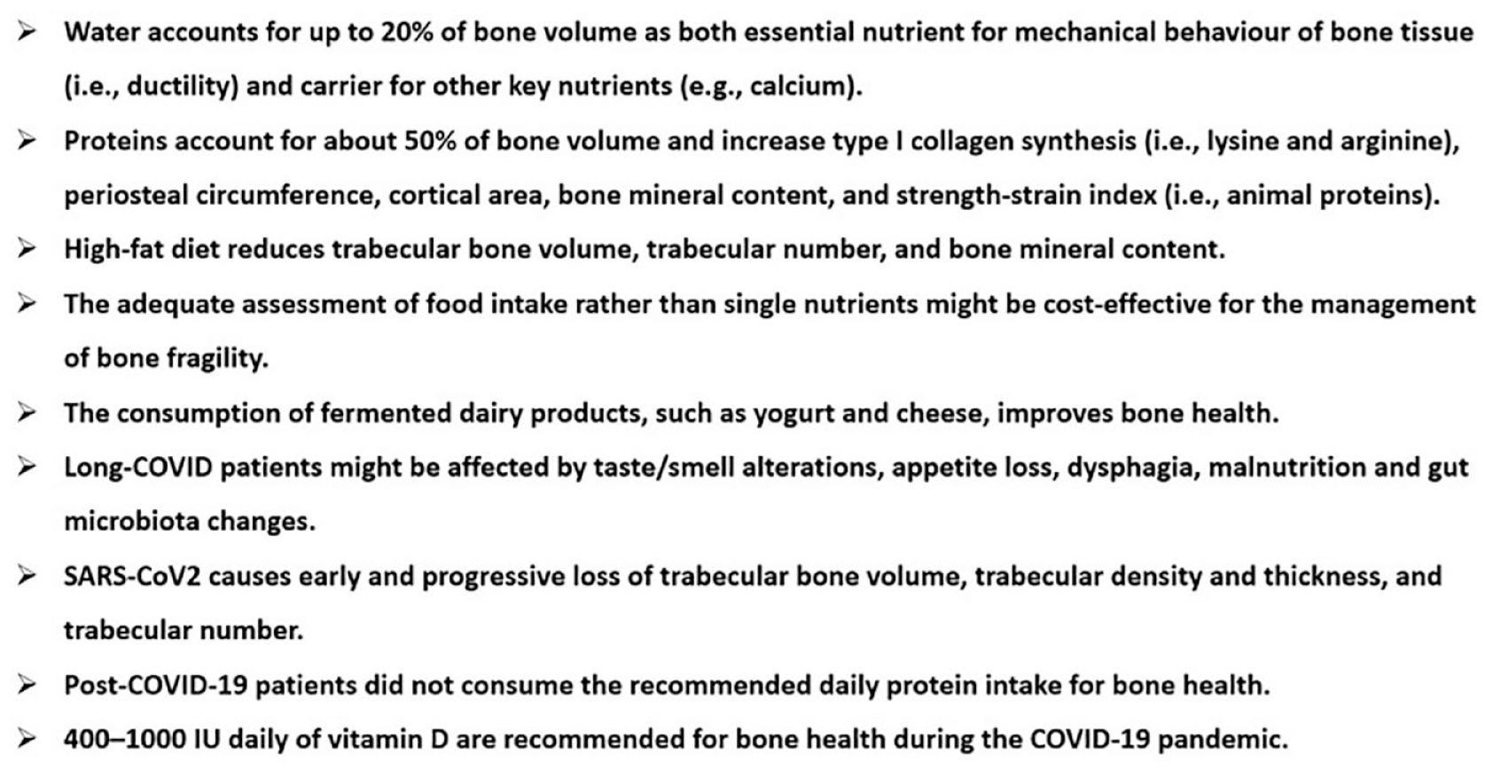
Bone fragility, nutrition, and COVID-19: highlights.
Conclusion
Several macro- and micronutrients play pivotal roles in the homeostasis of bone health, and their adequate intakea, such as food or dietary supplements, have biological plausibility for the management of bone fragility. The COVID-19 pandemic upsets lifestyle habits, including nutrition and dietary patterns, increasing the risk of skeletal fragility. In this context, the comprehensive management of COVID-19-related complications, including bone fragility, should provide an adequate intake of nutrients, starting from waters rich in calcium and bicarbonate to macronutrients, such as proteins rich in lysine and arginine, monounsaturated fatty acids (MUFAs), and water-soluble fibers containing inulin, and micronutrients, such as calcium, magnesium, vitamin C, vitamin D, vitamin K, copper, Si, and strontium, although, for some of these nutrients, no evidence is available so far.
On the other hand, during the COVID-19 pandemic, the number of consumers of dietary supplements significantly increased.
In conclusion, beyond the putative benefits of these substances, it is necessary to carefully consider that their safety profile is not systematically monitored and that not all nutraceuticals are approved by regulatory agencies, while advertising for these products is often not based on strong evidence and sometimes misleading. Finally, some dietary supplements are claimed as effective (e.g. to cure COVID-19) despite a lack of consistent data or negative findings drawn from clinical trials.