
Abstract
Background:
Head and neck squamous carcinoma (HNSCC) is caused by different exogenous risk factors including smoking cigarettes, alcohol consumption, and HPV infection. Base excision repair (BER) is the frontline to repair oxidative DNA damage, which is initiated by the DNA N-glycosylase proteins (OGG1) and other BER factors including DNA polymerase β (POLB).
Objective:
Explore whether BER genes’ (OGG1, POLB) overexpression in HNSCC alters genomic integrity, immunogenicity, and its role in prognostic value.
Design:
RNA sequencing (RNA-Seq) and clinical information (age, gender, histological grade, survival status, and stage) of 530 patients of HNSCC were retrieved from the Cancer Genome Atlas. Patients’ data are categorized HPV positive or negative to analyze the tumor data including the tumor stage, POLB, and OGG1 gene expression.
Methods:
RNA-Seq of HNSCC data retrieved and mutation count and aneuploidy score were compared using an unpaired t-test. The TIMER algorithm was used to calculate the tumor abundance of six infiltrating immune cells (CD4+ T cells, CD8+ T cells, B cells, neutrophils, macrophages, and dendritic cells) based on RNA-Seq expression profile data. The correlation between the POLB, OGG1, and immune cells was calculated by Spearman correlation analysis using TIMER 2.0.
Results:
Our data analysis reveals that BER genes frequently overexpressed in HNSCC tumors and increase mutation count. In addition, OGG1 and POLB overexpression are associated with low infiltration of immune cells, low immune checkpoint gene expression (PD-1, cytotoxic T-lymphocyte antigen 4, program death ligand 1, and program death ligand 2), and innate immune signaling genes. Furthermore, dysregulated BER factors in Human papillomavirus (HPV) positive tumors had better overall survival.
Conclusion:
Our analysis suggests that dysregulation of the BER genes panel might be a potential prognosis marker and/or an attractive target for an immune checkpoint blockade in HNSCC cancers. However, our observation still requires further experimental-based scientific validation studies.
Introduction
Head and neck carcinoma (HNSCC) is a heterogeneous tumor located at the nasopharynx, oropharynx, hypopharynx, larynx, and oral cavity. 1 HNSCC accounts for about 4% of all cancers in the United States and an estimated 66,920 people (49,190 men and 17,730 women) will be diagnosed in 2023. 2 Over 90% of HNSCC are squamous cell carcinomas and are caused mainly by environmental risk factors.3 –6 In addition, alternation of the genetic landscape of DNA damage repair enhances susceptibility to HNSCC.7,8
Interestingly, HPV-positive HNSCCs have distinct clinical, pathological, and molecular features compared to HPV-negative HNSCCs.9,10 HPV-induced HNSCCs are triggered by E6 and E7 onco-viral proteins. E6 promotes the degradation of the cellular proteins that lead to cell proliferation, and E7 promotes genomic instability and resistance to apoptosis. 11 Furthermore, E6 and E7 cause dysregulation of the cell cycle and DNA repair pathways, resulting in a dramatic increase in genomic instability.12–14 HPV-positive HNSCCs harbored oxidative stress associated with DNA damage with impaired DNA repair.12,15
Base excision repair is one of the key pathways to repair oxidative and alkylating agents and dysregulated in the tumor.16,17 In the first step of BER, reactive oxygen species-associated DNA damage, including oxidation of guanine to 8-dihydro-7,8-oxoguanosine (8-oxodG), is processed by OGG1 and apurinic/apyrimidinic endonuclease I. 18 This leads to a DNA gap, which is then filled with one or more nucleotide by DNA polymerase β (Pol β) and the DNA nick is sealed by DNA ligase III. 19 The DNA glycosylase not only recognizes and removes the damaged base but also protects cells from mutagenic and/or cytotoxic effects of DNA base lesions while still maintaining genomic integrity.20,21 Aberrant expression of BER creates an imbalance in the BER pathway, which leads to saturation of downstream repair enzymes and/or accumulation of genotoxic BER intermediates.18,22,23
BER expression is deregulated in several types of cancer, which leads to genomic instability, ultimately impacting treatment outcomes.24–27 To date, many studies have tried to establish prognostic signatures, including gene sets 28 as promising predictors of prognosis for HNSCC. There is a significant need to explore potential prognostic predictors in HNSCC. Therefore, it is crucial to identify new DNA repair markers that predict an antitumor immune response to achieve personalized treatment. In this study, the expression of OGG1 and Pol β in HNSCC tumors and the relationship between overexpression of POLB and OGG1 with genomic instability and tumor immune infiltration were investigated.
Methods
Data acquisition
RNA sequencing (RNA-Seq) and clinical information (age, gender, histological grade, survival status, and stage) of HNSCC (n = 530) were retrieved from the Cancer Genome Atlas [TCGA pan-cancer data sets: cBioProtal (http://cbioportal.org)]. Patients’ data are categorized HPV positive or negative to analyze the tumor data including the tumor stage, POLB and OGG1 gene expression, and patient characteristics.
Exclusion and inclusion criteria
Since BER (POLB and OGG1) is the primary source of interest for this analysis, only individuals with valid RNA Seq V2 RSEM data for BER were included. Individuals with a z-score of ⩽−0.5 were placed into the low-expressing group while individuals with a z-score of ⩾0.5 were placed into the high-expressing group. Individuals falling between −0.5 and 0.5 were excluded from the analyses.
Mutation count and aneuploidy score
The mutation counts and aneuploidy score for each individual were extracted from the TCGA data set for HNSCC. Values were grouped according to low and high BER gene expression for a given cancer type and an unpaired t-test was used to assess the statistical differences between the two groups. Data analysis was done using a Graph Pad prism.
Immune cell infiltration in HNSCC
The TIMER algorithm was used to calculate the tumor abundance of six infiltrating immune cells (CD4+ T cells, CD8+ T cells, B cells, neutrophils, macrophages, and dendritic cells) based on RNA-Seq expression profile data. The correlation between the POLB, OGG1, and immune cells was calculated by Spearman correlation analysis using TIMER 2.0.
Estimation of stromal and immune cells in tumor tissues
The ESTIMATE algorithm-generated matrix, immune scores, and stromal scores were used to estimate the level of infiltrating matrix and immune cells in HNSCC tissue and tumor purity through expression profiles. Results for the tumor-infiltrating immune component were yielded with data extracted from the TCGA database, which was analyzed by the CIBERSORT algorithm.
Statistical analysis
Group comparisons for continuous data were conducted using t-tests, and quantitative variables were analyzed with the Wilcoxon signed-rank test or the Spearman rank correlation test. Kaplan–Meier analysis was used to assess overall survival (OS). Uni-variable associations between BER overexpression and clinicopathologic variables were tested using nonparametric tests. Statistical significance was set at p < 0.05.
Results
Patient data analysis and clinical characteristics of BER factors in HNSCC patients
Data were accessed from cBioPortal (www.cBioPortal.org). TCGA PanCancer RNA-seq data and corresponding clinic information for HNSCC types were retrieved for validation. TCGA pan-cancer data sets were analyzed based on the expression of OGG1 and POLB from 523 HNSCC patients. The mRNA expression of OGG1 and POLB was significantly high in tumor versus normal adjacent tissues [Figure 1(a); p < 0.001]. In addition, POLB and OGG1 overexpression enhanced in patients infected with HPV [Figure 1(a); p < 0.001]. Further high expression of POLB has a positive lymph node status in HNSCC [Figure 1(b), *p < 0.05], and a higher histological grade tumor (G3/4) [Figure 1(b), **p < 0.01].

Overexpression of OGG1 and POLB in HNSC cancer. (a) The comparison of expression of mRNA in normal tissue adjacent to tumor versus tumor with and without HPV infection; HNSCC (n = 520); HNSCC-normal (n = 44); HNSCC-HPV+ (n = 97); and HNSCC-HPV− (n = 420). (b) Clinical characteristics of tumor stages, histological grades, and lymph node status.
BER overexpression correlated with overall survival of HNSCC patients
To investigate whether the aberrant expression of the BER in tumors alters patient survival, Kaplan–Meier survival curves were generated based upon the designation of individuals as either HPV negative or HPV positive with a high level of BER expression (z-score > 0.5) to assess the impact of survival for each gene set of HNSCC patients. The results showed that high OGG1 and POLB expression levels were significantly associated with better survival outcomes in stage III/IV versus low POLB-expressed tumors [Figure 2(a)–(f)]. In this study, we confirmed that the OGG1 and POLB overexpressed tumors on advanced pathologic stages III and IV tumors with high OGG1 overexpression achieve better overall survival outcomes. However, patients with stages II, III, and IV have no significant difference in survival, indicating that the pathological stage has certain defects in predicting the survival of HNSCC patients. OGG1 overexpressing tumors associated with better overall survival outcomes in HPV-positive patients [Figure 2(c); p < 0.001]. However, no significant change in the overall survival of patients associated with POLB overexpressed tumors was found with and without HPV infections [Figure 2(d)–(f)]. However, POLB overexpression slightly enhances the overall survival of HPV-positive HNSCC patients [Figure 2(d); p > 0.05]. Our data show that as BER mRNA levels increased, the outcomes for the HPV-positive HNSCC cancer patients became better, and the relative risk of death decreased.

Kaplan–Meier survival analysis of POLB and OGG1 expression. Time-dependent analysis of OGG1 and POLB gene expression (low and high groups) with and without HPV infection. (a) Overall survival of patients with Stage III/IV in OGG1 gene expression in tumor with high versus low. (b) The cumulative overall survival of HNSCC patients with mRNA of OGG1 high versus low expression. (c) The cumulative overall survival of HNSCC patients with OGG1 high expression versus low with HPV infection. (d) Overall survival of patients with Stage III/IV in POLB gene expression in tumor with high versus low. (e) The cumulative overall survival of HNSCC patients with mRNA of POLB high versus low expression. (f) The cumulative overall survival of HNSCC patients with high POLB mRNA expression versus low HPV infection. For Kaplan–Meier curves, p values and HR with 95% CI were generated by log-rank tests and univariate Cox proportional hazards regression. p < 0.05 was considered statistically significant.
Aberrant BER gene expression is associated with aberrant genomic instability in HNSCC tumor
BER plays a significant role in maintaining the genomic stability of cells. To examine whether aberrant BER gene expression is associated with genomic instability of HNSCC, we examined whether POLB and OGG1 expression changes in HNSCC with and without HPV infection are associated with mutation and chromosomal aberration. We found that the overexpression of OGG1 and POLB overexpression was significantly associated with the increased mutation count of the HNSCC tumors [Figure 3(a)–(d)]. Particularly, mutation count significantly increased in OGG1 and/or POLB overexpressed tumors [Figure 3(a) and (c); *p < 0.5]. In addition, OGG1 overexpressing tumors harbor significantly lower aneuploidy scores in HNSCC patients versus OGG1 low-expressing groups [Figure 3(b); *p < 0.05]. By contrast, POLB overexpression did not alter the level of aneuploidy score [Figure 3(d)].

BER overexpressed tumors accumulate genomic instability. (a) Mutation counts in OGG1 high expressed tumor (z-score > 0.5) versus low expressed tumor (z-score <−0.5). (b) Chromosomal instability (aneuploidy score) of OGG1 high expressed versus low expressed tumor groups. (c) HNSCC cancer mutation counts in POLB high versus low expressed tumor. (d) Chromosomal instability in POLB high versus low expressed tumor. The Mann–Whitney U test was used to compare the mutation count in high OGG1 and POLB-expressed tumors versus low OGG1 and POLB-expressed tumors, respectively.
Aberrant expression of BER genes is associated with changes in immune cell infiltration
Increasing evidence suggests that cancer progression is strongly influenced by the host’s immune response, which is represented by immune cell infiltrates. We compared changes in immune and stromal scores between high expression versus low expression of BER genes (OGG1 and POLB) tumor samples. Intriguingly, high OGG1 and/or POLB expression was associated with reduced innate and adaptive immune cell abundance, including CD8+ T cells, B cells, and dendritic cells (DC) [Figure 4(a)–(g)]. However, OGG1 overexpressed tumors were associated with a favorable higher immune score and ESTAMATE score but not stromal score [Figure 4(c); ****p < 0.001 and ***p < 0.01]. Interestingly, a high immune score was exhibited in neoplasm histological grade 3 and 4 tumors [Figure 4(d); *p < 0.5]. By contrast, POLB high-expressed tumors are associated with low immune, stromal, and ESTIMATE scores versus POLB low-expressed tumors [Figure 4(e)–(g); *p < 0.05; **p < 0.01; and ***p < 0.001]. In addition, the association of histological grades with immune scores as well as stromal scores is weak in POLB overexpressed tumors [Figure 4(g)]. To determine whether there is a correlation between immune cell infiltration and BER gene expression, the tumor infiltration with multiple immune cells was analyzed by TIMER 2.0 methods. Our heatmap data analysis shows that infiltration of four immune cells is decreased in OGG1 overexpressed HNSCC, HNSCC-HPV−, and HNSCC-HPV+ tumors. Particularly, CD8+ T cells [Figure 5(a) and Supplemental Figure 1(A); correlation HNSCC = −0.34, p = 1.43e−14; HNSC-HPV− = −0.28 p = 2.2e−08; HNSCC-HPV+ = −0.42, p = 5.99e−05] and B-cell infiltration significantly low, regardless of the HPV status off HNSCC [Figure 5(a) and Supplemental Figure 1; HNSCC correlation = 0.29, p = 1.28e−11; HNSCC-HPV− = 0.32, p = 7.7e−11; HNSCC-HPV+ = −0.24, p = 2.39e−02]. In addition, POLB overexpressed tumors have low infiltration of CD8+ T cells (HNSCC correlation = −0.39, p = 1.65e−04; HNSCC-HPV− = −0.03, p = 1.15e; HNSCC-HPV+ = 0.39, p = 1.65e−04); B cells [HNSCC correlation = −0.2; p = 5.53e−06; HNSCC-HPV− = −0.2; p = 6.79e−05; and HNSCC-HPV+ = −0.289, p = 6.06e−03; Figure 5(b) and Supplemental Figure 1(B)].

Landscape of tumor immune microenvironment impact on HNSCC histological tumor grades. (a) Immune, stromal, and ESTIMATE score of the tumor with high versus low OGG1 mRNA expression. (b) OGG1 high mRNA expression with immune score impact on histologically high-grade G3/4. (c) OGG1 high mRNA expression with stromal score impact on histologically high-grade G3/4. (d) Immune, stromal, and ESTIMATE score of the tumor with high versus low POLB mRNA expression. (e) OGG1 high mRNA expression with immune score impact on histologically high-grade G3/4. (f) High POLB mRNA expression with stromal score impact on histologically high-grade G3/4. The Mann–Whitney U test was used to compare the expression of OGG1 and POLB versus immune score as well as stromal score.
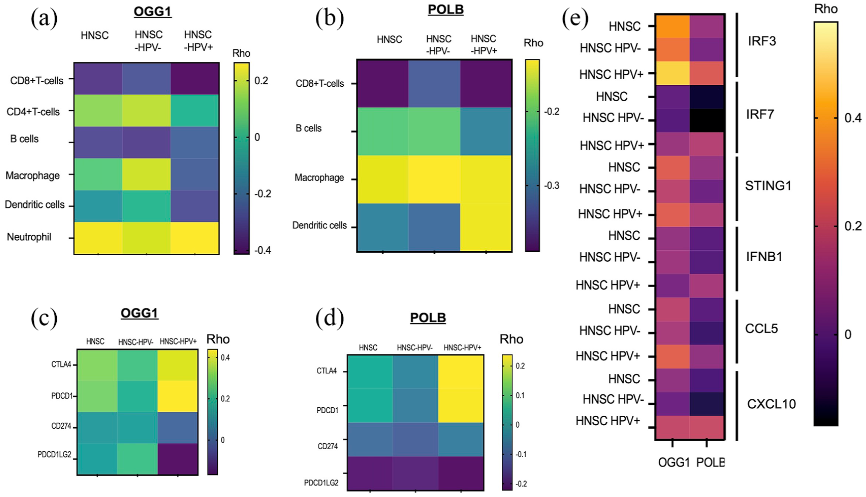
Overexpression of OGG1 and POLB is associated with a low level of infiltration of immune cells to the tumor microenvironment. (a) The expression of OGG1 mRNA versus immune cell infiltration negatively correlated on CD8+ T cells, B cells, and DC cells. (b) The correlation of POLB expression versus immune cells (CD8+ T, CD4+ T, B cells, macrophages, and DC cells). (c) Heatmap of innate immune signaling gene expression versus OGG1 and POLB mRNA expression in HNSC with and without HPV infection. (d, e) Landscape of immune checkpoint genes in OGG1 and POLB overexpressed tumor. (d) Heatmap of OGG1 mRNA expression versus immune checkpoint gene in HNSCC. (e) Heatmap of POLB mRNA expression versus immune checkpoint gene in HNSCC. The Spearman’s correlation was performed to analyze the relationship between the OGG1 and POLB expression versus immune checkpoint genes (CTLA-4, CD274 PDCD1, and PDCD1GL2).
Association of BER with immune checkpoint genes and innate immune signaling landscape in HNSCC
We also analyzed the correlation between OGG1 and POLB genes in innate immune gene expression as well as immune checkpoints in HNSC with and without HPV infection. The expression of OGG1 and POLB positively correlated with the expression of immune checkpoints including CTLA-4 and PDCD1 expression [Figure 5(c) and (d)]. Interestingly, the positive correlation is more pronounced in HPV-positive tumor groups. However, expression of CD274 and PDCD1GL2 negatively correlated with overexpression of OGG1, POLB [Figure 5(c) and (d)]. In addition, in HNSC patents with and without HPV infection, the innate immune signaling genes (IFNB1, IRF3, IRF7, STING1, CCL5, and CXCL10) negatively correlated with POLB and OGG1 expression [Figure 5(e)]. However, we have also noticed that OGG1 expression is positively correlated with interferon regulatory factor-3 (IRF3) expression in HPV-positive tumors [Figure 5(e)].
Discussion
POLB and OGG1 are key enzymes for the protection of the genome against DNA damage via their role in BER. 29 In this work, we report that OGG1 and POLB are overexpressed in a majority of HNSCC. The overexpression of OGG1 and POLB in tumors may contribute to the repair of oxidative DNA damage and or play a significant role in cellular proliferation. Our results show that OGG1 and POLB overexpression significantly increase the overall survival of HPV-infected HNSC patients (Figure 1), suggesting that the high DNA repair capacity of the infected cells likely repairs oxidative DNA damage. Our data agree with previous studies that have demonstrated that HPV-positive HNSCC patients had improved survival compared with HPV-negative patients. 30 Interestingly, our in silico analysis shows that high OGG1 expression favors overall survival of Stage III/IV HNSCC supporting that an important feature of a specific HNSCC with better prognosis. By contrast, Yousaf et al. 31 ’s study showed that the dysregulation of OGG1 expression may promote tumorigenesis. OGG1 overexpression might stimulate cell proliferation by increasing the activity of cell cycle-related proteins. 32
Notably, high expression levels of cellular BER factors (OGG1 and POLB) could be a protective response that occurs in cancer to manage HPV oncogenes-dependent DNA damage. 33 However, BER genes commonly mutated or overexpressed in cancer lead to the accumulation of mutations or chromosomal instability. 34 Our in silico study shows that OGG1 and POLB overexpressed tumors harbor significantly high mutation frequency that likely contributes to genomic instability mediated tumor initiation. 34 The mechanism of inducing mutation loads due to overexpression of OGG1 and POLB is likely associated with frameshift mutation. 35 By contrast, in vitro studies elucidated that OGG1 expression promotes genomic stability via the repair of oxidative DNA damage. 36
It is known that the tumor immune microenvironment has great implications for cancer progression and susceptibility to immunotherapy. 37 Recently, we have shown that defective BER induces innate immune signaling and Type I interferon response cytokines. 38 Moreover, sustained signaling through interferon-induced pathways has also been associated with resistance to immune checkpoint inhibitors through upregulation of program death ligand 1 (PD-L1), program death ligand 2 (PD-L2), CTLA-4, CIITA, IDO1, CXCL12, and nitric oxide production in tumor cells.39–42 We found that antitumor cells including CD8+ T cells and CD4+ T cells were less abundant in the tumor samples, while several types of immunosuppressive cells were more abundant in the low-immune score patients. Interestingly, overexpression of OGG1 and POLB is associated with low immune cell infiltration and low expression of Type I interferon response genes that are responsible for cytokine-driven immune cell recruitment and inflammation. Several studies have shown that Type I interferon responses result in upregulation of immune checkpoint proteins such as PD-L1, 43 resulting in inhibition of T-cell-mediated cancer cell killing.44–46 Furthermore, CTLA-4 (CD152) is a receptor expressed by both CD4+ T and CD8+ T cells, which inhibit T-cell activation, positively correlated in OGG1 and POLB overexpression. PDCD1 expression, which is a cell surface receptor on T cells and B cells that has a role in regulating the immune system positively correlated with OGG1 and POLB overexpressed tumors. By contrast, the tumors had low expression of PD-L1 and PD-L2 associated with OGG1 and POLB overexpressed tumors suggesting that the BER overexpression likely contributes to immunosuppressive tumor microenvironments. The data demonstrate that OGG1 and POLB negatively correlated with innate immune gene expression and immune cell infiltration, suggesting that OGG1 and POLB may be involved in regulating an antitumor immune response.
Conclusion
Taken together, our silico-data-driven observations suggest that tumors with dysregulated BER factors may enhance tumor immunogenicity that likely enhances immune checkpoint blockade therapy response. In addition, using POLB and OGG1 overexpression may likely contribute to identifying reliable biomarkers to stratify patients for appropriate immune-based treatment. Nevertheless, there is a clear need to understand the mechanisms through which aberrant BER contributes to the immunogenicity of the tumor either through increased tumor mutation burden or concurrent activation of the innate immune signaling via STING pathway that may lead to enhanced T-cell-mediated cancer cell death. Overall, the data analysis from the TCGA dataset suggests dysregulated BER factors in tumors appear to avoid host immune-mediated elimination through activation of immune checkpoints, raising the possibility that checkpoint blockade may represent an effective treatment strategy for BER dysregulated HNSCC tumors. Furthermore, our data suggest that a systemic understanding of the DNA repair landscape in a tumor may help assess the tumor’s susceptibility to immunotherapy. We recognized that this study has some limitations that excluded patients with a z-score ranging between −0.5 and 0.5 which may impact the interpretation of the result. Furthermore, future in vitro and in vivo study required to provide scientific evidence to further explore the molecular mechanism of how dysregulated BER regulates HNSCC tumor immunogenicity.
Supplemental Material
sj-jpg-1-tam-10.1177_17588359241248330 – Supplemental material for Dysregulation of base excision repair factors associated with low tumor immunogenicity in head and neck cancer: implication for immunotherapy
Supplemental material, sj-jpg-1-tam-10.1177_17588359241248330 for Dysregulation of base excision repair factors associated with low tumor immunogenicity in head and neck cancer: implication for immunotherapy by Zackary Shpilman and Dawit Kidane in Therapeutic Advances in Medical Oncology
Supplemental Material
sj-jpg-2-tam-10.1177_17588359241248330 – Supplemental material for Dysregulation of base excision repair factors associated with low tumor immunogenicity in head and neck cancer: implication for immunotherapy
Supplemental material, sj-jpg-2-tam-10.1177_17588359241248330 for Dysregulation of base excision repair factors associated with low tumor immunogenicity in head and neck cancer: implication for immunotherapy by Zackary Shpilman and Dawit Kidane in Therapeutic Advances in Medical Oncology
Footnotes
References
Supplementary Material
Please find the following supplemental material available below.
For Open Access articles published under a Creative Commons License, all supplemental material carries the same license as the article it is associated with.
For non-Open Access articles published, all supplemental material carries a non-exclusive license, and permission requests for re-use of supplemental material or any part of supplemental material shall be sent directly to the copyright owner as specified in the copyright notice associated with the article.