
Abstract
Introduction
Urolithiasis is a multifactorial condition affecting about 10% of the population worldwide, making it one of the most common diseases affecting the urinary tract. 1 Without appropriate management, this can lead to considerable morbidity, underscoring the importance of accurate diagnosis and optimal treatment strategies. In the modern world, the latter aspects are usually carried out through the prism of personalized and evidence-based medicine, which emphasize the need to consider individual patient factors and adhere to established clinical guidelines to maximize treatment efficacy while minimizing complications and the need for repeat interventions.2,3 The latest version of the European Association of Urology (EAU) guidelines makes a clear distinction between patients depending on the localization of the stone.4,5 Moreover, when choosing a surgical strategy for stones located in the lower pole, the guideline recommends considering infundibular length (IL), width (IW) and lower pole-based angle (LPA) to guide the selection of the most appropriate treatment modality. 6 Until 2025, EAU guidelines included specific cut-off values for these parameters to stratify patients, whereas the latest 2025 update omits such thresholds, reflecting the urological community’s recognition of the poor reproducibility of the original measurements. For example, when measuring IL and IW, no standardized criteria exist for defining the start and end points of measurements. The ambiguity is even greater for LPA, which is typically defined as the angle between two manually drawn lines and is often considered the most critical predictor of treatment outcomes for stones in this location.7,8 Moreover, manual measurement of LPA, IL, and IW is subject to significant intra- and inter-observer variability, limiting the clinical utility of fixed cut-off values. This issue is longstanding, and Knoll et al. 9 reported poor interobserver agreement for LPA (correlation coefficient < 0.44), and only modest agreement for IL (0.49) and IW (0.63). 9
This editorial aims to propose a conceptual semi-automated method for measuring these anatomical features. The outlined approach, based on the anatomical contours of the lower pole, is presented as a proof-of-concept that could potentially be integrated into standard imaging viewers with further development and validation.
Description of proposal
Currently, we have developed a prototype overlay software to illustrate how the proposed approach could be implemented on top of existing medical image viewers. After displaying the non-contrast computed tomography (NCCT) images and selecting the optimal plane, the program can be overlaid to set the boundaries of the frame and calibrate the measurements. A visual demonstration of this proof-of-concept, named EndoRad, is available as a video example using the freely available, open-source Horos viewer for macOS (HorosTM; https://youtu.be/J6PDgrXezWo?si=wow2d_MztggfNiYP).
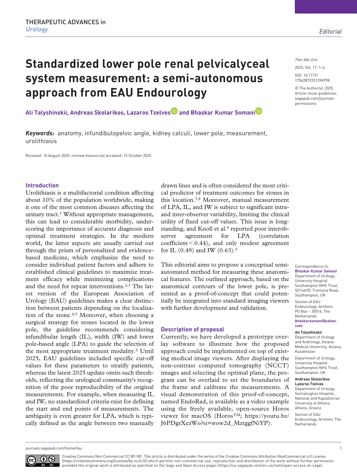
It is necessary to perform multiplanar reconstruction (MPR) of NCCT images to display them in all planes. Next, an oblique MPR should be performed in the frontal plane, taking into account both the spatial configuration of the pelvicalyceal system (PCS) and the orientation of the kidney itself. The aim is to align the imaging plane with the kidney’s frontal axis, not the patient’s, to visualize the lower calyces, infundibulum, and renal pelvis within a single slice. We propose using a quadrangular reference frame aligned with the medial edge of the kidney and spanning from the upper to the lower pole. The ideal frontal tilt is defined as the first slice where the lower calyces, infundibulum, pelvis, and ureteropelvic junction (UPJ) are simultaneously visualized, with the UPJ intersecting the medial border of the frame (Figure 1).

MPR of NCCT images to display them in all planes. Then, oblique multiplanar reconstruction in the frontal plane aligned to the kidney’s frontal axis, with a quadrangular reference frame from upper to lower pole. The optimal slice shows the lower calyces, infundibulum, pelvis, and UPJ, with the UPJ intersecting the frame’s medial border.
Once the plane is defined and the reference frame is constructed, the next step is to identify key anatomical points used to calculate IL and LPA. The initial and final points are placed at the intersection of the UPJ with the medial edge of the frame, and at the transition between the papilla and the most inferior visible minor calyx, respectively. Between these two landmarks, a series of at least four intermediate points are plotted along the PCS contour. A continuous line is then generated through these control points using a dedicated command. This approach enables simultaneous calculation of two parameters: IL, as the total length of the constructed line, and LPA, as the mean curvature angle derived from the line’s trajectory using Bézier curves. Then, points are placed along the opposite margin of the infundibulum or calyx (in cases where the lower calyces drain directly into the pelvis and no distinct infundibulum is present). The initial point is set at the junction between the renal pelvis and the infundibulum or calyx, while the final point is placed at the junction between the most proximal visible minor calyx and the papilla, or at the corresponding location if the calyx drains directly into the pelvis. Intermediate points are then positioned along the infundibular or calyceal contour, following the same methodology as in the previous step. Once all points are marked, a command is used to generate a second line representing the opposing boundary of the measured PCS segment. The narrowest distance between the two lines is then calculated automatically and corresponds to the IW (Figure 2).

Semi-automated calculation of IL, LPA (mean curvature from Bézier curve), and IW using fixed start/end points and margin-based contouring.
The type of scope and its defectivity can also influence the accessibility to the lower pole. 10 Given that ureteroscopy is amongst the most frequently performed procedures, improving patient selection through standardized lower pole measurements has the potential to reduce healthcare expenditure. 11
Conclusion
This editorial presents a conceptual approach for semi-automated measurement of IL, IW, and LPA by leveraging functionalities already available in most medical imaging viewers and standardizing the placement of start and end points and aligning measurements with the true anatomical contours of the PCS. While promising, the approach remains a methodological concept; future validation studies will be required to confirm its reproducibility, accuracy, and clinical utility.