
Abstract
Background:
Diuretics are current antihypertensive drugs since they reduce blood pressure and cardiovascular risk. Increased vascular tone is modulated in a relevant way by the RhoA/Rho-kinase (ROCK) pathway, by acting on vascular smooth muscle cell contraction. This pathway has also proremodeling vascular effects. There are few data on the role of diuretics on both vascular ROCK activation and on proremodeling effects. We assessed the effects of hydrochlorothiazide (HCTZ) and spironolactone (spiro) alone and in combination with the ROCK inhibitor fasudil (FAS) on ROCK activation, gene expression of proremodeling markers and on hypertrophy in the aortic wall of hypertensive rats.
Methods:
Deoxycorticosterone acetate (DOCA)-salt hypertensive rats (male, Sprague–Dawley) were randomized to the specific ROCK inhibitor FAS, HCTZ, spiro or the combinations of FAS/HCTZ or FAS/spiro for 3 weeks. At the end of the study, ROCK activation (by western blot), gene expression of proremodeling markers (by reverse transcription polymerase chain reaction, RT-PCR) and vascular hypertrophy (by morphometry) were determined in the aortic wall.
Results:
All treatments significantly reduced blood pressure. In the DOCA rats the p-myosin phosphatase target protein-1 (MYPT1)/t-MYPT1 ratio, index of ROCK activation was higher by 2.8 fold (p < 0.05) compared with control rats. All treatments reduced ROCK activation in the aortic wall to control levels (p < 0.05). Besides, significantly increased protein levels of transforming growth factor β1 (TGF-β1), gene expression of TGF-β1, connective tissue growth factor (CTGF), p22 phox and gp91 phox subunits of nicotinamide adenine dinucleotide phosphate (NADPH) oxidase, as well as increased media thickness and aortic media area/lumen area (AM/LA) in the untreated hypertensive rats were significantly reduced (p < 0.05) to control levels by all treatments. Similar effects were observed using both diuretics alone or in combination with FAS.
Conclusions:
In the aortic wall, both HCTZ and spiro in antihypertensive doses reduce ROCK activation, subsequent expression of genes that promote vascular remodeling and hypertrophy in this experimental model of hypertension. These effects could explain some of their clinical benefits in hypertensive patients.
Keywords
Introduction
Current antihypertensive treatment, by targeting specific pathophysiological and cellular pathways and mechanisms, normalizes high blood pressure (BP), slows down the progression of target organ damage, and reduces the incidence of heart and renal failure. Those favorable clinical effects are consequences of the effects of the antihypertensive agents on arterial vasoconstriction, intravascular volume and on cardiovascular remodeling.
One novel intracellular mechanism that triggers vasoconstriction and cardiovascular remodeling is the small guanosine triphosphatase Rho and its target Rho-kinase (ROCK). Both enzymes have important roles in BP regulation, vascular smooth muscle contraction, and also in cardiovascular and renal remodeling [Jalil et al. 2005, 2010; Shi and Wei, 2013; Loirand and Pacaud, 2010; Surma et al. 2011; Satoh et al. 2011; Shimokawa and Satoh, 2015].
Rho is activated by the agonists of receptors coupled to the cell membrane G protein, such as angiotensin II (Ang II) and noradrenaline. Activated Rho translocates to the cell membrane, where it activates ROCK. Activated ROCK has an important role in mediating various cellular functions, such as vascular smooth muscle cell contraction, actin cytoskeleton organization, adhesion and motility, cytokinesis, and expression of genes involved in cardiovascular and renal remodeling. ROCK also mediates the upregulation of several proinflammatory, thrombogenic and fibrogenic molecules, and the downregulation of endothelial nitric oxide (NO) synthase (eNOS) [Jalil et al. 2005, 2010; Shi and Wei, 2013; Loirand and Pacaud, 2010; Surma et al. 2011; Satoh et al. 2011; Shimokawa and Satoh, 2015]. Thus, when ROCK is activated, inflammation, thrombosis and tissue fibrosis are accelerated, whereas endothelial NO production is inhibited. In addition, ROCK activation promotes the activation of vascular nicotinamide adenine dinucleotide phosphate (NADPH) oxidase and oxidative stress [Rivera et al. 2007].
In hypertensive patients, the ROCK inhibitor fasudil (FAS) induced a larger vasodilator response in the arm compared with control subjects, whereas the vasodilator response to nitroprusside was similar in both groups [Masumoto et al. 2001], the first clinical evidence about the role of the RhoA/ROCK pathway in the pathogenesis of increased systemic vascular resistance in hypertensive patients. In untreated hypertensive patients, there is higher ROCK activity in peripheral blood leukocytes compared with healthy individuals [Hata et al. 2011; Gabrielli et al. 2014]. Besides, in patients with hypertension treated with antihypertensive agents, ROCK activity was lower in patients using calcium channel blockers compared with groups treated with renin-angiotensin system inhibitors, diuretics, or beta-blockers [Hata et al. 2011].
Diuretics, particularly thiazides, are relatively old antihypertensive agents. However, currently they are first line antihypertensive drugs [James et al. 2014; Mancia et al. 2013] since at common doses they produce a similar reduction in BP that is comparable with other antihypertensives [Law et al. 2009] and evidence from subsequent analyses of ALLHAT and other clinical outcome trials confirms that in hypertensive patients neither alpha-blockers, angiotensin-converting enzyme (ACE) inhibitors, nor calcium channel blockers surpass thiazide-type diuretics as initial therapy for reduction of cardiovascular or renal risk [Wright et al. 2009]. Thiazides are superior in preventing heart failure and new-onset diabetes mellitus associated with thiazides does not increase cardiovascular disease outcomes [Wright et al. 2009].
However, there are few studies assessing the role of diuretics on vascular ROCK activation in hypertension. In aortic rings from normotensive Wistar rats, both hydrochlorothiazide (HCTZ) and chlortalidone inhibited agonist-induced vasoconstriction in a concentration-dependent manner without changing intracellular calcium [Zhu et al. 2005]. In those in vitro conditions, the inhibitory effects of both diuretics were similar to the Rho-kinase inhibitor Y27632, whereas both RhoA and Rho-kinase mRNAs were significantly reduced in cultured vascular smooth muscle cells after administration of both diuretics [Zhu et al. 2005]. In aldosterone/salt-induced hypertensive rats increased cardiac expression of RhoA, Rho-kinase mRNA and myosin light chain (MLC) phosphorylation were decreased by spironolactone (spiro) or Y27632 [Nakano et al. 2005]. In the abovementioned study, spiro also suppressed upregulated expression of cardiac ACE, epidermal growth factor receptor (EGFR), LOX-1, of NAD(P)H oxidase subunits and of p44/p42ERK phosphorylation [Zhu et al. 2005]. In the renal cortex in Dahl salt-sensitive hypertensive rats eplerenone, an analogue of spiro, reduced Rho-kinase higher expression and also decreased expression levels of LOX-1, ICAM-1, and VCAM-1 [Kobayashi et al. 2005].
We hypothesized here that in experimental hypertension, diuretics, common antihypertensive agents, reduce ROCK activation and the consequent vascular expression of genes that promote remodeling in the vascular wall. We also assessed in this experimental context the effect of combining a diuretic with the direct ROCK inhibitor FAS on hypertension and on ROCK activation.
Materials and methods
This investigation complied with the Guide for the Care and Use of Laboratory Animals published by the United States National Institutes of Health (NIH publication N885 to 23, revised 1996), and it was approved by the Research Commission from the School of Medicine, Pontificia Universidad Católica de Chile. The deoxycorticosterone acetate (DOCA)-salt hypertensive model was used in Sprague–Dawley male rats, aged 5–6 weeks (150 ± 10 g) as previously described [Ocaranza et al. 2011]. Under anesthesia with ketamine HCl and xylazine (35/7 mg/kg intraperitoneally (ip), respectively), a left nephrectomy was performed. Afterwards, DOCA (100 mg/kg, subcutaneous; Steraloids Inc., Newport, Rhode Island, USA) once a week was administered starting immediately after recovery of surgery. The animals received 1% NaCl and 0.4% KCl in the drinking water [Ocaranza et al. 2011]. As controls, uninephrectomized rats were used (sham group). The hypertensive DOCA-salt rats were randomized into 6 groups to receive the specific ROCK inhibitor FAS (50 mg/kg/day), or HCTZ (6 mg/kg/day), or spiro (100 mg/kg/day), by gavage, for 3 weeks respectively) as well as the combinations of FAS/HCTZ (25/4 mg/kg/day) and FAS/spiro (25/50 mg/kg/day), all by gavage for 3 weeks, starting on the third week after surgery. Doses of HCTZ and spiro were selected by previous dose response pilot observations on BP in the same experimental model. Combination doses were selected empirically by combining half of the single doses from each drug.
Systolic BP (SBP) was measured by the tail-cuff method once a week. At 6 weeks after surgery, the rats were euthanized by deep anesthesia (ketamine HCl/xylazine 35/7 mg/kg ip). The aorta was removed, carefully washed with saline buffer and homogenized in cold lysis buffer (Tris HCl 50 mmol/l, NP40 1%, NaCl 150 mmol/l, Na-deoxycolate 0.25%, EDTA 1 mmol/l, SDS 0.1%, aprotinin 1 µg/ml, leupeptin 1 µg/ml and PMSF 1 mmol/l) [Rivera et al. 2007]. Samples were centrifuged at 4ºC and the protein content of supernatants was determined by Bradford assay using bovine serum albumin as standard. Soluble fractions were heated at 95ºC with 0.33 vol. of 4 × SDS-PAGE sample buffer for western blot analysis.
Myosin phosphatase target protein-1 determination
The levels of aortic phosphorylated myosin phosphatase target protein-1 (phospho-MYPT1) [Rivera et al. 2007; Ocaranza et al. 2011], downstream target of Rho-kinase, an index of ROCK activation, were assessed by western blot. Aorta extracts were matched for protein, separated by SDS-PAGE on 6% polyacrylamide gels, and electrotransferred to nitrocellulose using a Trans-blot unit (Bio-Rad) for 1.5 h at 300 mA. Membranes were blocked with 7% nonfat milk in PBS containing 0.05% Tween-20 (PBST) at room temperature. Antiphospho-thr853-MYPT1 (CY-P1025 CycLex Co., Ltd) or anti-MYPT1 (BD Transduction Laboratories) primary antibodies were diluted in blocking solution (1:715 and 1:1000, respectively). Nitrocellulose membranes were incubated with primary antibody overnight at 4°C. After washing in PBST, blots were incubated during 1 h at room temperature with a secondary antibody. After washing in PBST blots were incubated 1 h at room temperature with horseradish peroxidase-linked tertiary antibody. Blots were washed again in PBST, and specific binding was detected using enhanced chemiluminescence with exposure to Kodak film. Each blot was quantified by scanning densitometry with the Un-Scan-It software.
Western blot analysis
The protein levels of transforming growth factor β1 (TGF-β1 were assessed by western blot as previously described [Rivera et al. 2007; Ocaranza et al. 2011]. Each blot was quantified by scanning densitometry with the Un-Scan-It software.
RT-PCR
DNAase-treated total RNA (1.5 μg), isolated from the thoracic aorta with Trizol reagent, was quantified by ultraviolet spectroscopy. The reverse transcription polymerase chain reaction (RT-PCR) assay was performed using the primers for TGF-β1, connective tissue growth factor (CTGF), gp91 phox and p22 phox described previously [Rivera et al. 2007; Ocaranza et al. 2011]. The RT-PCR assay for TGF-β1, CTGF, gp91 phox and p22 phox was performed using published primer sequences and amplification conditions [Rivera et al. 2007; Ocaranza et al. 2011; Jalil et al. 2010a]. After PCR, the amplification products were fractionated on a 1.5% agarose gel and visualized by staining with ethidium bromide. Band intensities were quantified by computerized densitometry and normalized with respect to 18S RNA.
Aortic wall hypertrophy
After the aortas were removed and carefully washed with saline buffer, the ascending aorta was fixed in Bouin’s solution for 12 hours and then included in paraffin blocks. Aortic cross sections (5 μm thick) were stained with hematoxylin-eosin and visualized by light microscopy (Nikon Eclipse E400). The images were captured with a Nikon DS Fi1 camera and projected in a monitor for further analysis using the NIS-Element software BR. Aortic wall hypertrophy was assessed by measuring the media thickness (MT) in μm and the total aortic media area to lumen area (AM/LA) ratio [Ocaranza et al. 2011]. A total of three slices of the ascending aorta were analyzed per animal, and 40 measurements were measured in every aorta by only one blinded investigator.
Statistical analysis
Results (mean ± standard error of the mean; SEM) were compared by one-factor analysis of variance (ANOVA) followed by the Student–Newman–Keul test. A p value ⩿ 0.05 was considered statistically significant.
Results
Systolic blood pressure and cardiac mass
At the end of the experiments, SBP was higher in the DOCA-salt rats by 60 mmHg compared with the Sham rats, whereas it was significantly reduced with FAS, HCTZ and with spiro (Table 1). At the current combined doses of FAS with HCTZ or with spiro the reduction in SBP was similar (Table 1).
Systolic blood pressure at 3 weeks and at the end of the experiments, body weight and relative cardiac weight.
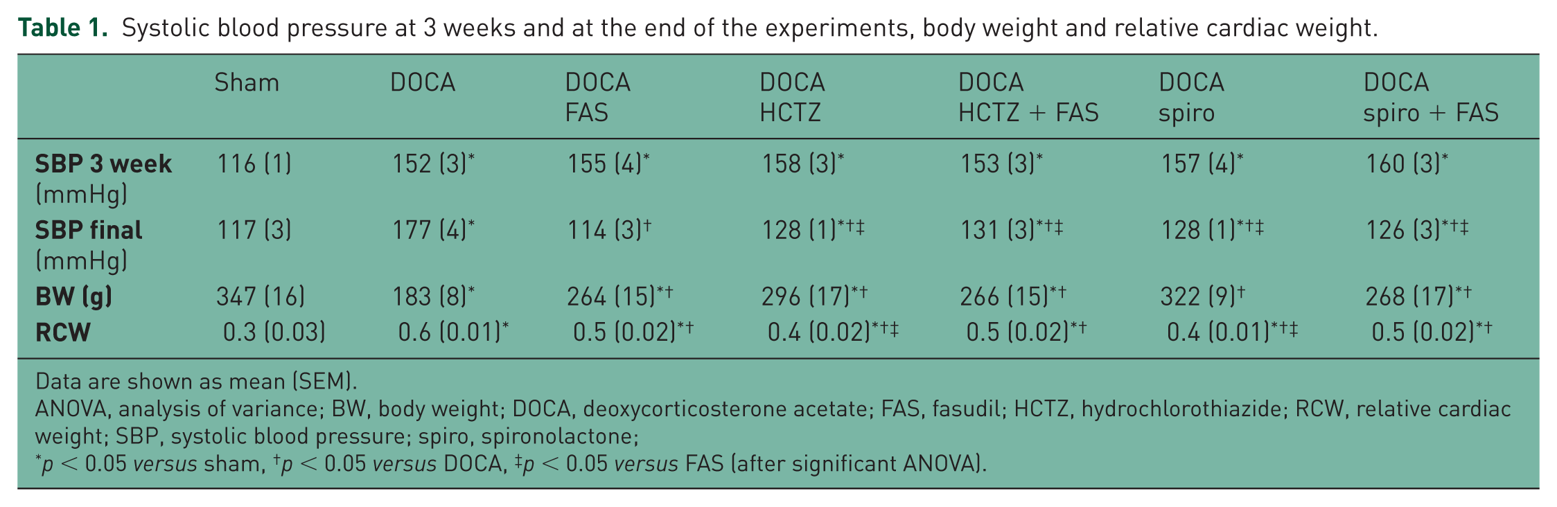
Data are shown as mean (SEM).
ANOVA, analysis of variance; BW, body weight; DOCA, deoxycorticosterone acetate; FAS, fasudil; HCTZ, hydrochlorothiazide; RCW, relative cardiac weight; SBP, systolic blood pressure; spiro, spironolactone;
p < 0.05 versus sham, †p < 0.05 versus DOCA, ‡p < 0.05 versus FAS (after significant ANOVA).
Cardiac hypertrophy, assessed by relative cardiac weight (RCW; cardiac weight × 100/BW) was observed in the DOCA-salt rats compared with sham animals, by 100% and it was significantly reduced with FAS, HCTZ and with spiro (as well as with their combinations; Table 1).
ROCK activation in the aortic wall
The p-MYPT1/t-MYPT1 ratio, an index of ROCK activation, was higher by 2.8-fold (p < 0.05) in the aortic wall of the untreated DOCA hypertensive animals as compared with the sham control animals (Figure 1). FAS, HCTZ and spiro (as well as their combinations) reduced the aortic p-MYPT1/t-MYPT1 ratio to levels observed in the sham control group (Figure 1).

ROCK activation in the aortic wall: effects of HCTZ, Spiro and FAS in hypertensive DOCA salt rats.
Protein levels of TGF-β1
Protein levels of TGF-β1 in the aortic wall were significantly higher in the DOCA-salt rats by 80% compared with sham rats (p < 0.05; Figure 2). In the hypertensive animals, FAS, both diuretics (and their respective combinations with FAS) significantly reduced the protein levels of TGF-β1 in the aortic wall to similar levels as those observed in the sham rats (p < 0.05; Figure 2).

Effects of FAS, HCTZ and Spiro and their combinations administered during 3 weeks on aortic protein levels of TGF-β1.
Expression of genes associated with aortic wall fibrosis and remodeling
mRNA gene expression of TGF-β1, CTGF, gp91 phox and p22 phox in the arterial wall was significantly increased in the untreated hypertensive DOCA-salt rats by 3.1-fold, 4-fold, 2.4-fold, and 1.8-fold respectively compared with the sham rats (p < 0.05, Figure 3A–C).

Gene expression of CTGF, TGF-β1, gp91 phox and p22 phox in the aortic wall (by RT-PCR) in DOCA salt hypertensive rats: effects of FAS, HCTZ and Spiro and their combinations administered during 3 weeks.
In the hypertensive animals, the administration of FAS, of both diuretics (and their respective combinations with FAS) significantly reduced the protein levels of TGF-β1 in the aortic wall as well as the levels of mRNA of TGF-β1, CTGF, gp91 phox and p22 phox to similar levels as those observed in the sham rats (p < 0.05; Figure 3A–C).
Aortic wall hypertrophy
The mean media thickness was 96 µm in the sham group and it was significantly increased in the untreated hypertensive DOCA salt rats by 57% (p < 0.05, Figure 4). In the hypertensive animals, the administration of FAS, of both diuretics (and their respective combinations with FAS) significantly reduced the media thickness in all groups. A larger effect was observed here with both diuretics (p < 0.05, Figure 4A–B).

Aortic wall hypertrophy in DOCA salt-hypertensive rats: effects of FAS, HCTZ and Spiro and their combinations administered during 3 weeks.
The total AM/LA ratio was 0.37 in the sham group and it was significantly increased in the untreated DOCA salt-hypertensive rats by 43% compared with sham rats (p < 0.05, Figure 4C). In the hypertensive animals, the administration of FAS, of both diuretics (and their respective combinations with FAS) significantly reduced the media thickness ratio in all groups. A larger effect was observed here with both diuretics (and their respective combinations with FAS) as compared with FAS alone (p < 0.05, Figure 4C).
Discussion
The main findings in this study were that both diuretics, HCTZ and spironolacone significantly reduced ROCK activation, over expression of profibrotic and pro-oxidative genes and also hypertrophy in the aortic wall in DOCA salt-hypertensive rats in a similar extent as the direct ROCK inhibitor, FAS. In this study, by using a combination of FAS with either diuretic, similar effects were observed.
Thiazides have been used in hypertension treatment for a long time. Their antihypertensive mechanism is due to inhibition of the sodium and chloride cotransporter at the distal convoluted tubule. With their chronic use peripheral resistance also decreases [Conway and Lauwers, 1960; van Brummelen et al. 1980]. In normotensive rats, thiazide-like diuretics inhibit agonist-induced vasoconstriction by calcium desensitization in smooth muscle cells which is linked to the ROCK pathway [Zhu et al. 2005]. The efficacy of thiazide diuretics as first-line therapy for preventing mortality and morbidity in patients with hypertension has been established [Wright et al. 2009]. Thiazide diuretics are as effective as many other classes of BP-lowering medications in preventing coronary heart disease and stroke for a given reduction in BP [Law et al. 2009]. HCTZ is one of the most commonly prescribed antihypertensive drug worldwide. Despite the data suggesting stronger and more consistent lowering of BP and lower risk for adverse CV outcomes for chlorthalidone compared with HCTZ, the latter agent continues to be the most prescribed diuretic in the United States [Hebert et al. 2007].
Spironolactone works through competitive inhibition of aldosterone receptors in the cortical segment of the collecting tubules. Activation of aldosterone receptors in the collecting tubules of the kidneys results in salt and water retention and wasting of potassium. Spironolactone is effective in the treatment of patients with resistant hypertension, irrespective of their aldosterone/renin ratios [Nishizaka et al. 2003]. Recently, the double-blind randomized trial Pathway II has shown that spironolactone at doses of 25–50 mg/day compared with bisoprolol and doxazocin was the most effective add-on drug for the treatment of resistant hypertension [Williams et al. 2015].
Our current observed effects of both HCTZ and spironolactone on BP, ROCK activation and subsequent vascular pro-remodeling gene expression were similar to those observed by direct ROCK blockade with FAS. There are no previous observations on the effect of diuretics on arterial wall expression of pro-remodeling genes through the ROCK pathway in experimental hypertension. Since in this study, we did not evaluate doses of HCTZ and spironolactone (as well as FAS) able to produce smaller reductions in BP, we were not able to split the antihypertensive from the ROCK-inhibitor effect of both diuretics. However, in normotensive brown Norway rats, Rivera and colleagues observed that the same dose of FAS administered for 7 days did not modify SBP [Rivera et al. 2007], but in the aortic wall it significantly reduced ROCK activity along with TGF-β1, plasminogen activator inhibitor-1, and monocyte chemoattractant protein-1 mRNA levels as well as NADPH oxidase activity and O2- production [Rivera et al. 2007].
In our study only antihypertensive doses of both HCTZ and spironolactone were used in order to better understand their clinical role and mechanisms as antihypertensive drugs. On the other hand, by combining FAS with each diuretic, compared with monotherapy, similar effects were observed, which were associated with reduced expression of both profibrotic genes TGF-β1 and CTGF as well as with reduced expression of the pro-oxidative genes gp91 phox and p22 phox in the aortic wall. In this respect, a combination of FAS with the ACE inhibitor imidapril compared with monotherapy has been used to improve renal interstitial fibrosis in an experimental model of renal fibrosis without hypertension [Takeda et al. 2010]. In the aforementioned study, the combination of the ROCK inhibitor and the ACE inhibitor was more effective than monotherapy in prevention of kidney interstitial fibrosis, inflammation, cytokine production and oxidative stress, whereas BP reduction was similar across both groups that received the ACE inhibitor [Takeda et al. 2010].
Most interesting here was the observed effect of both HCTZ and spironolactone on aortic wall hypertrophy. At the current antihypertensive doses, the effect of both diuretics alone on media thickness and on aortic media to lumen area ratio was significant and more important than of FAS alone, even though BP reduction at the current doses was larger with the direct ROCK inhibitor FAS. In the case of both diuretics, this effect on aortic wall hypertrophy was associated with BP reduction, ROCK inhibition and inhibition of ROCK-dependent pro-remodeling and pro-oxidative gene expression in the aortic wall. Concerning HCTZ, there are no previous studies assessing its effect on vessel wall hypertrophy, which is related to both BP reduction and to the consequences of ROCK inhibition and may explain in part, the observed benefits of thiazides as antihypertensive drugs [Wright et al. 2009; Messerli et al. 2011; Al Badarin et al. 2011]. On the other hand, spironolactone does improve hypertensive vascular hypertrophy and remodeling in Ang II-overproducing transgenic mice [Sakurabayashi-Kitade et al. 2009] and recently it was observed in untreated hypertensive patients that eplerenone improves flow-mediated vasodilation (endothelial function) and inhibits ROCK activity [Fujimura et al. 2012].
In conclusion, both diuretics HCTZ and spironolactone in antihypertensive doses reduced ROCK activation, over expression of profibrotic and pro-oxidative genes as well as hypertrophy in the aortic wall in DOCA salt-hypertensive rats to a similar extent as the direct ROCK inhibitor FAS.
Footnotes
Acknowledgements
The authors thank Soledad Veliz, for her dedicated technical assistance.
Funding
This research was supported by Fondo Nacional de Desarrollo Científico y Tecnológico, FONDECYT, Chile 1085208 and 1121060.
Conflict of interest statement
The authors declare that there is no conflict of interest.