
Abstract
Background:
Immunodeficiencies (IDs) are conditions caused by immune system dysfunctions which predispose to chronic infections. Cystic fibrosis (CF) patients are characterized by the presence of bronchiectasis filled with hyper-viscous secretions that constitute the ideal environment for infections. Although CF and IDs might share similarities in the pathophysiological mechanism of bronchiectasis development, they each offer different treatment options. We hypothesize that the introduction of a bundle of tests would increase the number of ID diagnoses among adults with Cystic Fibrosis Transmembrane conductance Regulator (CFTR) dysfunction.
Objectives:
The primary objectives of this study were (1) assessing the prevalence of IDs in CF and (2) defining clinical characteristics of adults with both CF and IDs. The secondary objectives were: (1) assessing the prevalence of IDs in CFTR-Related Disorder (CFTR-RD) patients; (2) comparing the prevalence of IDs in CF and CFTR-RD; (3) comparing the prevalence of treatable IDs in CF and CFTR-RD.
Design:
We conducted an observational, prospective, consecutive study on a cohort of 190 adult patients affected by CF or CFTR-RD.
Methods:
Blood samples underwent a standardized immunological screening, including complete white blood count, IgG, IgA, IgM, IgG subclasses, total IgE, lymphocyte subsets, and HIV test. Comprehensive clinical history was assessed to identify risk factors for secondary IDs.
Results:
We identify a high prevalence of immunodeficiencies among the entire cohort: 34 (20.1%) CF patients and 10 (47.6%) CFTR-RD patients are diagnosed with IDs via a blood screening. No statistically significant difference in terms of clinical characteristics was found between immunocompromised and immunocompetent CF patients.
Conclusion:
We identify a high prevalence of immunodeficiencies in both CF and CFTR-RD.
Background
Cystic fibrosis (CF) is the most common life-limiting genetic disease in Caucasians. 1 It is characterized by dysfunctional chloride transport across epithelial membranes, thus resulting in the formation of hyper-viscous secretions in the airways, exocrine glands, and the gastrointestinal tract.2,3 Despite multiorgan involvement, mortality is mainly secondary to end-stage lung disease. 4 Chronic bacterial infection is accountable for irreversible structural airway damage causing the formation of bronchiectasis, which are, in turn, the optimal substrate for further pathogen proliferation. 5 This process generates a self-sustaining vicious vortex of infection and local inflammation.6,7 The term CF Transmembrane conductance Regulator (CFTR)-Related Disorder (CFTR-RD) defines a clinical entity associated with CFTR dysfunction that does not fulfill diagnostic criteria for CF. 8 CFTR-RD includes different CFTR-related clinical manifestations, including disseminated bronchiectasis. 8 Although no specific CFTR variation is directly associated with bronchiectasis, an increased incidence of CFTR gene variations has been found in these patients. Moreover, the wide spectrum of CFTR variations identified in bronchiectasis is likely to result in residual CFTR function, as in the case of the IVS8-5T allele. 9 Thus, the variety of CFTR variations associated with bronchiectasis likely reflects the heterogeneous nature of this condition, and other etiologies need to be investigated. 8
Immunodeficiencies (IDs) are defined as any dysfunction of the immune system responsible for an impaired response to infections and encompass a wide spectrum of heterogeneous disorders: innate and adaptive immune system defects, phagocytic and complement defects, syndromic disorders, as well as secondary and acquired immunodeficiencies.10,11 IDs display defective mechanisms of pathogen clearance from the bronchial epithelia. These syndromes are among the most prevalent etiologies of diffuse bronchiectasis in both adults and children.12,13 However, studies conducted in the last 30 years have highlighted that the hyper-response of the immune system, supported by a chronic infection, represents a risk factor for the development of severe disease in CF.14,15
Although CF, CFTR-RD, and IDs might share similarities in the pathophysiological mechanism of bronchiectasis development, they each offer different treatment options. The diagnosis of IDs, especially in the case of CF and/or CFTR-RD overlapping, might lead to the presence of a double component, which sustains the previously mentioned vicious vortex inflammatory mechanism favoring the clinical phenotype to manifest. The diagnosis of IDs, especially in these patients, can lead to specific treatments.12,16 However, data on ID prevalence and clinical characteristics in adults with CF and CFTR-RD bronchiectasis are currently poor.
We hypothesize that the introduction of a minimum bundle of etiological tests for bronchiectasis among adults with CF and CFTR-RD would increase the number of ID detections in these groups and potentially lead to the optimization of bronchiectasis treatment in these patients. To answer this question, we analyzed a large cohort of adults with CF and CFTR-RD via a standardized bundle of immunological tests. The primary objectives of this study were (1) assessing the prevalence of IDs in CF and (2) defining clinical characteristics of adults with both CF and IDs.
The secondary objectives were: (1) assessing the prevalence of IDs in CFTR-RD patients, (2) comparing the prevalence of IDs in CF and CFTR-RD (3) comparing the prevalence of treatable IDs in CF and CFTR-RD.
Materials and methods
Study design and populations
We conducted an observational, prospective, consecutive study on 190 adults (⩾18 years) followed at the Adult CF Center of IRCCS Ca’ Granda Ospedale Maggiore Policlinico from January 2018 to December 2019. The enrolled patients were either diagnosed with CF (169 patients) or CFTR-RD (21 patients). All subjects underwent a bundle of blood tests for ID diagnosis during clinical stability. Patients who received gamma-globulin therapy when obtaining the sample were excluded from the cohort. The present study protocol adheres to the Strengthening the Reporting of Observational Studies in Epidemiology (STROBE) Statement. 17 The STROBE checklist is reported in the Supplemental Materials.
Study procedures
Blood samples underwent a standardized immunological screening, including complete white blood count, IgG, IgA, IgM, IgG subclasses, total IgE, lymphocyte subsets, and HIV test. Comprehensive clinical history was assessed to identify risk factors for secondary IDs. Patients with at least one positive result from the immunological screening repeated the test at the next clinical encounter 3 months later. If confirmed, patients underwent a specialist immunological evaluation to confirm the diagnosis.
Study definitions
CF and CFTR-RD were defined according to published diagnostic criteria,8,18 while IDs were defined according to the European Society for Immunodeficiencies (ESID) criteria. 17 Selective IgA deficiency was defined in the presence of undetectable serum level of IgA (when measured with nephelometry <0.07 g/L) and normal level of other immunoglobulins. Common variable immunodefiency (CVID) was defined in the presence of low total serum concentrations of immunoglobulin G (IgG, at least 2 SD below the mean for age), as well as low IgA with or without low IgM levels and low switched memory B cells (<70% of age-related normal value). Severe combined immunodeficiency was defined by the presence of at least two of the following T-cell criteria fulfilled: low or absent CD3 or CD4 or CD8 T cells; reduced naïve CD4 and/or CD8 T cells; elevated gamma-delta (g/d) T cells; reduced or absent proliferation to mitogen or T-cell receptor (TCR) stimulation. These criteria were identified in the context of invasive bacterial, viral or fungal/opportunistic infections within the first year of life. Combined immunodeficiency was defined by the presence of at least two of the following T-cell criteria fulfilled: reduced CD3 or CD4 or CD8 T cells (using age-related reference values); reduced naïve CD4 and/or CD8 T cells; elevated g/d T cells; reduced proliferation to mitogen or TCR stimulation. These criteria were identified in the context of at least one severe infection (requiring hospitalization) and/or one manifestation of immune dysregulation (such as autoimmunity, inflammatory bowel diseases, severe eczema, lymphoproliferation, granuloma) and/or malignancy and/or affected family member. DiGeorge Syndrome was defined by the presence of documented microdeletion 22q11 or 10p and recurrent or severe infections. Hyper-IgE syndrome was defined by the presence of IgE > 10 times the normal limit of age and pathologic susceptibility to infectious diseases and no evidence of T-cell or B-cell deficiency. IgG subclass deficiency was defined by persistently low levels of one or more IgG subclasses and a normal total IgG, IgA, and IgM serum levels and exclusion of T-cell defect. Selective IgM deficiency was defined by low IgM plasma levels and normal IgG and IgA plasma levels and exclusion of T-cell defect. Unclassified antibody deficiency was defined by marked decrease of at least one of total IgG, IgG1, IgG2, IgG3, IgG4, IgA, or IgM levels and no clinical signs of T-cell related disease and did not fit any of the other definitions (excluding unclassified immunodeficiencies). Unclassified immunodeficiency was defined by at least one numeric or functional abnormal finding upon immunological investigation and does not fit any of the other working definitions.
Secondary immunodeficiencies that could lead to hypogammaglobulinemia and/or lymphopenia included AIDS; organ transplantation or graft-versus-host disease; splenectomy; bone marrow aplasia; hematological malignancies (lymphoma, leukemia, multiple myeloma); immunosuppressive agents (chemotherapy, long-term steroids, immunomodulatory agents, and monoclonal antibodies).
Definitions of treatable immunodeficiencies
Potential candidates for treatment with immunoglobulin intravenous (IGIV) or subcutaneous immunoglobulins were patients suffering from either primary immunodeficiency syndromes with impaired antibody production or secondary immunodeficiency with proven specific antibody insufficiency or serum IgG level <4 g/L plus at least one of the following criteria: (1) ⩾3 annual exacerbations; (2) ⩾1 systemic infection occurred during the previous year; (3) ⩾1 hospitalization due to bacterial infection in the previous year; or (4) poor quality of life due to recurrent infections. 18
Study endpoints
The primary endpoints were: The primary objectives of this study were (1) assessing the prevalence of IDs in CF and (2) defining clinical characteristics of adults with both CF and IDs. The secondary objectives were (1) assessing the prevalence of IDs in CFTR-RD patients and (2) comparing the prevalence of IDs and treatable IDs in CF and CFTR-RD.
Statistical analysis
Qualitative variables were summarized using absolute and relative (percentage) frequencies. Quantitative variables were summarized with means (standard deviations, SD) and medians [interquartile ranges (IQR)] depending on a normal and non-normal distribution. Qualitative variables were compared using chi-squared and Fisher exact tests when appropriate. ANOVA and Kruskal–Wallis were used to compare quantitative variables with a normal and non-normal distribution. Sidak correlation was adopted for multiple comparisons. A two-tailed p-value less than 0.05 was considered statistically significant. The sample size was based on prevalence scenarios from published studies in CF pediatric patients.19,20 Sample size requirements were also calculated for detecting an association between CF and IDs using the following parameters: confidence level of 95%, power of 90%, ID, and relative risk of 2.0. This calculation requires 106 patients. We performed statistical computations using IBM SPSS, Statistics for Windows, Version 22.0. Armonk, NY:IBM Corp.
Results
Study cohort
A total of 190 adults [median age 30 years (IQR 23–34.25); 42% females] were included. The enrolled study population consisted of 169 CF patients and 21 CFTR-RD patients.
Demographic and clinical characteristics of the CF study cohort are reported in Table 1. A total of 24 (14.2%) CF patients had F508del/F508del, while 52 (30.8%) had at least one residual function mutation. Male sex was prevalent (n = 93, 55%), the median age was 27 (IQR 22–33), and the median body mass index (BMI) was 22.2 kg/m2 (IQR 20.3–23.9). All CF patients had evidence of bronchiectasis on computed tomography scan. Chronic respiratory infections were diagnosed in 140 (82.8%) patients in our cohort. Among the causative agents of chronic infection, the predominant pathogen was Pseudomonas aeruginosa, which was present in 91 (53.8%) patients, while the second most frequently isolated organism was MSSA which was identified in 67 (39.6%) patients. The median number of exacerbations in the year before the enrollment was 1 (IQR 1–3). However, a total of 83 (49.1%) patients had at least 2 exacerbations in the previous year, and 56 (33.1%) patients had 3 or more exacerbations. A total of 58 (34.3%) patients had more than one hospital admission, and the median antibiotic course per year was 2 (IQR 1–4).
Demographics, clinical, functional, and microbiological characteristics of the CF study population.
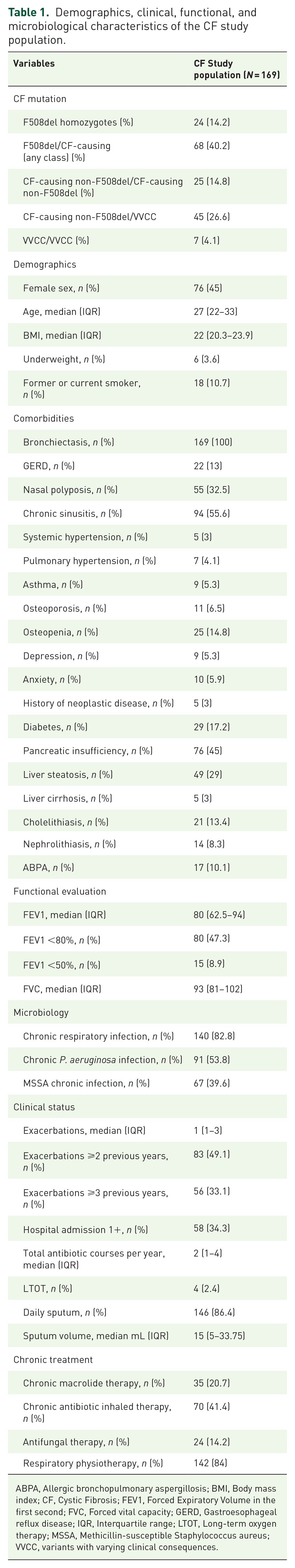
ABPA, Allergic bronchopulmonary aspergillosis; BMI, Body mass index; CF, Cystic Fibrosis; FEV1, Forced Expiratory Volume in the first second; FVC, Forced vital capacity; GERD, Gastroesophageal reflux disease; IQR, Interquartile range; LTOT, Long-term oxygen therapy; MSSA, Methicillin-susceptible Staphylococcus aureus; VVCC, variants with varying clinical consequences.
Immunodeficiency diagnosis in CF. A total of 34 (20.1%) patients affected by CF had at least one immunological alteration. Table 2 shows the most common IDs identified in our cohort. Primary immunodeficiencies accounted for the majority and were diagnosed in 33 (19.5%) patients. Of these, unclassified immunodeficiencies were detected in 23 (13.6%) patients, while isolated IgG subclass deficiency was found in 9 (5.3%) patients. Specifically, both isolated IgG1 subclass deficiency and isolated IgG3 subclass deficiency were identified in 3 (1.8%) patients. Isolated IgG4 subclass deficiency was diagnosed in 2 (1.2%) patients, while IgG1 + IgG4 subclass deficiency was diagnosed in 1 (0.6%) patient. Unclassified antibody deficiency was diagnosed in 1 (0.6%) patient. Lastly, 1 (0.6%) patient had a diagnosis of secondary immunodeficiency.
Prevalence of CF adults (N = 169) with any, primary, secondary, and potentially treatable ID.

Data are presented as n (%).
CF, Cystic Fibrosis; ID, immunodeficiency.
Clinical characteristics of ID-CF and not-ID CF patients. The comparison between immunodeficient and immunocompetent patients is reported in Table 3. No statistically significant difference in terms of respiratory and extra-respiratory involvement, microbiology, comorbidities, and chronic treatment were found.
Comparison of demographics, clinical, functional, and microbiological characteristics of the CF study population based on ID status.

BMI, Body mass index; CF, cystic fibrosis; FEV1, Forced Expiratory Volume in the first second; FVC, Forced vital capacity; GERD, Gastroesophageal reflux disease; ID, immunodeficiency; IQR, Interquartile range; LTOT, Long-term oxygen therapy; MSSA, Methicillin-susceptible Staphylococcus aureus; VVCC, variants with varying clinical consequences.
Immunodeficiency diagnosis in CFTR-RD. The CFTR-RD cohort was characterized by the presence of bronchiectasis in 18 (85.7%) patients, recurrent pancreatitis in 2 (9.5%) patients, and congenital bilateral absence of the vas deferens in 1 (4.8%) patient. According to the CFTR-2 database, 12 (57.1%) patients carried a class II mutation (of whom 10 patients with an F508del) associated with a variant of varying clinical consequence (VVCC), 6 (28.6%) patients carried a CF-causing non-class II mutation associated with a VVCC, and 3 (14.3%) patients carried a VVCC on each allele. Table 4 shows the prevalence of IDs among the CFTR-RD group. Primary immunodeficiencies were detected in 10 (47.6%) patients, while secondary immunodeficiencies were not present. Among the primary IDs, the most frequently detected were isolated IgG subclass deficiency and unclassified antibody deficiency, each accounting for 3 (14.3%) patients. Isolated IgG1 subclass deficiency, isolated IgG4 subclass deficiency, and IgG3 + IgG4 subclass deficiency were detected in 1 (4.8%) patient each. Lastly, unclassified immunodeficiencies were detected in 4 (19%) patients.
Prevalence of CFTR-RD adults (N = 21) with any, primary, secondary, and potentially treatable ID.

Data are presented as n (%).
CFTR-RD, Cystic Fibrosis Transmembrane conductance Regulator - Related Disorder; ID, immunodeficiency.
Comparison of IDs and IDs prevalence in CF and CFTR-RD. IDs were more frequently identified in the CFTR-RD (10 patients, 47.6%) cohort compared to the CF (34 patients, 20.1%) cohort, p = 0.005. Among the entire cohort, 16 (8.4%) patients with immunodeficiency met the pre-specified criteria for treatment with IGIV or subcutaneous immunoglobulins. Patients with treatable IDs were more frequently identified in the CF group (15 patients, 8.9%) compared to the CFTR-RD group (1 patient, 4.8%), p = 0.049.
Discussion
This is the first study evaluating the prevalence and clinical characteristics of ID status in an adult cohort of CF patients. Till now, the only data available on IDs and CF came from single-center and pediatric cohorts.14,21–25 The results of our study show that the prevalence of IDs among CF patients should not be underestimated, as it can account for up to 23.2% of CF patients and 47.6% of CFTR-RD patients. In the past decades, case reports have shown that IDs can be associated with CF in both adult and pediatric patients.22,24 However, the vast majority of studies on immunological function among CF patients have been conducted in children. Results from a systematic screening in pediatric patients with CF for IDs found abnormal immunoglobulin levels, particularly hypogammaglobulinemia IgG, in a single-center study from the UK. 21 In this study, 19 (14%) patients had low levels of IgG1, and 40 (29%) patients had low levels of IgG2. A recent study conducted in Belgium identified hypo-IgG in 15.2% of a CF pediatric cohort. 22 However, the longitudinal assessment of IgG levels since diagnosis demonstrated that hypo-IgG was transient in at least one-third of patients. The progressive increase in mean IgG since diagnosis, according to age group, supports immunological maturation increases with age, thereby possibly explaining transient deficiency in pediatric patients with CF. Moreover, the oldest study published in CF patients demonstrates that hypogammaglobulinemia is associated with significantly less severe lung disease compared to CF patients with normal or elevated IgG levels. 25 Similarly, another study conducted 40 years ago highlighted that children with CF with persistent hypogammaglobulinemia had better clinical status and slower deterioration in pulmonary function compared to age-matched patients with normal or elevated immunoglobulin G values. 26 This evidence might lead to the hypothesis that the progression of CF lung disease may be due in part to a hyper-immune response. However, treatments and outcomes of CF lung disease have changed considerably in the last decades, and the presence of hypergammaglobulinemia is no longer observed in the majority of CF subjects.21,22 Thus, the advantage of subjects with a poor immune response is no longer so evident. This is also in line with our data, showing no difference in clinical characteristics and severity of disease of CF patients with or without IDs.
Our study identifies a prevalence of IDs in 23.2% of CF adult patients. A possible explanation for this high prevalence in an adult might be related to the extended life expectancy of CF subjects that can promote senescence, which is early activated in patients with CF. 25 Indeed, it has been hypothesized that the resulting vicious cycle of infection and inflammation in the CF lung may play a deleterious role in suppressing immune function.26,27
However, the comparison between CF patients with and without IDs was unable to highlight any difference in terms of respiratory and extra-respiratory involvement, microbiology, comorbidities, and chronic treatment. Thus, extensive screening of the immune status might be limited to selected CF cases that clinically suggest IDs or to CF patients with recurrent respiratory exacerbations or extensive bronchiectasis damage. Indeed, it could be hypothesized that patients with both ID and CF might have an earlier and more pronounced bronchiectasis extensive pattern following a ‘double hit’ theory.
Although CF is common in the Caucasian population, the prevalence of CFTR-RD is lower, partially due to the underdiagnosis. The bronchiectasis manifestation of CFTR-RD is more common in the case of the coexistence of IDs. The results of our study show that IDs are common in CFTR-RD patients, and for this reason, IDs should be actively looked for, especially in this category of patients.
The association between CFTR-RD and IDs may reflect subtle changes in the host’s immune and inflammatory response, which may accentuate the development and progression of bronchiectasis. Recognizing CFTR variants as part of an ID diagnosis may help to identify individuals who would benefit from interventions that have the goal of reducing disease progression. Recently, a large European cohort of ID patients was analyzed using genome sequence data. 28 Carriage of several pathogenic CFTR gene variants was increased in ID associated with lung damage, compared to ID patients without lung damage. These data reinforce the hypothesis that there are subjects with IDs and bronchiectasis in which the presence of variants of the CFTR gene acts as modifiers.
In our study, 5% of the CFTR-RD population had potentially treatable ID. For this reason, IDs should be screened in CFTR-RD patients since they might lead to significant changes in the management and prognosis of these patients.
The study limitations are related to the single-center design and the tertiary care setting which could hinder the reproducibility of the findings. Different prevalences of IDs across different settings could be found.12,29 Thus, our epidemiological analysis needs external validation in a multicentric cohort of CF patients. Another limitation is the missing follow-up to evaluate outcomes of patients with ID. Moreover, third-level functional studies concerning B and T cells, such as proliferation to mitogen or T-cell stimulation, were not performed in our center. Finally, the study was performed before CF modulator therapy availability.30–33 Thus, this might lead to an overestimation of treatable ID prevalence in the CF population.
The evaluation of a large cohort of adult CF and CFTR-RD patients who underwent the same comprehensive immunological work-up is a strength of our manuscript. Furthermore, the classification of IDs we used in our study followed the latest ESID criteria.
Conclusion
A high number of adults with CFTR dysfunction have received a diagnosis of IDs via an extensive bundle of tests. Since IDs can represent a relevant treatable trait, they should be properly assessed in this patient group. A multidisciplinary team, including an experienced clinical immunologist and a CF clinician, should evaluate patients with abnormal results. Management and follow-up of each patient should be individualized to determine the need for further tests and identify candidates for immunoglobulin replacement therapy.
Supplemental Material
sj-docx-1-tar-10.1177_17534666241253945 – Supplemental material for Immunodeficiencies and CFTR dysfunction: results from a systematic screening in a cohort of adults with cystic fibrosis and CFTR-related disorders
Supplemental material, sj-docx-1-tar-10.1177_17534666241253945 for Immunodeficiencies and CFTR dysfunction: results from a systematic screening in a cohort of adults with cystic fibrosis and CFTR-related disorders by Francesco Amati, Gloria Leonardi, Martina Contarini, Letizia Corinna Morlacchi, Anna Stainer, Giovanna Pizzamiglio, Stefano Aliberti, Francesco Blasi and Andrea Gramegna in Therapeutic Advances in Respiratory Disease
Footnotes
References
Supplementary Material
Please find the following supplemental material available below.
For Open Access articles published under a Creative Commons License, all supplemental material carries the same license as the article it is associated with.
For non-Open Access articles published, all supplemental material carries a non-exclusive license, and permission requests for re-use of supplemental material or any part of supplemental material shall be sent directly to the copyright owner as specified in the copyright notice associated with the article.