
Abstract
The type I interferon family of cytokines are rapidly produced following innate pattern recognition receptor engagement and establish a critical early state of host defense. Type I interferons act in antiviral immunity as transcriptional activators and the binding of any type I interferon to the common IFNAR receptor triggers the transcription of
Keywords
Introduction
The type I interferon cytokine system represents the central pillar of the early innate immune response to viral infections and is essential for the control and/or clearance of most viral pathogens.1–4 Within the context of viral infection, type I interferons (IFN) increase cell intrinsic resistance to infection by functioning as transcriptional regulators to increase the expression of hundreds of interferon stimulated genes (ISGs). Many of the ISGs are known to encode proteins with directly antiviral functions.5,6 The molecular pathway by which IFNs positively regulate ISG expression has been extensively studied and is well-reviewed elsewhere. 7 Briefly, extracellular binding of any type I IFN to the common type I interferon receptor (IFNAR) activates proximal intracellular factors comprising the Ser/Thr kinase Janus Kinase 1 (JAK1) and the Tyrosine kinase, Tyk2. JAK1 and Tyk2 activation results in the recruitment and subsequent phosphorylation of the transcription factors Signal Transducer and Activator of Transcription 1 and 2 (STAT1 and STAT2). Phosphorylated STAT1 and STAT2 physically associate and, along with the Interferon Regulatory Factor 9 (IRF9) protein, form the interferon stimulated gene factor 3 (ISGF3) complex. Subsequent ISGF3 nuclear trafficking initiates the transcription of ISGs through binding to Interferon Stimulated Response Element (ISRE) motifs within ISG promoter regions driven by RNA pol II recruitment and activity.7,8 (see Figure 1). A critical requirement for each of the ISGF3 subunits in the positive transcriptional regulation of ISGs and for the establishment of a resulting antiviral state has been confirmed in genetic models of infection.9–13 Given the importance of ISGs in anti-viral host defense, the identification and characterization of ISGs has been the subject of hundreds of studies using transcriptomic approaches and as a result, comprehensive annotated lists of ISGs across cell types and species have been generated.5,14,15

Model of positive and negative gene regulation by type I interferons. (a) type I interferon activation of interferon stimulated gene (ISG) transcription requires the IFNAR induced assembly of the hetero-trimeric ISGF3 transcription factor. ISGF3 recognizes the ISRE sequence motif proximal to the promoter of ISGs and drives increased RNApolII dependent gene transcription. (b) Interferon dependent transcriptional suppression may proceed in a gene direct manner through ISGF3 dependent or independent transcriptional regulators including DNA binding transcriptional repressors recognizing a target motif within suppressed promoters and/or histone modifying enzymes. Alternatively, the product(s) of one or more upregulated ISGs, such as immunosuppressive cytokines, may function to suppress subsequent gene transcription.
While positive transcriptional regulation by IFNs has been extremely well described, transcriptomic studies of IFNs have also noted that IFN signaling simultaneously leads to diminished mRNA expression of a number of distinct genes in human and mouse.16,17 We here refer to these Type I Interferon Inhibited Genes as “TIIGs” in order to unambiguously differentiate this population from the positively regulated ISGs. TIIGs therefore comprise an additional aspect of transcriptional regulation by IFNs. However, perhaps because of the difficulties inherent in studying the functional consequences of transcriptional suppression, TIIGs have been significantly understudied when compared to the positively regulated ISGs. As a result, a curated and validated list of core TIIGS is lacking in the field, as is a consensus molecular model of the signaling elements required for IFN transcriptional suppression.
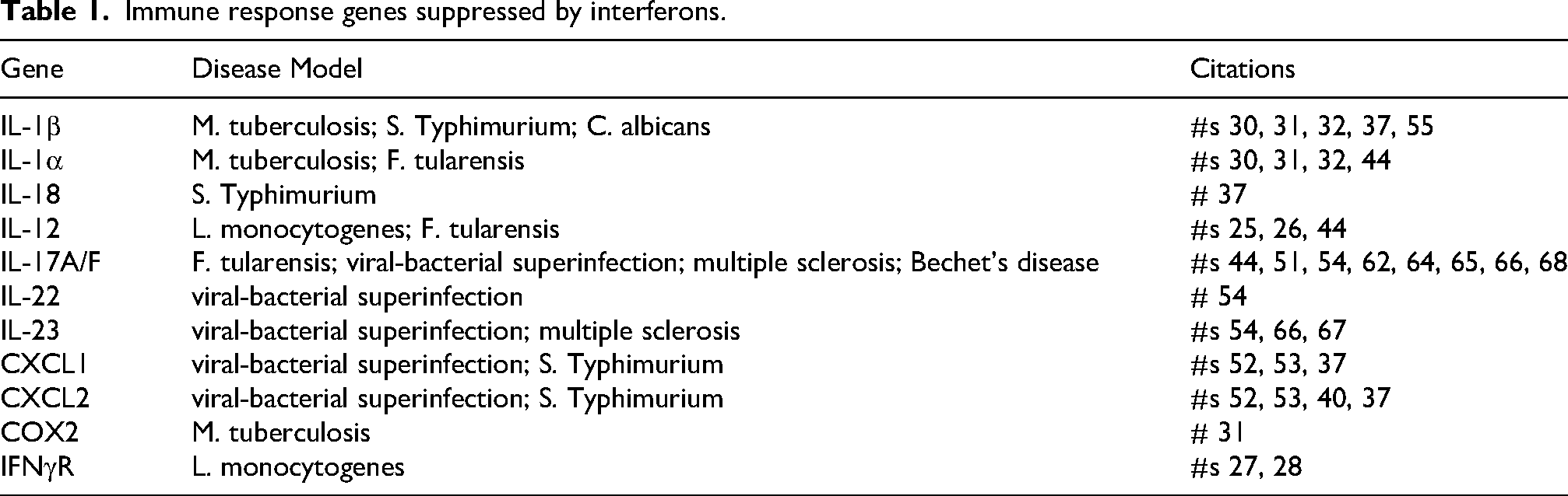
Despite the paucity of research focused on TIIGs specifically, these suppressed genes may be key to understanding how IFNs impact several important human microbial and autoimmune diseases. A GO Pathway analysis of a previously published 18 and publicly available RNA-Seq dataset (GEO: GSE61055) generated using control and IFNβ treated mouse BMDMs, identifies IFN downregulated genes within an array of cell biological processes (See Figure 2). A survey of published literature reveals that a significant number of genes identified as TIIGs encode proteins involved in the immune response (see Table 1), suggesting that transcriptional repression by IFN may be relevant to understanding the systemic immunomodulatory activity of IFNs specifically in contexts beyond acute viral infections.

Type I IFN suppresses expression of genes from a variety of biological processes. GO pathway analysis of publicly available RNA-Seq data (GEO: GSE61055) generated using IFNβ treated mouse BMDMs. List of IFN down-regulated genes generated by comparing untreated and IFNβ treated samples. Differentially expressed genes identified using GEO2R (adjusted p < 0.05, Fold change < −1). GO analysis was performed using the enrichGO function from the clusterProfiler R package with ontology set to biological processes. Genes listed in each category represent top 3 nonredundant downregulated genes in that category.
Immune response genes suppressed by interferons.
In this review, we will focus on the available literature within this underexplored area of negative transcriptional regulation by type I IFNs, with particular emphasis on suppressed immune and immune-adjacent genes. We will identify disease models in which IFN transcriptional suppression has been proposed to contribute prominently to immune suppression and the potential molecular mechanisms underlying IFN gene suppression. Finally, we highlight the open questions that remain in this area of IFN research with an eye toward stimulating renewed discussion of this second arm of IFN action.
Disease models evidencing IFN transcriptional suppression of immune associated genes
Disease relevance for negative transcriptional regulation by type I interferons has been found in studies using a broad variety of models involving both infectious disease and autoimmune contexts. In these models, IFN transcriptional suppression has most often been studied in the context of the reduced expression of immune and immune adjacent genes as downregulation leads to either deleterious or palliative effects for the host depending upon the specific disease model. Despite the wide variety of in vivo and in vitro models involved, a core population of IFN suppressed genes have been repeatedly identified as important across models suggesting some specificity in the genes targeted by IFN for suppression. Defining the commonalities among disease models in which IFN expression is immunosuppressive may help to shed light on how these differ from autoimmune diseases in which constitutive expression of type I interferons promotes damaging systemic inflammation (a group collectively known as the interferonopathies 19 ) often associated with dermatological and neurological pathologies caused by chronic myeloid cell activation.
Below we summarize the relevant literature in which IFN suppressed immune modifying genes have been identified.
Bacterial infection
While far more often studied in the context of viral infections, IFN expression is also triggered during many bacterial infections. 20 Paradoxically, unlike the protective effects in viral infections, IFN expression has been shown to be deleterious to the host in many if not the majority of mouse models of acute bacterial pathogenesis.21–24 In these bacterial models, type I IFNs are often correlated with a poorly defined state of immunosuppression, involving reduced transcription of key immune mediators which contributes to enhanced pathogenesis. While IFN expression is known to be detrimental in many bacterial infection models, only a limited number of studies have defined the immune defects underlying this phenotype. Investigations of the immunosuppressive effects of IFN in in vivo bacterial infections have repeatedly identified a small number of common genes encoding systemic inflammatory mediators as having reduced expression. Specific transcriptional suppression of many of these genes can also be observed in reductionist tissue culture models of IFN exposure suggesting that there may be a core transcriptional signature of type I IFN mediated immune suppression.
Listeria monocytogenes
Listeria monocytogenes was the first pathogenic bacterial infection shown to be promoted by IFNs in an in vivo mouse model. In concurrent reports, two groups independently reported reduced lethality of L. monocytogenes infection in IFNAR−/− animals compared to WT, with IFNAR−/− animals being up to 1500-fold more resistant to L. monocytogenes infection.25,26 Despite similar early colony forming units (CFUs) (24–48hrs) in tissues between the WT and IFNAR−/− animals, profiling of serum cytokine expression by ELISA identified specific elevation of IL-12 p70 protein in IFNAR−/− animals. No significant difference in IL-6 protein levels were observed at the same timepoints, suggesting that IFNs can blunt expression of immune mediators in a gene specific manner.25,26
Subsequent in vitro studies aimed at defining the mechanism of the IFN action in L monocytogenes infection found that IFNβ secreted by L. monocytogenes infected cells was capable of acting in an autocrine and paracrine fashion to downregulate expression of the interferon gamma receptor (IFNγR) specifically on myeloid cells. 27 The suppression of ifngr occurred at the transcriptional level and could be replicated in vitro following treatment of mouse bone marrow derived macrophages (BMDMs) with recombinant IFNβ alone. 27 Additional experiments identified the IFN induced assembly of a regulatory complex at an Early Growth Response Factor (Egr) DNA binding site in the proximal ifngr1 promoter 28 as the mechanism of IFNγR suppression.
Mycobacterium tuberculosis
The most extensive literature describing transcriptional suppression of immune response mediators by IFNs in bacterial pathogenesis has been generated utilizing Mycobacterium tuberculosis (M. tb) infection.29,30
In vitro, M. tb infection of healthy human monocytes was shown to induce robust release of IFN-β and IFN-α. 31 Transcriptomic analysis of the cellular response to M. tb infection of human monocytes following addition of anti-IFNAR blocking antibodies revealed significantly greater mRNA expression of IL-1β, IL-1α and COX2 when IFN responses were inhibited, indicating that IL-1 cytokine family members are targets of IFN mediated suppression in M. tb. This impact of IFN on IL-1β was independent of changes in expression of IL-1 receptor or Caspase1 enzyme activity. Importantly, addition of type II IFN (IFNγ) during in vitro M. tb infection did not lead to similar suppression of IL-1 family members. 31
Subsequent M. tb infection of in vivo mouse models confirmed that endogenous or exogenous IFNs can suppress both IL-1β and IL-1α expression during in vivo M. tb infection, further supporting the connection between IFN and the regulation of IL-1 family cytokines.32,33
A transcriptomic approach focused on identifying systemic immunologic effects of M. tb identified a dominant ISG signature in M.tb infected mouse BMDMs when compared to uninfected BMDMs. 34 Interestingly, the magnitude of this IFN transcriptomic signature was greater for more pathogenic M. tb strains and correlated with increased IFN protein production by these strains. 34 Analysis of global gene expression changes caused by IFN induction during in vitro M. tb infection identified several IFN suppressed genes known to be involved in the glycolytic metabolic pathway, including the genes encoding glucose-6-phosphate isomerase and aldolase-C. Transcriptional suppression of these mitochondrial genes may directly explain the role of IFNs in promoting M. tb pathogenesis in vivo as a programmatic shift toward aerobic glycolysis is critical for inflammatory activation of macrophages.35,36 This data suggests that, during M. tb infection, IFN transcriptionally suppresses secreted cytokines which mediate a systemic inflammatory response (e.g., IL-1α and IL-1β), as well as metabolic genes required for an effective cell intrinsic host defense. 34
Salmonella typhimurium
The Gram negative intracellular pathogen Salmonella enterica serovar typhimurium (St) elicits a robust innate inflammatory cytokine response during in vitro or in vivo infections, a response which includes the production of type I interferons through Toll Like Receptor 4 (TLR4) activation.37–41 In vivo production of IFNs following oral St infection of mice enhances pathogenesis and increases lethality.37,39,42 Analysis of innate cytokine transcription during infection of mouse macrophages with St following exposure to recombinant IFN-β identified inflammatory mediators whose rapid transcription was inhibited by IFN. These inhibited genes included IL-1 family members IL1-β and IL-18. 37 Significantly, three described mouse neutrophil attracting chemokines induced by St infection, CXCL1 (KC), CXCL2 (MIP2), and CXCL5 (LIX) were transcriptionally repressed by type I IFN. 37 This enrichment of suppressed genes within a common functional immune pathway suggests that IFN suppression in the context of oral St infection may preferentially target select immune functions.
Francisella tularensis
Francisella tularensis is an intracellular Gram negative bacterial pathogen and is the causative agent of tularemia in humans. Francisella is remarkable for its immune evasive qualities in comparison to other human bacterial pathogens, failing to elicit rapid transcription of inflammatory mediators during in vitro and in vivo 43 infections. As observed for several other Gram negative pathogens, infection of IFNAR−/− mice with the Francisella novocida strain reveals that animals with defective type I IFN responses are largely protected from F. novocida lethality. 44 Immuno-phenotyping of WT and IFNAR−/− infected animals identified higher levels of IL-1α and IL1-2p70 protein in the serum of IFNAR−/− animals compared to WT. The observed reduced expression of these cytokines in F. novocida infection of WT mice is consistent with IFN suppression of IL-1 family and IL-12 cytokines in other bacterial infection models. 44 Importantly, IFN signaling during F. novocida infection was also associated with a marked reduction in serum levels of IL-17 cytokine family members IL-17A and IL-17F. 44 IL-17 A and IL17F are critical mediators of resistance to many mucosal bacterial infections through mobilization of neutrophils and the amplification of mucosal barrier functions.45,46 Inhibition of IL-17A and IL-17F gene transcription by IFNs has been also reported during non-bacterial infections as well as in sterile autoimmune models (see section below) indicating Th17 responses may be a common target for IFNs across disease models. Additional evidence for IFN dependent functional suppression of immune related genes in Francisella infection is provided by Bauler et al. 47 This study compared the inflammatory transcriptional responses triggered in infected human dendritic cells (hDCs) by the avirulent Francisella Live Vaccine strain (LVS) and the hypervirulent ShuS4 strain. Virulent ShuS4 induced 5–6 fold greater amounts of IFN protein release in comparison to the avirulent LVS strain. 47 This elevated ShuS4 induced IFN response could specifically suppress production of IL-12p70 protein by hDCs in agreement with previous reports, 44 suggesting that IL-12 encoding genes are a significant IFN suppressive target and that reduced IL-12 is likely relevant for understanding the role of IFN in exacerbating Ft pathogenesis.
Viral & bacterial co-infections
The data summarized in the above sections argues that IFNs produced directly in response to infection by intracellular bacterial pathogens drive gene specific immune suppression causing more severe disease phenotypes. IFN production during bacterial infections can, however, be dramatically enhanced by the presence of an initial confounding viral co-infection. 48 Clinically, such viral-bacterial superinfections are common following severe human influenza infections and account for most influenza associated mortalities.49,50 On a molecular level how collaboration between an initial viral and subsequent bacterial infection occurs is subject of intense ongoing study. In several co-infection models, viral induction of high levels of IFNs has been shown to impair innate and adaptive immune responses to subsequent bacterial infection leading to greater microbial growth and tissue damage.
A mouse model of Gram positive bacterial-viral superinfection identified important gene targets of IFN resulting in immunosuppression. 51 In this model, the X31 (H3N2) mouse adapted influenza strain was delivered intranasally and followed 4 days later by a second intranasal inoculation with Streptococcus pneumoniae (Sp). Prior influenza infection increased IFN production during Sp infection by an order of magnitude and IFN production induced by influenza infection was sufficient to promote Sp pathogenesis. 51 Analysis of mRNA expression in whole lung tissue of superinfected animals identified specific defects in expression of IL-17A and IL-17F following Sp infection only in conjunction with influenza. This IL-17 suppression was gene specific, as transcription of other pro-inflammatory cytokines was not similarly affected. γ/δ T cells were found to produce the majority of IL-17 in this superinfection model and transfer of IFNAR−/− γ/δ T cells to WT animals was sufficient to retore IL-17A and IL-17F expression and reverse multiple disease sequelae, 51 suggesting that IFN action on γ/δ T cells was responsible for IL-17 inhibition and increased pathology in this superinfection model.
The importance of IFNs in shaping the immune transcriptome through suppression was confirmed in a second model of Gram positive superinfection. 52 In this model the PR8 (H1N1) mouse adapted influenza strain was administered intratracheally followed 5 days later by intra-tracheal inoculation with S. pneumoniae. 52 Analysis of cytokine protein levels by ELISA in whole lung tissue homogenates found that prior influenza infection significantly reduced S.p. induced CXCL1 and CXCL2 neutrophil chemokine protein levels. The expression of other inflammatory cytokines such as IL-6 and CXCL10 remained unchanged and the effect on CXCL1 and CXCL2 was largely reversed in the IFNAR−/− animals. IFN suppression of CXCL1 and CXCL2 has also been reported using a distinct strain of S.pneumo in influenza superinfection. 53 Importantly the suppressive effect of IFN on CXCL1 and CXCL2 transcription could be recapitulated in a reductionist in vitro model using BMDMs pre-treated with recombinant IFNα followed by stimulation with TLR2 ligand Pam3Cys. 52
A complimentary mouse model of Gram negative superinfection also identified IFN dependent transcriptional suppression underlying immunosuppression. 54 In this model the PR8 strain of influenza was administered intra-nasally followed 6 days later by intranasal inoculation with the DH5a strain of E. coli. Analysis of mRNA expression of immune relevant genes in whole lung tissue during the first 24hrs of superinfection with Influenza and E. coli identified specific reduction in expression of IL-17A, IL-22, IL-23, ROR-a and RORgt, but no loss of IL-6, CXCL1 or G-CSF expression. 54 As with Gram positive superinfection, inhibition of cytokine expression was not observed in superinfected IFNAR−/− animals.
An additional model of Gram negative superinfection focused on enteric pathogenic bacteria corroborated tissue specific immune response genes suppressed by IFN. 40 In this model, the PR8 influenza strain was administered intranasally followed 5 days later by intragastric gavage inoculation with Salmonella enterica serovar typhimurium. Both tissue dissemination of St and lethality were enhanced by PR8 induced IFN and superinfection of IFNAR−/− animals reversed these phenotypes. Importantly comparison of mRNA expression in the cecum of superinfected animals identified IFN dependent defects in the mRNA expression of neutrophil chemokine CXCL2 and the mucosal specific antimicrobial peptides S100A9 and LCN2. The identification of gut specific suppressed genes suggests that IFN may effect specific negative transcriptional regulatory programs at such mucosal surfaces impairing resistance to enteric pathogens. 40
Candida Albicans
In a study exploring the molecular mechanisms of immune suppression by type I IFNs during fungal infection, 55 WT mice were treated by intravenous injection with synthetic dsRNA (poly IC) to induce systemic IFN expression followed 5 h later by intravenous infection with Candida albicans to mimic a disseminated fungal infection. Poly IC treatment significantly increased C. albicans lethality and colony counts in the sterile organs, effects which were lost in IFNAR−/− and STAT1−/− animals. 55 Importantly, the in vivo production of both pro-IL-1β protein and secreted, processed IL-1β were strongly inhibited by IFNs, 55 an observation which is likely to be medically relevant as IL-1 is critical for controlling C. albicans. 56 Similar IFN dependent immunosuppression driving C. albicans infections has also been reported by other groups.57,58
Auto-Immune models
In addition to their role in shaping immune-related transcriptional responses in infectious disease, IFNs have emerged as central regulatory circuits in the inflammatory gene expression characteristics of several systemic autoimmune diseases. In select sterile autoimmune conditions characterized by destructive unrestrained inflammation, expression of endogenous type I interferons can be palliative and correlated with reduced expression of inflammatory genes. 59 For multiple autoimmune conditions, the immunosuppressive actions of IFNs are leveraged pharmaceutically, with several formulations of IFNα and IFNβ currently approved for therapeutic use. 59 Notably, administration of IFN is effective in reducing immune pathology and is associated with reduced expression of many of the same genes also identified as IFN targets in infectious disease contexts.
Multiple sclerosis
Multiple Sclerosis is a chronic autoimmune condition affecting the central nervous system. In the western hemisphere, Multiple Sclerosis (MS) is the most well described autoimmune disease for which type I IFNs are used therapeutically and associated with reduced inflammatory sequalae. 59 The palliative actions of endogenous and exogenous IFNs in MS are strongly correlated with diminished mRNA expression of select key inflammatory mediators.60–63 In the experimental autoimmune encephalomyelitis (EAE) mouse models of MS, and in human patients with relapsing remitting MS given IFNs therapeutically, IFNs act to specifically reduce expression of IL-17 family cytokines by unknown mechanisms.62,64–66 This finding is clinically significant as Th17 responses are associated with severe pathology in MS. In addition to potent antagonism of IL-17, IFNs are also reported to antagonize expression of IL-23 in MS.66,67
Bechet's disease (BD)
Bechet's disease is a chronic, relapsing, multisystemic inflammatory autoimmune condition with clinical features including mucocutaneous lesions, as well as frequent ocular, urogenital and gastrointestinal involvement likely resulting from severe vasculitis of unknown etiology. 68 Given the immune involvement in BD, IFNs have been used clinically to treat BD with great success.69,70 As reported for MS treatment, use of IFNs to treat BD is associated with decreased TH17 responses, which may underlie the observed therapeutic benefit. 68 Additionally, IL-1 family members IL-1β and IL-18 are both implicated in the pathology of BD and are known to be suppressed at the transcriptional level by IFNs in other infectious disease models.37,55
Proposed molecular mechanisms
Given the demonstrated importance of IFN mediated immune suppression across disease models and the common set of immune response genes identified as transcriptionally suppressed by IFN in these divergent contexts, understanding the molecular mechanism(s) by which IFN mediates gene specific transcriptional suppression is critical. Several molecular mechanisms/mediators have been proposed (see Figure 1) although the extent to which each might contribute to the totality of gene suppression by IFN remains to be tested.
Immunosuppressive cytokines. IL10 & IL27
IL-10 is a secreted cytokine and a component of the innate response to microbial infection or injury. IL-10 acts as an immune response repressor on most cell types to suppress inflammation through a variety of mechanisms which are still being elucidated.71,72 It is clear that IL-10 can function to broadly and potently suppress the inducible transcription of classical inflammatory genes (e.g., IL-1, TNF, IL-6…etc) downstream of innate pattern recognition receptors, 73 and the existence of IFN induced negative feedback circuits have been described using transcriptomics and in silico analysis. 74 Expression of type I interferons synergistically amplifies the production of IL-10 by immune cells such as macrophages and dendritic cells in multiple infection models and in response to purified pattern recognition receptor ligands in vitro.37,75–77 In one model, genetic ablation of IL-10 protein was reported to partially but incompletely reverse suppression of IL1β protein expression by IFNs during LPS stimulation of BMDMs suggesting that for IL-1β at least, IL10 can contribute to the suppressive effect, but that other mechanisms remain to be described. 55
IL-27 is a secreted innate cytokine with demonstrated immunosuppressive properties in various bacterial infections. 78 like IL-10, IL-27 transcription is stimulated by IFNs. 79 IL-27 can suppress expression of innate immune cytokines and promote pathogenesis in bacterial infections as well as suppressing autoimmune inflammation in MS models.64,80–82 IFN induced IL-27 can also cooperatively enhance IL-10 production from macrophages suggesting multiple pathways by which IL-27 might mediate IFN transcriptional suppression. 79
DNA binding transcriptional repressors
Type I IFNs have been demonstrated to regulate the expression and activity of multiple proteins with sequence dependent transcriptional suppressive capacities. Promoter specific targeting of these negative transcriptional regulators may therefore contribute to the activities of IFNs.
EGR3/Nab1
Induced exposure to IFNs either alone or in the context of Listeria monocytogenes infection down regulates expression of ifngr at the transcriptional level. 27 Reporter analysis of regulatory sequence elements in the TATA proximal promoter region of the ifngr gene identified a single Early Growth Response (EGR) protein binding motif as essential for IFN mediated transcriptional suppression. 28 Subsequent biochemical analysis determined that IFN treatment induced recruitment of EGR3 protein specifically to the EGR motif within the ifngr1 promoter and that EGR3 additionally recruited the co-repressor NGFI-A binding protein 1 (Nab1). Abolition of either EGR3 binding or Nab1 expression was sufficient to reverse IFN inhibition of IFNγR expression. 28 Whether EGR3 mediates IFN suppression of additional promoters remains to be experimentally determined.
CREM
Experiments conducted to identify the mechanisms by which IFNs regulate proliferation of T cells determined that exposure to IFNs inhibited transcription of IL-2 by CD4+ T cells in response to antigen non-specific activation with either PMA/ionomycin or anti CD3 beads. 83 Type I IFNs suppressed CD4+ IL-2 transcription in a cell intrinsic fashion and in a manner which was promoter specific, as IFNγ and IL-4 were transcribed at normal levels in the presence of IFNs. 83 The mechanism(s) of IL-2 suppression was reported to involve IFN induced expression of the transcriptional regulator protein CREM. CREM (cAMP response element modulator) is a transcription factor first identified as a regulator of gene expression in response to signaling pathways involving cyclic AMP. 84 CREM acts as a transcriptional repressor or activator, depending on the presence of co-regulatory proteins and the cellular context. CREM has been reported to negatively regulate IL-2 and other immune gene expression in other contexts 84 SiRNA Knockdown of CREM in CD4+ T cells reversed IFN suppression of IL-2.
DAXX
DAXX (Death-domain associated protein) is a multifunctional protein that plays a critical role in transcriptional regulation. DAXX acts primarily as a transcriptional repressor through interaction with various transcription factors, chromatin remodeling complexes, and co-repressors to modulate gene expression. Daxx protein expression can be induced by type I interferons in a Tyk2 dependent fashion.85,86 DAXX has been reported to suppress the transcription of immune mediators such as IL-6 and TNF, although the regulation of Daxx targeting to specific promoters is still being established.87,88
BLIMP1/PRDM1
BLIMP1 (B lymphocyte-induced maturation protein 1), also known as PRDM1, is a key transcriptional repressor involved in the regulation of cellular differentiation, particularly in immune cells. BLIMP1/PRDM1 exerts its function by binding to the promoters of target genes, which leads to repressed transcription. BLIMP1 gene expression is IFN regulated in both B and T cells 89 and functional BLIMP1/PRDM1 can suppress RNA pol II dependent transcription at the promoters of genes coding for critical innate immune mediators.90,91
Epigenetics. Chromatin Remodeling
Post-translational modification of histones can regulate local chromatin structure and gene specific transcription by modifying DNA accessibility independently of DNA sequence.92,93 Epigenetic regulation of chromatin structure through histone modifications allows precise control of gene transcription influencing immune and inflammatory responses.94,95 The contribution of individual cytokines in establishing gene specific epigenetic modifications, particularly for repressive chromatin marks, remains an ongoing area of study.96,97 However, both type I and type II interferons induce genome wide changes in chromatin accessibility in responding cells which is associated with both increased accessibility at ISG loci and more robust transcription of inflammatory cytokines downstream of PRRs.1,98,99 Demonstrated type I interferon regulated epigenetic memory marks include increased deposition of histone 3 variant H3.3 at ISGs and increased deposition of di-H3K9 and tri -H3K27 methylation.99,100 To date no molecular model explicating the breadth of IFN transcriptional suppression through individual histone modifying enzymes has been proposed.
Setdb2
Two contemporaneous reports found that, in screens for IFN regulated chromatin modifying enzymes, expression of Setdb2, a SET family histone methyltransferase, was uniquely induced by type I interferon stimulation.101,102 Subsequent viral infection experiments found that IFN elevated Setdb2 expression suppressed both Cxcl1 and Cxcl2 chemokine transcription in vitro and in vivo.101,102 Mechanistically, Setdb2 catalyzes H3K9me3 histone methylation, a repressive histone modification. 103 ChIP-seq experiments conducted in murine BMDMs identified direct Setdb2 binding to the Cxcl1 promoter region and exon 1, correlating with increased H3K9me3 histone marks at these locations following viral challenge. 102 In viral and bacterial animal infection models increased SETDB2 expression led to more severe tissue damage associated with type I IFN driven H3K9me3 deposition at ISGs such as Mx1, and NF-κB target genes such as Cxcl1 and Cxcl2.101,102 This effect required JAK STAT signaling but was unique to type I interferons as IFNɣ and IFNλ stimulation produced only a modest increase in H3K9me3 signature or no increase, respectively. 102 In a Setdb2 knockout mouse model, Cxcl1 levels were elevated after viral-bacterial superinfection resulting in increased neutrophil trafficking and reduced tissue pathology. Another report found that in a mouse model of influenza-associated encephalopathy (IAE), SETDB2 expression levels correlated with H3K9 methylation at the Cav-1 promoter. 104 Setdb2 repressed Cav-1 transcriptional expression and disrupted the blood-brain barrier. 104 Therefore, Setdb2 acts as an IFN induced chromatin modifying enzyme capable of epigenetically suppressing transcription at select genes, although the full scope of Setdb2 target genes is currently unknown.
Egr sequence binding proteins
Type I interferons have been shown to suppress the response to IFNɣ in pathogenic Listeria monocytogenes infection models by reducing Ifngr1 transcription leading to a loss of IFNɣR1/2 surface expression. 28 This suppressed expression of IFNɣR, is observed in primary BMDMs, the RAW26.7 cell line and human primary PBMCs following stimulation with IFNβ. Transcriptional suppression at the Ifngr1 promotor was a result of decreased RNA pol II binding and correlated with deacetylation of histones H3Ac and H4Ac. Promoter mapping studies found that a functional Egr binding motif in the Ifngr1 promoter region was required for transcriptional suppression. Egr transcription factors suppress target genes by preventing recruitment of transcriptional activators or increasing recruitment of transcriptional repressors such as NCDI-A binding (Nab) proteins to form transcriptional repressor complexes. 28 These complexes are a common mechanism of transcriptional suppression through recruitment of HDACs which reduce acylation associated with open chromatin. Interestingly other previously reported interferon suppressed genes (ICAM-1, Jagged-1, and Il-1β) may also contain functional Egr sequences. 28
While there have been few proposed epigenetic mechanisms explaining transcriptional suppression by type I interferons, a contribution by HDACs has been proposed in multiple models. Whether transcription suppression by IFNs absolutely requires HDAC involvement or if there is an epigenetic modification associated with TIIGs remains an open question.
Closing remarks
Transcriptional suppression represents the ‘other half’ of gene regulation by type I interferons occurring both during the low level, constitutive, ‘tonic’ IFN action which establishes cellular homeostatic states and during inflammatory diseases. Despite the substantial evidence that this targeted gene suppression underlies the immune suppression associated with the actions of IFNs in several diseases, we yet know remarkably little about negative gene regulation by type I IFNs. Several outstanding questions in this field remain to be answered before we can move forward with pharmacologically leveraging this immunomodulatory aspect of IFN action.
Due to a lack of focused transcriptomic analyses in this area, there is at present no annotated description of all the TIIGs across distinct cell and tissue types, as has been generated for ISGs. As a result, we do not yet know how many TIIGs there may be or whether a core set of TIIGs exists. Relatedly, it is not known whether distinct subtypes of type I interferon (e.g., IFNα4, α6,…etc) which have distinct affinities for the IFNAR receptor might differentially trigger TIIG repression relative to ISG induction. It is also not clear whether the type III IFNs, which largely share a common ISG transcriptional program with type I IFNs, also induce a similar pattern of gene repression.
Most of the TIIGs which have been identified to date in the disease models discussed above (e.g., IL-1β and IL-17 A and F) encode critical immune modulators which are not actively transcribed by immune cells in the basal state. This argues that a full transcriptomic identification of many TIIGs may be confounded by the need to measure differential transcription associated with IFN expression during immune cell activation and/or in the context of an inflammatory milieu. Once this can be accomplished, computational analysis of the relevant immune related pathways into which TIIGs fall may indicate whether specific immune pathways and/or functions are enriched in suppressed genes, giving insight into the overall logic of IFN transcriptional suppression. A detailed enumeration of TIIGs will also permit motif analysis using TIIG DNA sequences to determine whether there exist common regulatory sequences which denote TIIGs, analogous to the role of ISRE motifs within ISG promoters.
Beyond identifying and categorizing the TIIGs, more work needs to be done to understand the molecular determinants of transcriptional repression by IFNs. Specifically, it will be important to determine whether the well described ISGF3 transcription factor which is essential for ISG transcription is also required for IFN mediated transcriptional suppression indicating that an intermediating ISG may be required for IFN to target genes for suppression.
Footnotes
Author contributions
Ella Brunsting: conceptualization, writing and figure making. Darren Perkins: conceptualization, writing and figure making.
Funding
The authors disclosed receipt of the following financial support for the research, authorship, and/or publication of this article: The completion of this work was supported by NIH grants R21AI152051 and R21AI151352 (DJP) as well as funds through the Maryland Department of Health's Cigarette Restitution Fund Program CH-649-CRF. EB was supported by NIH T32 AI095190.
Declaration of conflicting interests
The authors declared no potential conflicts of interest with respect to the research, authorship, and/or publication of this article.