
Abstract
In the mammary glands, production of antimicrobial components and formation of less-permeable tight junctions (TJs) are important for safe milk production. Previously, we reported that local heat treatment of udders using disposable heating pad enhances the components of innate immunity in lactating goat mammary glands. Gingerol is a polyphenol present in ginger that can induce heat-like effects. However, oral administration of polyphenols causes a decrease in biological activity through conjugation and metabolic conversion. Here, we investigated the effects of gingerol on antimicrobial components and TJs by topically applying it to lactating goat udders. Gingerol application increased the somatic cell count, cathelicidin-2 concentration, and proportion of polymorphonuclear cells in the milk and interleukin-8 production. Moreover, gingerol treatment enhanced β-defensin-1 production in milk, cultured mammary epithelial cells, and cultured somatic cells. Contrastingly, gingerol treatment did not affect the concentrations of blood-derived components (Na+, albumin, and IgG) in the milk or the TJ barrier function of cultured mammary epithelial cells. These findings suggest that the topical application of gingerol, similar to local heat treatment, to udders enhances the components of innate immunity in mammary glands. These findings may be useful for the prevention of mastitis in milk-producing animals and, hence, safe and stable dairy production.
Introduction
Mastitis is one of the most widespread diseases in the dairy industry. Mastitis-causing pathogens, such as Escherichia coli and Staphylococcus aureus, or their cell wall components, such as lipopolysaccharide and lipoteichoic acid, cause inflammation in the mammary glands, resulting in decreased milk yield and quality. 1 To defend the host against invading pathogens, the mammary glands produce antimicrobial components 2 and form less permeable tight junctions (TJs). 3 [6]-Gingerol, a bioactive compound found in ginger that inhibits the production of inflammatory cytokines in various cell types, 4 exhibits anti-inflammatory effects against lipoteichoic acid-induced mastitis. 5 To our knowledge, the effects of gingerol on the production of antimicrobial components and TJ function remain largely unknown.
Mammary epithelial cells (MECs) and leukocytes produce various antimicrobial components such as β-defensin-1 and lactoferrin. 2 β-defensin-1 shows antimicrobial activity by disrupting target microbe membranes, and lactoferrin shows bacteriostatic effects by binding iron.6,7 Cathelicidin-2, which also disrupts the target microbe membranes, 8 is mainly produced by neutrophils. 9 MECs and leukocytes produce the calcium-binding proteins S100A7 and S100A8, respectively.10–12 Immunoglobulins such as IgA and IgG are produced by the plasma cells differentiated from B cells and are responsible for bacterial agglutination and toxin neutralization. 13
TJs, another defense mechanism against pathogens, are composed of occludin and claudin, which are transmembrane proteins. 14 In the luminal epithelium including that of the intestines, lungs, and mammary glands, TJs are formed at the most apical regions of lateral membranes. The subtype of claudin in TJ regions determines the permeability of water, ions, and small molecules through paracellular pathways. 15 In the mammary glands, the expression and localization of claudin-3 and claudin-4 depend on the stage of pregnancy, lactation, and involution. 16 In the lactating MECs of rodents and ruminants, claudin-3 localizes at TJ regions, leading to the formation of less-permeable TJs called the blood-milk barrier.17,18 These less-permeable TJs prevent blood-derived components from leaking into the milk, regulate the migration of leukocytes into mammary alveoli, and prevent pathogens from invading the host body. 3 In contrast, mammary glands with mastitis have increased amounts of claudin-4, including in the TJ regions, which induces a loose TJ barrier and leakage of blood-derived components into the milk.19,20
In our previous study, we found that local heat treatment of udders with a disposable heating pad strengthened the components of innate immunity i.e., increased somatic cell count (SCC) and concentrations of cathelicidin-2 and IgA, in the mammary glands. 21 These enhanced components of innate immunity contribute to the prevention of pathogen invasion in the body. However, it is difficult to cover the udders on a daily basis. In organoids of mouse MECs, high temperature (41°C) treatment adversely affects alveolar luminal formation and alters the heat shock proteins, such as HSP27 and HSP90. 22 Gingerol regulates the heat shock proteins. In lung cancer cells, gingerol regulates cell growth and migration with altering HSP90 expression. 23 In addition, ginger powder extracts containing gingerol regulate cell migration and heat shock proteins in mouse fibroblasts. 24 Furthermore, ginger extract facilitates heat tolerance in mouse fibroblast cells and broilers.24,25 These observations indicate that gingerol has the potential to induce heat treatment-like effects. However, oral administration of polyphenols often causes a decrease in biological activities in target organs through conjugation and metabolic conversion.26,27 In this study, we, therefore, focused on percutaneous absorption of gingerol. Given the 500 Dalton rule for skin penetration of chemical compounds 28 and the molecular weight of [6]-gingerol being 294 Dalton, we hypothesized that the topical application of gingerol to udders would enhance the components of innate immunity in the mammary glands. The increased SCC and antimicrobial components will contribute to the elimination of a wide range of pathogens, including mastitis-causing pathogens, and prevent the occurrence of mastitis in all lactational stages. We aimed to investigate the effects of gingerol on antimicrobial component production and TJ barrier function using in vitro cell culture of goat mammary epithelial cells (GMECs) or somatic cells including leukocytes and lactating goat udders.
Materials and methods
Animals
All experiments were approved by the Animal Research Committee of Hiroshima University (No. C21-20) and conducted in accordance with the Guidelines for Animal Experiments prescribed by Hiroshima University.
Five Tokara goats (milk yield 40–240 mL/udder/d, 22.4–27.8 kg, 2–3 years, primiparous) and four Shiba goats (milk yield 80–180 mL/udder/d, 25.0–29.2 kg, 2–3 years, primiparous) at the mid-lactation stage and unafflicted by mastitis were used for the in vivo experiment (topical application). The goats were individually housed under a 14:10 h light: dark cycle, fed 0.6 kg hay cubes and 0.2 kg barley per day, and had free access to water and trace-mineralized salt blocks. The feed was offered twice daily at 08:00 and 15:00. The feed supply (energy, protein, and minerals) was calculated according to the Japanese feeding standard for sheep (Ministry of Agriculture, Forestry and Fisheries in Japan, 1996), as reported in a previous study. 29
Gingerol was purchased from Tokyo Chemical Industry ([6]-gingerol; #G0413, Tokyo, Japan) and dissolved in dimethyl sulfoxide (DMSO). The goats were manually milked daily at 08:00 h and the milk of each udder was separately collected in a bottle. The influence of gingerol on mammary glands in vivo in lactating goats was investigated by applying 1 mL of 100 μM gingerol, dissolved in 70% ethanol containing 0.2% DMSO, to half of an udder on days 0–6 after milking. Ethanol (70%), containing 0.2% DMSO, was then applied to the other half. At day 7 of treatment, two Tokara goats and two Shiba goats were subjected to deep sedation and anesthesia by slow intravenous injection of xylazine (Bayer HealthCare Pharmaceuticals Inc., Leverkusen, Germany) and pentobarbital (Somnopentyl; Kyoritsu Seiyaku, Tokyo, Japan) and euthanized by exsanguination for tissue collection.
Analysis of milk samples
The collected milk samples were centrifuged at 1000 × g for 10 min at 4°C. Milk fat and skim milk were separated from the somatic cell pellets. The cell pellets were resuspended in PBS to determine their SCC, which was measured using a Countess II FL Automated Cell Counter (Thermo Fisher Scientific, Waltham, MA, USA), as reported previously. 30 The skim milk was stored at − 30°C for later use in enzyme-linked immunosorbent assay (ELISA). The Na+ concentration in the milk was measured using a LAQUAtwin Na-11 pocket meter (HORIBA, Kyoto, Japan). Milk components (fat, protein, lactose, and solids) were measured using a LactoScope Filter (Delta Instruments LLC).
Culture of GMECs
We prepared an MECs culture model following a previously established model.18,31,32 In brief, mammary gland tissue was collected from Tokara or Shiba goats during mid-lactation following a previously reported method. 33 The minced mammary glands were incubated in Dulbecco's modified Eagle Medium/Ham's F-12 (DMEM/F12) medium containing 1.5 mg/mL collagenase (#032-22364, Wako, Osaka, Japan) for 2.5 h at 37°C while shaking horizontally. After centrifugation (645 × g, 1 min), the pellet was resuspended in DMEM/F12 medium containing 0.2% trypsin (Nacalai Tesque, Kyoto, Japan) for 5 min at room temperature. After centrifugation (645 × g, 1 min), the pellet was resuspended in DMEM/F12 containing 60% fetal bovine serum (MP Biomedicals, Irvine, CA, USA) and then centrifuged (20 × g, 5 min). Trypsin treatment and centrifugation with fetal bovine serum were repeated. The isolated GMECs were stored in Bambanker (Wako) at − 80°C until use.
The defrosted GMECs were cultured on a 24-well plate or a 12-well cell culture insert (0.4 µm pore size; BD Biosciences, Bedford, MA, USA) coated with collagen gel (Cellmatrix type 1A; Nitta Gelatin, Osaka, Japan) with a growth medium consisting of DMEM/F12 supplemented with 5% fetal bovine serum, 5 µg/mL ITS-X (1 mg/mL insulin, 0.55 mg/mL transferrin, 0.67 mg/mL selenium, 0.20 mg/mL ethanolamine; Wako), 10 ng/mL epidermal growth factor (BD Biosciences), and 5 mM sodium acetate (Nacalai Tesque) for 6 d until confluence. Subsequently, GMECs were cultured in a differentiation medium containing 1% fetal bovine serum, 5 µg/mL ITS-X, 1 ng/mL epidermal growth factor, 5 mM sodium acetate, 1 μg/mL prolactin (provided by A. F. Parlow, lot AFP7170E, NHPP, NIDDK, Torrance, CA, USA), and 1 µM dexamethasone (Sigma-Aldrich) in a DMEM/F12 medium. The upper chamber of the insert was filled with Hanks’ balanced salt solution (HBSS) (Thermo Fisher Scientific) during differentiation. After 2 d of culture, GMECs were treated with gingerol or 0.1% DMSO as the vehicle control.
The epithelial barrier was evaluated in terms of the transepithelial resistance (TEER) and the flux of fluorescein through it, as described in previous studies. 34 To measure the TEER, the electrodes of Millicell-ERS-2 (Millipore, Billerica, MA, USA) were placed in the upper and lower chambers of the insert, and the resistance was measured. To measure fluorescein isothiocyanate (FITC) permeability, the upper chamber of the insert was filled with HBSS, containing 10 μg/ml fluorescein sodium salt (molecular weight 376; Sigma-Aldrich), and the lower chamber was filled with the medium alone. The medium from the lower chamber was collected 3 h after the addition of fluorescein, and its paracellular flux was measured using a fluorometer (excitation 492 nm, emission 520 nm).
Culture of leukocytes
We prepared a culture model of somatic cells following a previously established model. 32 The collected milk was centrifuged at 500 × g for 3 min at 4°C. The precipitated somatic cells were washed with PBS and centrifuged at 500 × g for 3 min at 4°C; this step was repeated twice. The precipitated somatic cells were resuspended in DMEM/F12 medium supplemented with 1% fetal bovine serum and 50 μM gingerol or 0.1% DMSO and cultured at 5 × 10⁵ cells/mL in 500 μL per well for 24 h.
ELISA
A competitive ELISA was performed to measure lactoferrin, β-defensin-1, cathelicidin-2, S100A7, S100A8, interleukin (IL)-1β, IL8, and tumor necrosis factor (TNF)-α levels, based on previous reports.9,30,35 Rabbits were immunized with synthetic peptides against target proteins (Scrum, Tokyo, Japan). After whole blood collection, the target antibody was purified using a HiTrap Protein G high performance affinity column (Cytiva, Tokyo, Japan). The target peptides were conjugated to horseradish peroxidase (HRP) using the peroxidase labeling kit-NH2 or peroxidase labeling kit-SH (Dojindo Laboratories, Kumamoto, Japan). An anti-lactoferrin antibody (Life Laboratory Company, Yamagata, Japan), purified lactoferrin from goat milk, and lactoferrin conjugated with HRP were used for lactoferrin measurement.
A sandwich ELISA was performed to measure the levels of albumin, IgA, and IgG using an anti-albumin antibody (Life Laboratory Company), goat-albumin antibody-HRP (#A50-103P, Bethyl Laboratories, Montgomery, TX, USA), goat-IgA antibody (#A50-106A, Bethyl Laboratories), goat-IgA antibody-HRP (#A50-106P, Bethyl Laboratories), goat-IgG antibody (#A50-104A, Bethyl Laboratories), and goat-IgG antibody-HRP (#A50-104P, Bethyl Laboratories).
The mammary gland tissues, cultured GMECs, and somatic cells were lysed in a radioimmunoprecipitation buffer (25 mM Tris-HCl [pH-7.6], 150 mM NaCl, 1% NP-40, 1% sodium deoxycholate, 0.1% sodium dodecyl sulfate, and protease inhibitors) after washing with PBS to remove the extra milk or medium. Sample dilutions yielding optical density readings in the linear portion of the appropriate standard curve were used to quantify the concentration of each protein. Standard and sample dilutions were added to each ELISA plate in duplicate wells, and the optical density was measured using a microplate reader (Multiskan FC Microplate type 357; Thermo Fisher Scientific). Protein concentrations in mammary gland tissues, GMECs, and somatic cells, measured using ELISA, were normalized to the concentration of total protein lysed in radioimmunoprecipitation buffer. The ELISA performed in this study was specific to goats.
Western blotting
Western blotting was performed as previously reported. 36 The samples were lysed in a Laemmli sodium dodecyl sulfate-solubilizing buffer and then heated for 15 min at 70°C. The samples were separated on sodium dodecyl sulfate–polyacrylamide gels and transferred onto polyvinylidene difluoride membranes (Bio-Rad Laboratories, Hercules, CA, USA). Immunosignals were detected using claudin-3 (#34-1700, Thermo Fisher Scientific), claudin-4 (#PA5-32354, Thermo Fisher Scientific), α-tubulin (#GTX628802, GeneTex, Los Angeles, CA, USA), phospho-STAT3 (pSTAT3; Tyr705, #sc-8059, Santa Cruz Biotechnology, Santa Cruz, CA, USA), STAT3 (#sc-8019, Santa Cruz Biotechnology), phospho-STAT5 (pSTAT5; Tyr694, #9351, Cell Signaling Technology, Danvers, MA, USA), STAT5 (#94205, Cell Signaling Technology), secondary HRP-conjugated anti-rabbit antibody (Abcam, Cambridge, UK), anti-mouse antibody (Sigma-Aldrich), and Immobilon Forte Western HRP substrate (Millipore). Images of the bands were obtained using an Ez-Capture II (Atto, Tokyo, Japan). For quantification, the bands were analyzed using CS Analyzer 3.0 (Atto).
Immunohistochemistry and immunofluorescence
Immunohistochemistry and immunofluorescence were performed as previously reported.21,37 The mammary gland tissues were fixed, dehydrated, and embedded in paraffin. Sections of the tissues (3 μm thick) were air-dried on MAS-coated slides. After deparaffinization, the sections were immersed in 0.3% hydrogen peroxide in methanol to inactivate endogenous peroxidases for immunohistochemistry. After washing with PBS, antigen retrieval was performed by autoclaving the sections in a citric acid buffer (pH 6.0) for 20 min at 121°C. The tissue sections were washed with PBS for 10 min and incubated in PBS-T (PBS containing 0.05% tween-20) containing 5% bovine serum albumin (MP Biomedicals) for 1.5 h at room temperature. Next, the sections were incubated overnight at 4°C with either rabbit polyclonal antibodies against goat-IgA (#A50-106A, Bethyl Laboratories), cathelicidin-2 (homemade 9 ), claudin-3 (#34-1700, Thermo Fisher Scientific), and claudin-4 (#PA5-32354, Thermo Fisher Scientific) or mouse monoclonal antibodies against occludin (#sc-133256; Santa Cruz Biotechnology) diluted in PBS-T containing 2.5% bovine serum albumin.
To identify the immunoreaction products from the immunohistochemical analysis, the tissue sections were incubated with peroxidase-labeled goat anti-rabbit IgG and anti-mouse IgG antibodies (Histofine MAX-PO, Nichirei Bioscience, Tokyo, Japan) for 1 h at room temperature. Immunosignals of the sections were visualized by incubation with a diaminobenzidine reaction mixture. The sections were counterstained with hematoxylin, and then were dehydrated and covered. To evaluate their immunofluorescence, the sections were incubated with secondary antibodies (Alexa Fluor 488-conjugated goat anti-rabbit, #A32731; Alexa Fluor 555-conjugated goat anti-mouse, #A32727; Thermo Fisher Scientific), diluted with PBS-T containing 2.5% bovine serum albumin, for 1 h at room temperature. Immunohistochemical images were obtained using an Eclipse E400 microscope and Digital Sight DS-Fi1 camera (Nikon, Tokyo, Japan); immunofluorescence images were obtained using a fluorescence microscope (BZ-9000) and processed using an analysis software (Keyence, Osaka, Japan). Three different tissues per udder were collected, and four to six different images (0.47 mm × 0.63 mm) per section depending on collected section size of tissues were photographed, and used to measure the frequency of cathelicidin-2-positive cells and IgA-positive cells as in previous studies. 21 The average number of positive cells per udder was calculated using the photographed images. In the immunohistochemistry experiment, the number of samples was defined as n = 1 for a sample derived from an udder.
Quantitative polymerase chain reaction (qPCR)
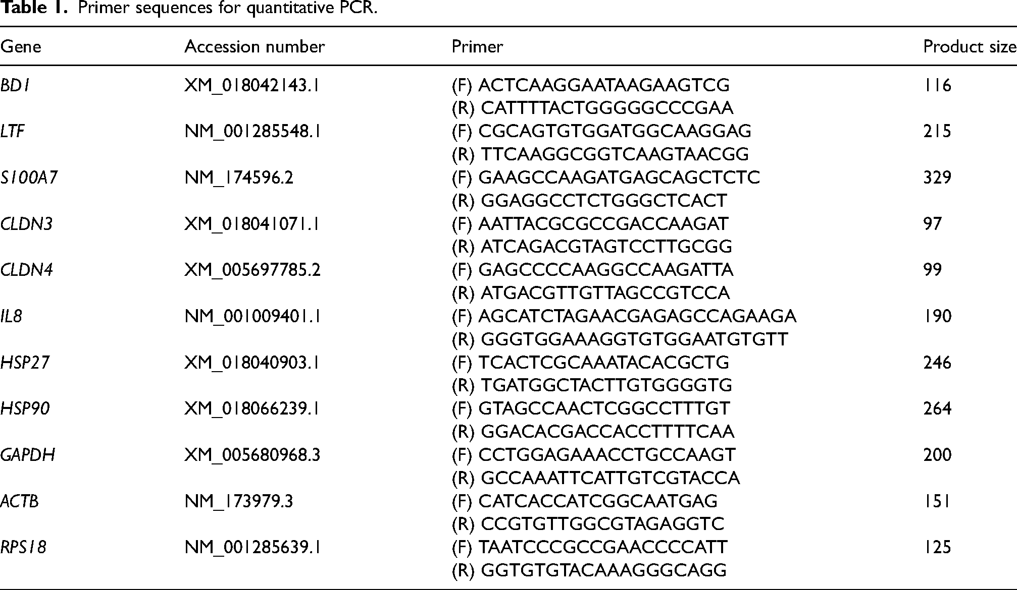
qPCR was performed as previously reported. 21 Total RNA from applied mammary gland tissues, cultured GMECs, and somatic cells was extracted using Sepasol-RNA I Super G (Nacalai Tesque). Reverse transcription was performed using the ReverTra Ace qPCR RT Master Mix (Toyobo, Osaka, Japan). qPCR was conducted on an AriaMx Real-Time PCR System (Agilent Technologies, Santa Clara, CA, USA) using Brilliant III Ultra-Fast SYBR QRTPCR (Agilent Technologies). The cycling conditions were 95°C for 1 min, followed by 45 cycles of 95°C for 15 s and 60°C for 1 min. The primers used are listed in Table 1. Ribosomal protein S18 (RPS18) was used as an internal control, which was selected by BestKeeper from among RPS18, actin beta (ACTB), and glyceraldehyde-3-phosphate dehydrogenase (GAPDH). qPCR data were analyzed using the 2−ΔΔCT method, and expression levels of the target gene were normalized against RPS18 expression, to calculate the relative levels of gene expression in each sample.
Primer sequences for quantitative PCR.
Statistical analysis
Data were expressed as mean ± SD. Statistical analyses were performed using the SAS software (version 9.4, SAS Institute Inc., Cary, NC, USA). Significant differences were calculated using a one-way analysis of variance and post-hoc Dunnett's test or Student's t-test. Differences were considered significant at p < 0.05. The relative values of milk yield, SCC, and other components were calculated based on the average of pretreatment levels (days − 1 and 0). Western blotting was performed three to four times using an acrylamide gel with two sets of GMEC samples from different wells. For the quantification of bands, the relative value of one set of GMECs samples was calculated based on the value of each control. In the in vivo immunohistochemical test (n = 4), the number of samples was defined as n = 1 for a sample derived from an udder. The in vivo experiments (n = 9) were performed using nine different udders, and the in vitro experiments (n = 6–12) were performed using three different GMECs or somatic cells derived from different goats. Each assay was performed twice or thrice to validate the results.
Results
Effects of topical application of gingerol on milk yield, SCC, and milk components
One mL of 100 μM gingerol, dissolved in 70% ethanol containing 0.2% DMSO, was applied to the udders of lactating goats for 7 d. The SCC in milk at 5 and 7 d significantly increased to approximately 3-fold compared with the SCC average of day − 1 and 0 (pre-treatment; control) (Figure 1B). There were no significant differences in milk yield or milk components (fat, protein, lactose, and solids) (Figure 1A and C). The 70% ethanol containing 0.2% DMSO (vehicle) did not affect the milk yield or SCC (Supplementary Figure 1).

Influence of topical application of gingerol to udders on milk yield and somatic cell count (SCC) in milk and milk components.
Influence of topical application of gingerol to udders on antimicrobial component production in milk
The concentrations of antimicrobial components in the milk were examined using an ELISA (Figure 2). The concentrations of β-defensin-1 and S100A7 were significantly higher at 3 and 7 d, respectively, than the pre-treatment levels. In addition, cathelicidin-2 concentration at 3–7 d was significantly higher (approximately 1.3-fold) than that before treatment. In contrast, there was no significant influence on the concentrations of lactoferrin, S100A8, and IgA.

Influence of topical application of gingerol to udders on the antimicrobial component concentration of milk.
Influence of gingerol on antimicrobial component production in GMECs and somatic cells
GMECs were treated with gingerol (0.5, 1, 5, 10, or 50 μM) or 0.1% DMSO as a vehicle control for 3 d, and the concentrations of β-defensin-1, lactoferrin, and S100A7 in the GMECs were examined using an ELISA (Figure 3A). The GMECs treated with 50 μM gingerol for 3 d showed significantly higher concentration of β-defensin-1, i.e., approximately 1.4-fold higher than that of the control group. The effects of gingerol on mRNA expression were examined using qPCR (Figure 3B). The GMECs treated with 10 μM gingerol for 3 d showed significantly higher, approximately 1.3-fold, expression of BD1 (β-defensin-1) than that in the control group. In contrast, the GMECs treated with 50 μM gingerol showed significantly lower expression of BD1, LTF (lactoferrin), and S100A7 by approximately 0.6-, 0.6-, and 0.4-fold, respectively, than that in the control group. To investigate the effect of gingerol on the secretion of antimicrobial components, the GMECs were treated with 50 μM gingerol for 4 d, and their concentrations in the upper chamber medium were examined using an ELISA (Figure 3C). Gingerol treatment significantly increased S100A7 concentrations by up to 2.5-fold compared to that in the control group.

Influence of gingerol on antimicrobial component production in cultured goat mammary epithelial cells (GMECs) and somatic cells.
The somatic cells were treated with 50 μM gingerol or 0.1% DMSO for 24 h, and the concentrations of β-defensin-1, lactoferrin, and cathelicidin-2 in somatic cells or culture medium were examined using ELISA (Figure 3D and E). Gingerol treatment significantly increased the concentration of β-defensin-1 by approximately 1.4-fold in the somatic cells compared to that in the control group.
Effect of gingerol on the number of cells positive for cathelicidin-2 or IgA in the mammary glands
The localization of cathelicidin-2 and IgA was observed using immunohistochemistry (Figure 4). In both the control and treatment groups, cathelicidin-2-positive and IgA-positive cells were observed in the mammary stromal region. The number of cathelicidin-2-positive cells in the mammary glands was significantly higher in the gingerol treatment group than in the control group (14.6 ± 2.9 cells/mm2 vs. 29.8 ± 4.5 cells/mm2). In contrast, the number of IgA-positive cells was not significantly different between the groups.

Influence of topical application of gingerol to udders on numbers of cathelicidin-2 and IgA positive cells in the mammary glands.
Influence of topical application of gingerol to udders on barrier function in the mammary glands
The topical application of gingerol to udders significantly increased the SCC in milk. To investigate the influence of gingerol on the barrier function of mammary glands, the concentrations of blood-derived components (Na+, albumin, and IgG) in the milk were examined (Figure 5A–C). However, gingerol treatment did not have a significant effect on these concentrations in the milk. In addition, the influence of gingerol on TJ protein localization was investigated using immunofluorescence (Figure 5D and F). Claudin-3 was localized in the TJ regions, as indicated by occludin, and claudin-4 was localized near the TJ regions in both the control and gingerol-treated groups.

Influence of topical application of gingerol to udders on tight junction barrier in mammary gland tissue.
Influence of gingerol treatment on TJs in GMECs
GMECs were treated with gingerol (0.5, 1, 5, 10, or 50 μM) or 0.1% DMSO for 3 d, and the levels of claudin-3 and claudin-4 in the GMECs were examined using western blotting (Figure 6A). Treatment with 50 μM gingerol significantly reduced the amount of claudin-4 by 0.7-fold compared to that in the control group. GMECs were treated with 10 μM gingerol for several days; after 9 d, the levels of both claudin-3 and claudin-4 in GMECs decreased to less than 0.75-fold compared to those in the control group (Figure 6B). In addition, the GMECs treated with 10 μM gingerol for 3 d showed significantly reduced mRNA expression of both claudin-3 and claudin-4 (Figure 6C). In contrast, treatment with 50 μM gingerol for 4 d did not influence TJ barrier function, as evaluated by measuring the TEER and FITC permeability of the barrier (Figure 6D and E).

Influence of gingerol on the tight junction barrier in goat mammary epithelial cells (GMECs).
Influence of gingerol on cytokine production
The concentrations of cytokines (IL-8, IL-1β, and TNF-α) in the collected milk samples were examined using ELISA (Figure 7A–C). The concentration of IL-8 at 7 d was significantly higher (1.8-fold) than the pre-treatment levels. In addition, IL8 concentration in mammary gland tissues treated with gingerol tended to be higher than that in the control group (p = 0.082) (Figure 7D). Subsequently, somatic cells in the milk were observed using Giemsa staining (Figure 7E). The proportion of polymorphonuclear cells (PMN) in the gingerol group was significantly higher than that in the control group. In addition, mRNA expression and intracellular or secreted concentrations of IL-8 in cultured GMECs or somatic cells were investigated (Figure 7F and G). In the GMECs treated with 50 μM gingerol for 3 d, the intracellular IL-8 concentration significantly increased to approximately 1.4-fold compared with that in the control, although gingerol did not affect IL-8 production in somatic cells.

Influence of gingerol on cytokine production and the proportion of polymorphonuclear cells (PMN) in milk.
Influence of gingerol on heat shock protein expression and STAT signal
Regarding the mRNA expression of heat shock proteins (HSP27 and HSP90), HSP27 expression in mammary gland tissues treated with gingerol tended to be lower than that in the control group (p = 0.052) (Figure 8A). The GMECs treated with 10 or 50 μM gingerol for 3 d showed significantly lower expression of both HSP27 and HSP90 than the control group. In contrast, gingerol treatment did not affect HSP27 or HSP90 expression in the cultured somatic cells.

Influence of gingerol treatment on heat shock protein (HSP) and STAT expression.
The effect of gingerol on STAT3 and STAT5 was examined using western blotting (Figure 8D and E). In the mammary glands treated with gingerol, the amount of pSTAT3 significantly decreased by 0.6-fold compared with that in the control. In addition, the amount of pSTAT3 in the GMECs treated with 50 μM gingerol for 3 d decreased by 0.7-fold compared to that in the control. Gingerol did not significantly affect STAT5 expression in either the mammary glands or GMECs.
Discussion
In this study, topical application of gingerol to udders enhanced cathelicidin-2 concentration in the milk, with an increase in SCC and the proportion of PMN. Our previous research indicated that cathelicidin-2 is mainly secreted by neutrophils. 9 Additionally, in the present study, gingerol application increased the number of cathelicidin-2-positive cells in the mammary glands. Furthermore, gingerol treatment increased IL-8 concentration in the milk and IL-8 production in cultured GMECs and showed a tendency toward higher concentrations of IL-8 in treated mammary glands. IL-8 is commonly considered as a chemotactic factor in neutrophils. 38 Therefore, these findings indicate that the topical application of gingerol to udders enhances cathelicidin-2 concentration by facilitating the migration of neutrophils into the mammary glands via IL-8 production by GMECs. In this study, gingerol treatment increased the concentrations of β-defensin-1 and S100A7 in the milk, the secretion of S100A7 and mRNA expression of β-defensin-1 in cultured GMECs, and the intracellular concentration of β-defensin-1 in both cultured GMECs and somatic cells. Cathelicidins and defensins can disrupt various microbial membranes,6,8 and S100A7 shows antimicrobial activity against E. coli. 39 Here, we found that gingerol application to udders enhanced the concentrations of antimicrobial components in the milk without influencing milk production, which may contribute to the prevention of mastitis.
In mammary glands with mastitis, the SCC in milk is considerably increased with an increase in the levels of blood-derived components, such as Na+, albumin, and IgG, due to disruption of the TJ barrier formed by MECs.19,20,40,41 However, gingerol application to udders did not affect the concentrations of Na+, albumin, and IgG in the milk, although SCC in the milk was significantly increased. In addition, alveolar structures and TJs in the mammary glands were found to be normal. Moreover, gingerol did not affect the TJ barrier function in cultured GMECs, as measured from the TEER and FITC permeability. These findings indicate that gingerol does not affect the passive movement of blood-derived components into the milk. In contrast, as mentioned above, gingerol increases IL-8 production in mammary glands, which promotes the migration of neutrophils into the milk. 42 Gingerol treatment reduced the mRNA and protein levels of both claudin-3 and claudin-4 in cultured GMECs. Thus, gingerol application may help in the active movement of neutrophils by regulating the amount of claudins in MECs. It has been reported that spermatogonial stem cells pass through the blood-testis barrier and express the same claudin as testis cells. 43 Thus, neutrophils may express the same claudin as MECs and pass through TJs in the mammary glands. Further research is needed to reveal how neutrophils pass through TJs in the mammary glands without damage.
Gingerol exhibits anti-inflammatory effects4,5 and heat stimulation-like effects. 24 In a previous study using cultured mouse MECs, a high temperature (41°C) led to a reduction in the amount of claudin-4 and pSTAT3 compared to the normal temperature (37°C), 44 which is consistent with the results of the present study. The lactating MECs during inflammation due to lipopolysaccharide treatment also show increased pSTAT3 and claudin-4 levels. 45 In addition, local heat treatment of udders increases the SCC and cathelicidin-2 concentrations in the milk, 21 which is consistent with the results of the present study. Moreover, gingerol affected the mRNA expression of HSP27 and HSP90. It has been previously reported that gingerol treatment reduces the protein levels of HSP90, with a decrease in hypoxia-inducible factor 1α as its transcriptional regulator. 23 These findings, in concert with ours, indicate that gingerol can exhibit potential anti-inflammatory effects and partly induce heat stimulation-like effects. In addition, heat shock proteins have been previously reported to regulate STAT signals.45,46 Further research is needed to reveal the relationship between anti-inflammatory effects or heat stimulation-like effects of gingerol and the signals of heat shock proteins or STAT in the mammary glands and the underlying mechanism.
Conclusions
We investigated the effects of topical application of gingerol on udders. The results showed that gingerol treatment increased the SCC and antimicrobial components in milk without influencing the TJ barrier and milk production. These findings may be useful for the prevention of mastitis in milk-producing animals and, hence, safe and stable dairy production. This study did have some limitations, such as the small sample size and some individual differences in the effects; therefore, further studies should use a greater number of animals, including dairy cows.
Supplemental Material
sj-docx-1-ini-10.1177_17534259231191252 - Supplemental material for Potential effects of gingerol topical application on components of the innate immunity in lactating goat mammary glands
Supplemental material, sj-docx-1-ini-10.1177_17534259231191252 for Potential effects of gingerol topical application on components of the innate immunity in lactating goat mammary glands by Yusaku Tsugami, Takahiro Nii, Ken Kobayashi and Naoki Isobe in Innate Immunity
Footnotes
Acknowledgments
We thank Yukinori Yoshimura, Masayuki Shimada, and Takashi Umehara, Graduate School of Integrated Sciences for Life, Hiroshima University, for their helpful advice and technical assistance with our experiments.
Authors’ contributions
Yusaku Tsugami: Conceptualization, Data curation, Investigation, Methodology, Formal analysis, Visualization, Writing – Original Draft, Funding acquisition, Project administration, Supervision.
Takahiro Nii: Conceptualization, Resources, Writing – Review & Editing.
Ken Kobayashi: Conceptualization, Resources, Writing – Review & Editing.
Naoki Isobe: Conceptualization, Resources, Writing – Review & Editing.
Declaration of conflicting interests
The author(s) declared no potential conflicts of interest with respect to the research, authorship, and/or publication of this article.
Funding
The author(s) disclosed receipt of the following financial support for the research, authorship, and/or publication of this article: This work was supported by the the Lotte Research Promotion Grant.
Supplemental material
Supplemental material for this article is available online.
References
Supplementary Material
Please find the following supplemental material available below.
For Open Access articles published under a Creative Commons License, all supplemental material carries the same license as the article it is associated with.
For non-Open Access articles published, all supplemental material carries a non-exclusive license, and permission requests for re-use of supplemental material or any part of supplemental material shall be sent directly to the copyright owner as specified in the copyright notice associated with the article.