
Abstract
Shiga toxin (Stx)-producing Escherichia coli (STEC) infections in cattle are asymptomatic; however, Stx impairs the initiation of an adaptive immune response by targeting bovine peripheral and intraepithelial lymphocytes. As presumptive bovine mucosal macrophages (Mø) are also sensitive to Stx, STEC may even exert immune modulatory effects by acting on steps preceding lymphocyte activation at the Mø level. We therefore studied the expression of the Stx receptor (CD77), cellular phenotype and functions after incubation of primary bovine monocyte-derived Mø with purified Stx1. A significant portion of bovine Mø expressed CD77 on their surface, with the recombinant B-subunit of Stx1 binding to >50% of the cells. Stx1 down-regulated significantly surface expression of CD14, CD172a and co-stimulatory molecules CD80 and CD86 within 4 h of incubation, while MHC-II expression remained unaffected. Furthermore, incubation of Mø with Stx1 increased significantly numbers of transcripts for IL-4, IL-6, IL-10, IFN-γ, TNF-α, IL-8 and GRO-α but not for IL-12, TGF-β, MCP-1 and RANTES. In the course of bovine STEC infections, Stx1 appears to induce in Mø a mixed response pattern reminiscent of regulatory Mø, which may amplify the direct suppressive effect of the toxin on lymphocytes.
Introduction
Shiga toxin (Stx)-producing Escherichia coli (STEC) are major zoonotic pathogens, causing severe diseases in humans, such as haemorrhagic colitis and haemolytic uremic syndrome. 1 Once proven to be pathogenic in humans, respective STEC strains are referred to as enterohaemorrhagic E. coli (EHEC). Pathogenesis of human EHEC-related disease is strongly dependent on the action of macrophages (Mø), which act as major sources of pro-inflammatory cytokines. Indeed, production and secretion of these is induced by Stx, the most important STEC virulence factor released from the bacteria upon intestinal colonization. 2 Subsequent inflammatory responses facilitate toxin absorption from the intestine. 3 Additionally, the released cytokines induce up-regulation of the Stx receptor [globotriaosylceramide (Gb3) syn. CD77] expression on the surface of endothelial cells in the glomeruli of kidneys and small blood vessels in other tissues. This renders endothelial cells, the main target cells of Stx, highly susceptible to the cytotoxic actions of Stx.4,5
Cattle are the most important reservoir for STEC worldwide; 1 however, clinical symptoms in bovines occur only rarely, usually manifesting as mild diarrhoea in young calves. Importantly, however, animals remain persistently infected and may shed STEC intermittently over long periods of time.6,7 Our earlier research showed that Stx may be implicated in the establishment of persistence of infection by attacking cells of the bovine immune system. Stx1 does not induce direct cytolethal effects but hinders the activation and proliferation of the cells in vitro,8,9 and affects cytokine gene expression in ileal intraepithelial lymphocytes.10–12 In contrast, other cell types, such as granulocytes exposed to Stx during enteric STEC infections seemed to be resistant to any impact by Stx. In these cells, Stx1 neither induced cell death nor impacted on the phagocytic and oxidative burst activity, which is in line with the observation that bovine granulocytes fail to express CD77. 13 In addition to the cell types mentioned above, we were also able to show that primary bovine colonic epithelial cells synthesized CD77 but barely expressed the receptor on the cellular surface and thus did not respond to Stx1. 14 Characterization of such epithelial cell cultures led to the discovery of a population of vimentin-positive cells of mesenchymal origin expressing substantial amounts of CD77 on their surface. 14 Supplementation of culture medium with Stx1 resulted in the rapid elimination of this cell type. A cell line generated from these cells expressed CD77 constantly on the surface and up-regulated cytokine and chemokine gene expression in response to Stx1. Based on their phenotype and function, this cell line was considered to be derived from mucosal Mø. 14
Therefore, it is tempting to assume that Mø also play an important yet undisclosed role in the course and duration of bovine STEC infections. The aim of the present study was to investigate potential changes in surface expression of CD77 by primary, non-immortalized monocyte-derived Mø exposed to purified Stx1, as well as their phenotype and cytokine expression profile after short-term exposure to Stx1 in vitro.
Materials and methods
Primary bovine monocyte-derived Mø culture
Primary bovine Mø were isolated as described elsewhere.15,16 Briefly, a citrated whole blood sample (5:1 diluted in 3.8% sodium citrate solution) was centrifuged (2380 g, 20 min) and the buffy coat was collected. After several washing steps with PBS-EDTA buffer (PBS supplemented with 5.4 mM EDTA; 800 g, 10 min), remaining erythrocytes were lysed by incubation of the re-suspended pellet in lysis buffer (8.26 g NH4Cl, 1.09 g NaHCO3, 0.037 g Na3EDTA and 1000 ml A. dest.; 10 min). The resulting buffy coat was washed three more times (300 g, 4℃, 10 min) and layered onto Ficoll Histopaque (GE Healthcare, Munich, Germany) for density centrifugation (800 g, 45 min at ambient temperature). Cells were collected by taking the interphase, centrifuged at 800 g, 4℃, for 10 min and washed twice with PBS buffer (300 g, 4℃, 10 min). Cells were adjusted to 4 × 10⁶/ml in cell culture medium 1 (IMDM without phenol red, 20% FCS, 1% penicillin/streptomycin, 1% amphotericin B, 0.05% 100 mM β-mercaptoethanol) and 25 ml of this cell suspension were transferred to Teflon bags (VueLife Bags, American Fluoroseal Corp., Gaithersburg, MD, USA) and incubated for 8 d (37℃, 5% CO2). At the end of the incubation period, cells were harvested and diluted to 2 × 10⁶/ml in cell culture medium 2 (IMDM without phenol red, 2% FCS, 1% penicillin/streptomycin, 1% amphotericin B, 0.05% 100 mM β-mercaptoethanol). Cells were seeded into Petri dishes, microtitre plates or onto glass slides, and incubated for a further 18 h. Subsequently, lymphocytes were removed by careful washing and adherent Mø were left within the vessels in cell culture medium 2 without or with supplementation with LPS from E. coli O111:B4 (100 ng/ml; Sigma-Aldrich, Taufkirchen, Germany), with purified Stx1 [200 verocytotoxic doses 50% (CD50/ml)] in the absence or after pre-incubation with purified mouse anti-StxB1 [immunoglobulin G1 (IgG1), clone 13C4; 1.5 µg/ml]. 17 Methods for Stx1 and anti-Stx1 subunit B (StxB1) purification were performed as previously described.8,9
Flow cytometric analysis
Cells were detached by incubation with Accutase (PAA Laboratories, Cölbe, Germany), transferred to V-shaped microtitre plates and pelletized by centrifugation (150 g, 4℃, 7 min). For detection of intracellular Ags, cells were fixed in paraformaldehyde (2% in PBS; 25℃, 30 min), washed with PBS and permeabilized with digitonin (0.005% in PBS, 25℃, 10 min; Sigma-Aldrich, Taufkirchen, Germany). Detection of cellular Ags and binding sites for the recombinant B-subunit of Stx1 (rStxB1) was performed as described previously.9,18 Briefly, pellets were re-suspended in washing buffer (PBS supplemented with 1% BSA, 0.01% sodium azide and 0.5% goat serum) as a negative control or with 50 µl of primary Ab solution. Primary Abs were produced as hybridoma supernatant (IL-A 30, anti-bovine surface IgM; IL-A24, anti-bovine CD172a; N32/52-3, anti-bovine CD80; IL-A190, anti-bovine CD86; CC108, anti-bovine MHC-II), or purchased from either VMRD (anti-bovine CD3 clone MM1A, anti-bovine CD11b clone MM12A, anti-bovine CD11c clone BAQ153A, anti-bovine CD14 clone CAM36A; Labor Diagnostik Leipzig, Leipzig, Germany) or AbDSerotech (anti-human CD77 clone 38-13; Puchheim, Germany). In assays assessing the impact of Stx on surface expression of Ags, rat IgM (2 µg/ml; Camon, Wiesbaden, Germany) and supernatants containing one of two Abs directed against Clostridium perfringens phospholipase C (IgG1 and IgG2, respectively) were included as isotype controls. Cells were incubated for 20 min on ice, washed with washing buffer and diluted in 50 µl washing buffer with secondary Abs [FITC-labelled α-rat IgM (Dianova GmbH, Hamburg, Germany); APC-labelled anti-mouse IgG1 (Southern Biotech c/o Biozol, Eching, Germany)] supplemented with 7-amino actinomycin D (7–AAD; f.c. 2 µg/ml; Sigma-Aldrich) and diluted.
To assess rStxB1 binding and for competition assays, cells were incubated with rStxB1 (30 µg/ml or dilutions thereof as indicated) in PBS for 30 min on ice. Afterwards, cells were washed once (400 g, 4℃, 4 min) and re-suspended in 50 µl anti-StxB1 (45 µg/ml), followed by another incubation on ice for 30 min. Cells were washed again and re-suspended in 50 µl APC-labelled anti-mouse IgG1 (Southern Biotech). Prior or after this procedure, cells were submitted to anti-CD77 staining as described above.
Finally, cells were washed with washing buffer and analysed on a BD FACSCalibur Analyzer (Becton-Dickinson, Heidelberg, Germany). Raw data were analysed using the FCS Express software package (version 2; DeNovo Software, Thornhill, Canada). Gates were defined according to the negative control (PBS) and isotype or secondary Ab control included in the test series, defining < 2% of the cells as positive as exemplified in Figure S1.
Immune fluorescence microscopy
Cells grown in 12-well culture plates were fixed and permeabilized as described for flow cytometry but omitting Accutase treatment. Labelling was carried out as described above except that phycoerythrin-conjugated anti-mouse IgG Ab (Sigma-Aldrich) was used to detect anti-StxB1 binding and DAPI (0.02%; Invitrogen, Karlsruhe, Germany) was used instead of 7-AAD. In addition, some samples were incubated with FITC-conjugated anti-pan cytokeratin Ab (clone C-11; Sigma-Aldrich) or anti-vimentin Ab (clone 3B4; Dako, Hamburg, Germany) and phycoerythrin-conjugated anti-mouse IgG Ab. Samples were mounted on glass slides and dried overnight at 4℃. Immune fluorescence microscopy was performed using a Leica DMRB Laborlux 12 microscope (Leica, Solms, Germany) equipped with an analogue camera.
Reverse quantitative PCR
Cells were detached from the plates with a cell scraper and washed twice (202 g, 7 min, 20℃) with PBS. Cells were counted and 1 × 10⁶ cells were lysed in 600 µl of RLT buffer (RNeasy mini-kit; Qiagen, Hilden, Germany) with 2.5% β-mercaptoethanol (Amersham Biosciences, Buckinghamshire, UK) and stored at –70℃. The RNA isolation procedure and reverse transcription were performed according to the method described by Moussay et al. 12 PCR amplification was done on an automated fluorometer (ABI PRISM 5700 sequence detection system; Applied Biosystems, Foster City, CA, USA) using 96-well optical plates. Sequences of primers and probes used to detect mRNA for GAPDH, IFN-γ, IL-4, IL-6, IL-8, IL-10, IL-12, growth regulated oncogene alpha (GRO-α), monocyte chemotactic protein 1 (MCP-1), regulated upon activation normal T cell expressed and secreted (RANTES), TGF-β and TNF-α proteins have been published previously.19–22
Statistical analysis
Statistical analysis was done using SPSS for Windows (version 15; IBM, Armonk, NY, USA) applying the t-test for paired samples, one- and two-way ANOVA, and Student–Newmans–Keuls test with Bonferroni correction. P-Values ≤ 0.05 were considered significant (*P ≤ 0.05; **P ≤ 0.01; ***P ≤ 0.001).
Results
Phenotype of primary bovine Mø in vitro
Teflon-bag-propagated monocytes formed a confluent monolayer of adhesive, round-to-polymorphic cells, positive for intracellular vimentin and negative for cytokeratin when further cultured on plastic surfaces thereby differentiating to Mø. Flow cytometric analysis (mean ± SD) of the cells after detachment showed that medium control cultures consisted of 80 ± 11% and 75 ± 12% Mø after 18 h of plastic adherence and further 4 h (n = 8 cultures) and 24 h of incubation (n = 7), respectively, as defined by light scatter characteristics (see Figure 4A for definition of cells). Cells of the respective population stained negative for CD3, and detection of sIgM and CD11c was on 5% and ≤ 30%, respectively. In contrast, surface expression of CD11b, CD14, CD172a and MHC-II was detected on ≥ 98% of the cells (Figure S2). Immunolabelling with anti-CD80 and anti-CD86 Abs showed a shift in the fluorescence intensities of all cells; however, it varied significantly between single cells, as well as between cell preparations.
CD77 and Shiga toxin receptor expression by primary bovine Mø
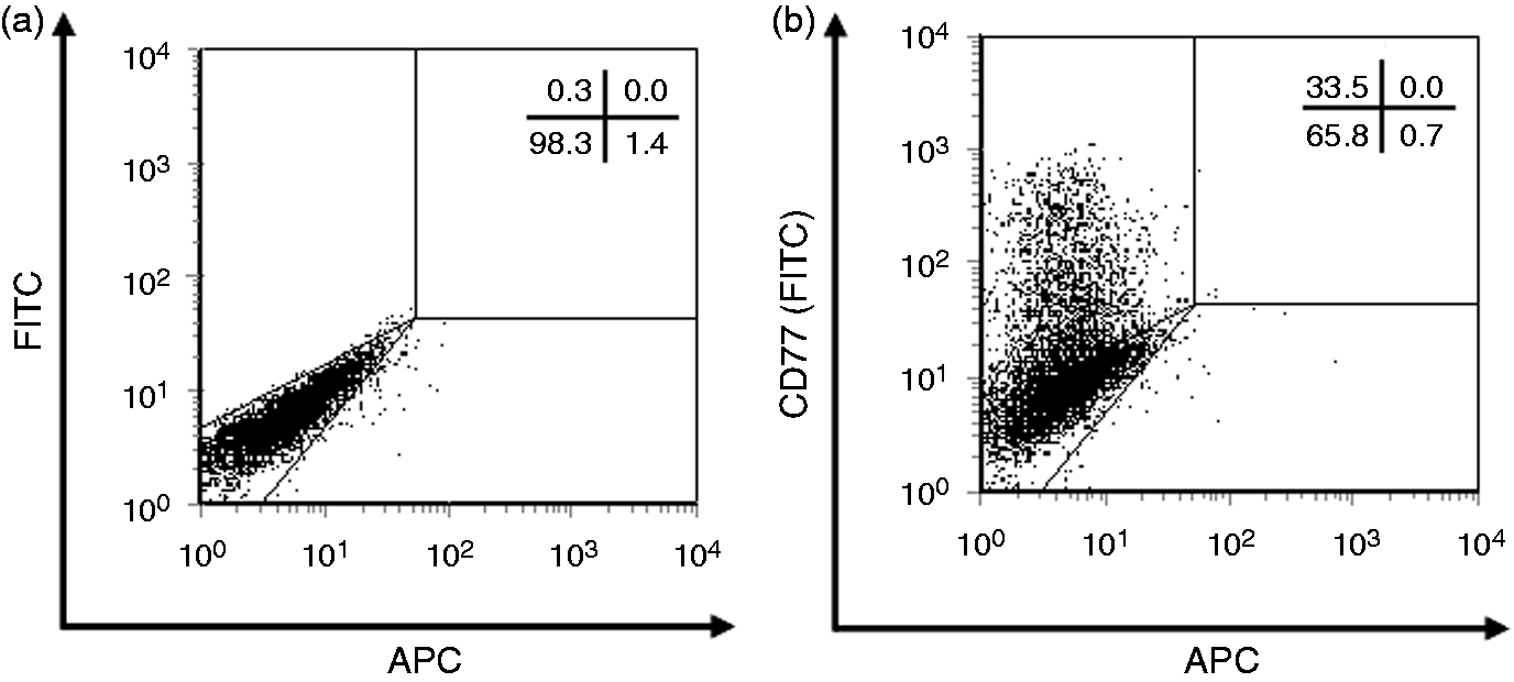
Immune fluorescence microscopy of Mø (n = 3 cultures) revealed surface expression of CD77 on a subset of cells (Figure 1). Permeabilization resulted in an evenly distributed anti-CD77 staining pattern (data not shown), indistinguishable from that obtained after binding of rStxB1 (Figure 1). When quantitatively analysed by flow cytometry, 31.9 ± 15.7% of Mø (mean ± SD of four independent experiments) stained positive for surface CD77 (Figure 2).
Localization of CD77 Ags on the surface (green; c) and binding sites for rStxB1 within primary bovine Mø (red; d). Teflon bag-propagated Mø were submitted to immunolabelling with (a, b) secondary Abs only or (c) with anti-CD77 Ab and (d) rStxB1, (a, c) without and (b, d) after permeabilizing pre-treatment, respectively. A subpopulation of cells strongly bound anti-CD77 at the surface (arrows). Nuclei were counterstained with DAPI (blue). Photographs are representative of three independent experiments. Detection of CD77 expression by primary bovine Mø. Teflon bag-propagated Mø were submitted to immunolabelling with (a) secondary Ab only or (b) with anti-CD77 Ab and secondary Ab. Dot plots are representative of four independent experiments. Percentages of positive cells among morphologically intact cells according to the light scatter characteristics are shown in the upper right corner of the plots.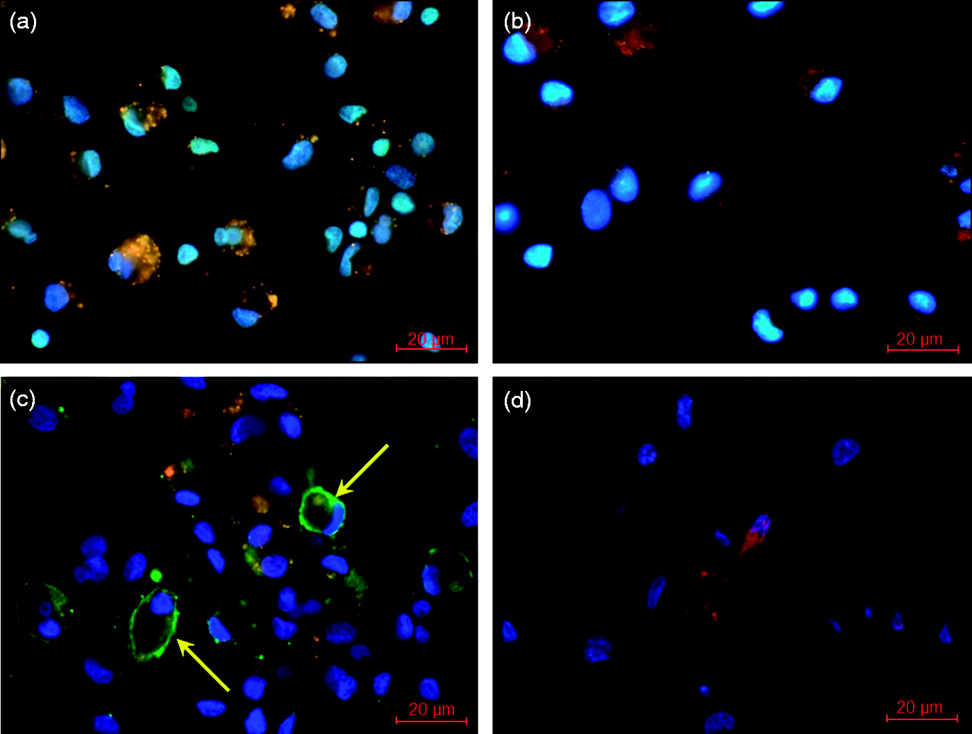
Having established that Mø express CD77 and are able to bind rStxB1, we assessed whether anti-CD77 Ab and rStxB1 compete for binding to Mø (Figure 3). Incubation of cells with increasing concentrations of rStxB1 prior to immunolabelling with the Ab led to a clear reduction in the number of CD77+ cells in a concentration-dependent fashion, while the number of cells having bound rStxB1 raised to >50% (Figure 3). Pre-incubation with rStxB1 at 30 µg/ml completely abolished CD77 binding (data not shown). A comparable level of inhibition was observed when cells were incubated with anti-CD77 prior to rStxB1, suggesting that rStxB1 was able to remove bound anti-CD77 from the cellular surface (data not shown).
Competition of rStxB1 and anti-CD77 for binding to primary bovine Mø. Dot plots represent analyses of Teflon bag-propagated Mø immunolabelled with (c–e) anti-CD77 after prior incubation with increasing concentrations of rStxB1 as indicated. Cells incubated in (a) the absence of anti-CD77 and rStxB1 and (b) in the presence of anti-CD77 only were included as controls. Dot plots are representative of three independent experiments. Percentages of positive cells among morphologically intact cells according to the light scatter characteristics are shown in the upper right corner of the plots.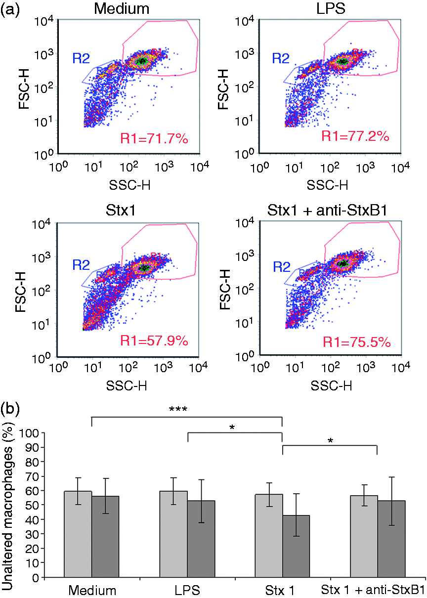
Effect of exposure to purified Stx1 on morphology, phenotype and cytokine expression profile of primary bovine Mø
Beginning 24 h after the addition of 200 CD50 purified Stx1/ml, a concentration sufficient to induce modulating effects reliably on bovine immune cells in vitro,
8
Mø showed a disruption of the monolayer by cell detachment (data not shown). Although supplementation of culture medium with a sub-lethal concentration of LPS (100 ng/ml) as stimulation control for the same time period also led to some reduction in the percentage of morphologically intact cells as defined by their light scatter characteristics in flow cytometry, the effect was significantly stronger in cultures supplemented with Stx1 (Figure 4). Percentages of unaltered cells in cultures additionally supplemented with neutralizing anti-StxB1 Ab 13C4 did not differ from medium controls.
Effect of purified Stx1 on the morphology of primary bovine Mø. (a) Representative scatter plots obtained with cells from cultures incubated for 24 h in the presence of purified Stx1 (200 CD50/ml as determined on Vero cells). Control cultures were established with medium only, with medium supplemented with LPS (100 ng/ml) and with Stx1 after pre-incubation with anti-StxB1 (1.5 µg/ml). Gate R1 defines morphologically intact Mø, gate R2 defines presumptive lymphocytes. (b) Percentage of morphologically intact Mø upon cultivation under the respective conditions for 4 (grey bars) and 24 h (black bars), respectively. Bars represent mean ± SD of cells in R1 relative to all cells included by R1 plus R2. Data were obtained from eight independent experiments. Asterisks indicate significant differences according to one-way ANOVA with repeated measurements and Bonferroni correction (*P ≤ 0.05; ***P ≤ 0.001).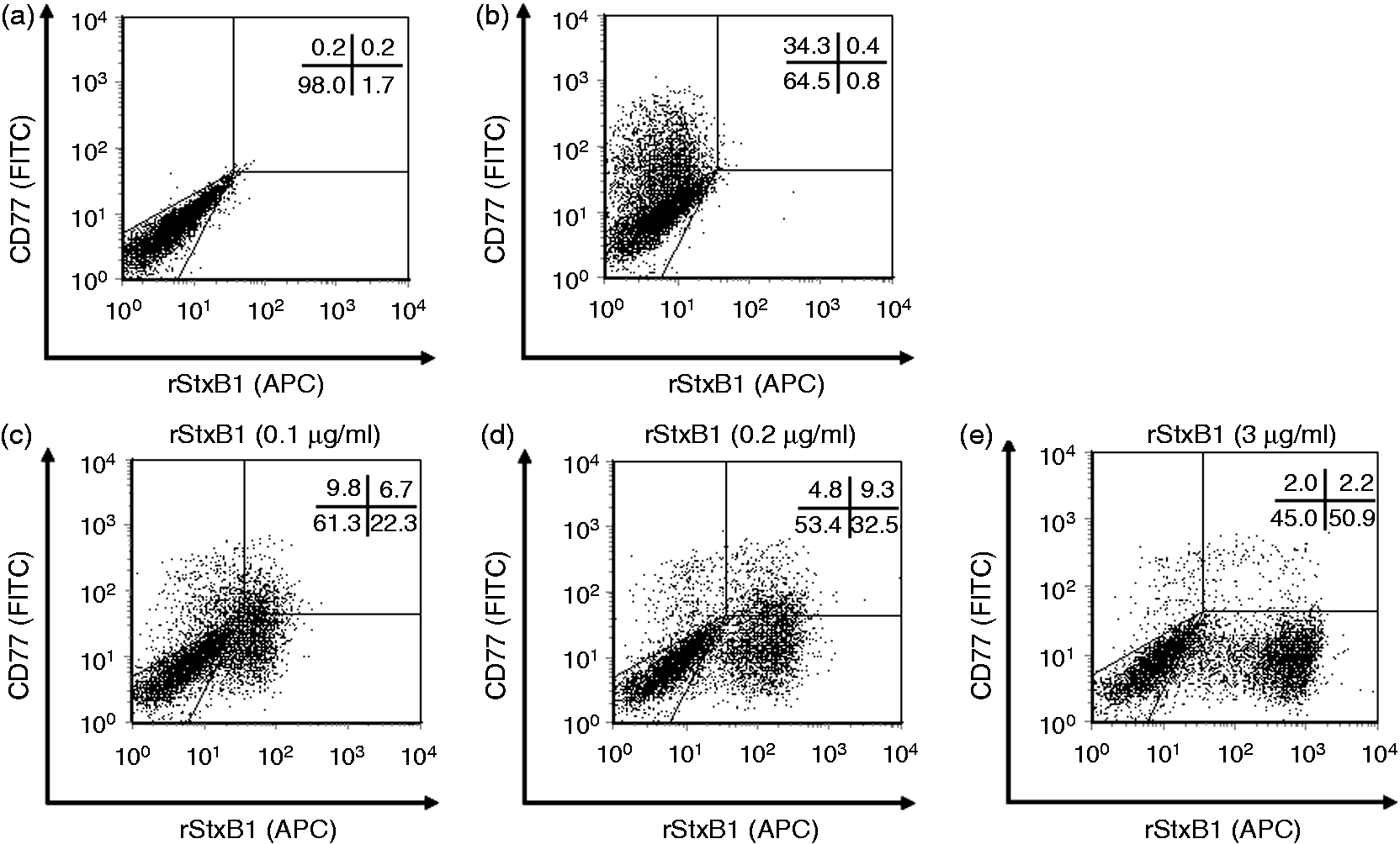
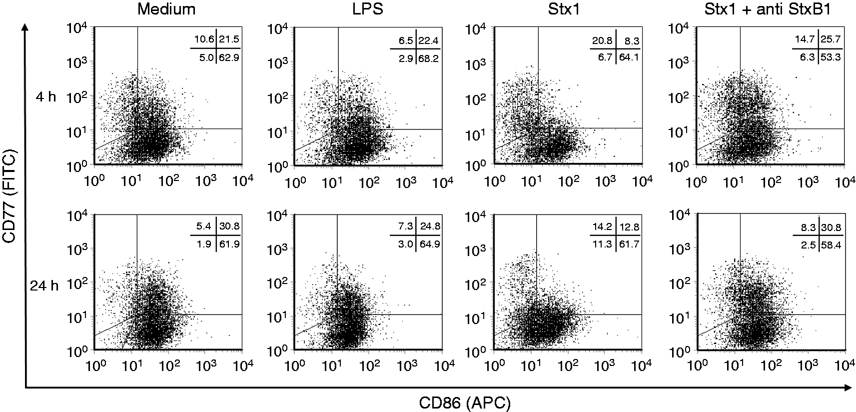
While it took 24 h for the cytopathic effect to develop, Mø responded to medium supplementation with Stx1 by down-regulation of surface-expressed Ags within 4 h (Figure 5). This effect was strongest in CD77-co-expressing cells (data for CD77− Mø not shown). Average MFI values for CD14, CD172a, CD80 and CD86 were all significantly reduced in Stx1-supplemented cultures compared with cultures supplemented with Stx1 plus anti-StxB1, or LPS. The effect was mainly based on a down-regulation of the respective Ags on the surface of cells rather than depletion of CD77+ cells, as exemplified for CD86 (Figure 6).
Expression of surface markers by primary bovine Mø in the presence of purified Stx1. Cells were incubated for 4 and 24 h with LPS (100 ng/ml; white bars) and with Stx1 (200 CD50/ml as determined on Vero cells) without (grey bars) or after pre-incubation with anti-StxB1 (1.5 µg/ml; black bars). Mean fluorescence intensities (MFLI) for the detection of the respective Ags on the surface of CD77 co-expressing cells were normalized to values obtained with cells cultured in non-supplemented medium (100% as visualized by the black line). Bars represent mean ± SD from data obtained in 7–8 independent experiments. Asterisks indicate significant differences according to one-way ANOVA with repeated measurements and Bonferroni correction (*P ≤ 0.05; **P ≤ 0.01; ***P ≤ 0.001). Expression of CD86 by primary bovine Mø in the presence of purified Stx1. Cells were incubated for 4 and 24 h in medium, in medium supplemented with LPS (100 ng/ml) or in medium supplemented with Stx1 (200 CD50/ml as determined on Vero cells) without or after pre-incubation with anti-StxB1 (1.5 µg/ml) as indicated in the figure. Dot plots are representative of 14–16 determinations from 7–8 independent experiments. Percentages of positive cells among morphologically intact cells according to the light scatter characteristics are shown in the upper right corner of the plots.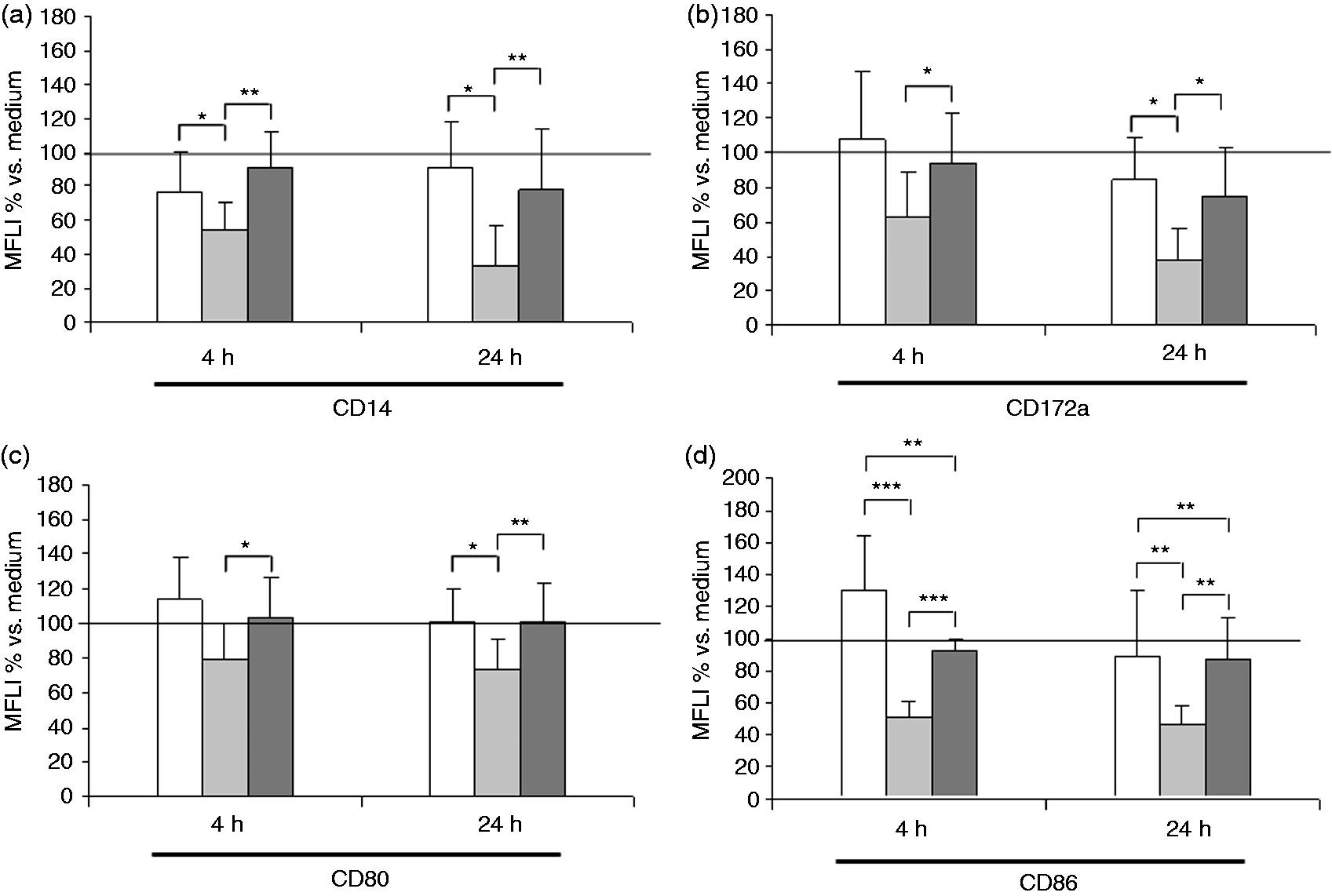
After 4 h of incubation with Stx1, increased amounts of mRNA transcripts for IL-4, IL-6, IL-10, IFN-γ and TNF-α, and the chemokine genes IL-8 and GRO-α were detected in Mø compared with cultures supplemented with Stx1 plus anti-StxB1 (Figure 7). In contrast, transcripts for IL-12, TGF-β, MCP-1 and RANTES remained unaffected. The mRNA pattern induced by Stx1 clearly differed from the pattern induced by LPS, which was characterized by an up-regulation of IL-12, IFN-γ, IL-8 and GRO-α.
Relative amounts of gene transcripts harboured by primary bovine Mø upon cultivation in the presence of purified Stx1. Cells were incubated for 4 h in medium supplemented with LPS (100 ng/ml; white bars) and in medium supplemented with Stx1 (200 CD50/ml as determined on Vero cells) without (grey bars) or after pre-incubation with anti-StxB1 (1.5 µg/ml; black bars). Subsequently, mRNA was reversely transcribed and quantified by real-time PCR. The transcription of the housekeeping gene GAPDH was used for normalization of the samples. Cells incubated with medium were used as a control. Amounts of mRNA equivalent to medium control were assigned a value of 100% and visualized by a black line. Data represent median and minimum / maximum of the results obtained with Mø preparations from seven different animals. Different lower case letters indicate significant differences with P ≤ 0.05 according to one-way ANOVA with repeated measurements and Bonferroni correction.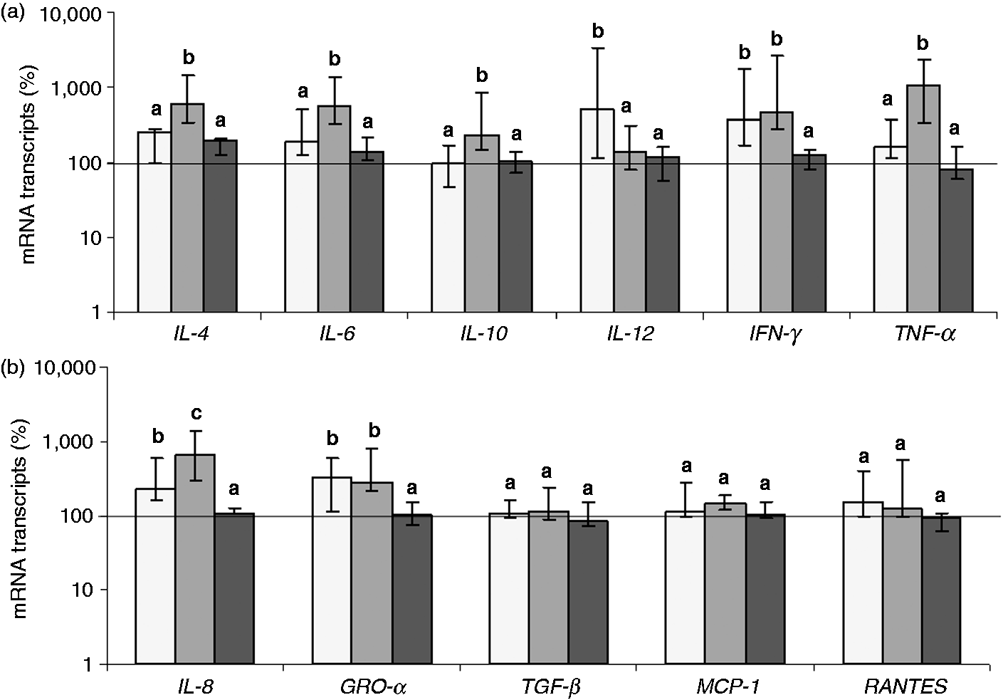
Discussion
STEC are persistently able to colonize the intestinal mucosa of bovines;6,7 however, inflammation is absent or mild, with tissue damage being only rarely observed and restricted to confined areas. 23 Cells of mesenchymal, that is, non-epithelial origin, in primary cultures of bovine colonic epithelial cells with Mø-like phenotype are highly sensitive to the cytokine-inducing activity of Stx1. 14 To confirm that cells of the monocyte lineage are target cells for Stx in cattle, the aim of the current study was to investigate primary monocyte-derived Mø in vitro, with particular emphasis on their role as cytokine secreting and T-cell-activating cells, orchestrating inflammatory and initiating specific immunological responses in the tissues. 24 Indeed, bovine Mø expressed the CD77 molecules on their surface, capable of binding StxB1. Additionally, exposure of these cells to Stx1 resulted in an altered phenotype and altered cytokine expression profile within hours, before cells eventually succumbed to cell death. In addition to cells of the adaptive immune system, with CD8+ T cells and B cells in particular,8,10 Mø thus also represent target cells for the immunomodulatory effects of Stx in cattle.
The anti-CD77 Ab (clone 38.13) used in the present study and StxB1 have been shown to bind to overlapping but not identical epitopes in Gb3 molecules. 25 However, even low concentrations of rStxB1 prevented anti-CD77 from binding to bovine Mø, confirming that CD77 functions as Stx receptor on bovine Mø as described earlier for bovine lymphocytes. 9
A prominent subset of Mø cultured in the absence of toxin expressed CD77 on the cellular surface, while nearly all cells seemed to harbour CD77 intracellular (data not shown). Gb3 can be detected biochemically in freshly cultured bovine PBMC, although surface expression of CD77 only occurs from day 1 of culture onwards. 26 This is similar to observations done in bovine BL-3 lymphoma B cells, which are CD77 surface negative but express CD77 intracellularly. Surface expression of CD77 can be induced in these cells by mitogenic stimulation, rendering them sensitive to Stx, 26 similarly to that described for human endothelial cells after activation with LPS, IL-1β and TNF-α.5,27 Activation either results in de novo synthesis of CD77 or in translocation of CD77 from intracellular stores to the surface. 28 The sensitivity of bovine Mø to Stx1 may thus not be confined to a particular subset of Mø but an inherent feature of many cells belonging to the monocyte lineage throughout the body, for example Mø situated next to the STEC colonization site in the intestinal mucosa of cattle. 14
The principal activity of Stx—a potent protein synthesis inhibitor—is induction of cell death in many cell types, including leukocytes from different species.29,30 As part of an endoplasmic stress response, Stx can induce both the intrinsic and extrinsic apoptosis pathways.29,31 However, intensive research in the last few decades revealed that Stx also activates signal transduction cascades and exerts a variety of modulating rather than cytolethal effects in cells.32,33 In a given cell model, the predominance of one of these two facets of Stx activity is closely linked to the cells’ level of differentiation. 34 In the present study, primary bovine Mø succumbed to cell death when cultured in the presence of Stx1 for several days, while control cells formed intact monolayers even after 6 d of culture. Considering that the early events detected after exposure of cells to stressors in vitro may be more significant for our understanding of the sequence of events in vivo, we refrained from further studying the mechanisms leading to induction of cell death by Stx1 in primary bovine Mø.
One of the immediate effects induced by Stx1 was the induction of changes in the surface expression of Ags implicated in the physiological role of Mø as Ag-presenting and T-cell-activating cells. The effects were considered specific for Stx1 as they did not appear in the presence of anti-StxB1 and differed from the effects induced by LPS. Bovine monocytes differentiate into CD14low, LPS-hyporesponsive Mø when cultured in the presence of RANTES, 35 the transcription of which was not affected by Stx1 in this study, implying that down-regulation of CD14 surface expression is a direct effect of Stx1 rather than mediated by chemokines. Differently from LPS, Stx1 also led to a significant decrease in CD86 expression primarily on CD77+ bovine Mø within 4 h that persisted for subsequent 20 h. A similar effect could be observed for CD80 at 4 h. The presence or absence of CD80 and CD86 on APC drives determination of T cells towards activation or anergy. 36 Whereas the expression of MHC-II molecules was not affected by Stx1 treatment of bovine Mø, the effects of Stx1 on co-stimulatory molecule expression would potentially indicate a direct suppressive effect of Stx on subsequent bovine T- and B-cell maturation.8,37
Primary bovine Mø generated in the present study were functional and sensitive to external signals. Indeed, exposure of Mø to LPS led to increased amounts of transcripts for IL-12, IFN-γ, IL-8 and GRO-α within 4 h. This pattern is consistent with classically activated Mø, also referred to as M1-Mø, occurring in the course of a Th1-biased immune response. M1 cells are effector cells highly efficient in eliminating microbes and this function is further characterized by production of pro-inflammatory cytokines such as IL-1, IL-6, IL-12, IFN-γ und TNF-α. 38 In contrast, Stx1-treated primary bovine Mø displayed a different pattern of cytokine expression. Addition of Stx1 significantly increased mRNA amounts for IL-4, IL-6, IL-10, IFN-γ, TNF-α, IL-8 and GRO-α. Such a mixed pattern consisting of Th1- and Th2-associated cytokines is reminiscent of the pattern expressed by alternatively activated M2-Mø. 39 M2 cells develop following activation in the presence of IL-4 and consist of several functionally different subtypes. 38 The cytokine pattern induced by Stx1 most closely resembles the phenotype of type II-activated Mø, also referred to as regulatory Mø. These cells typically express both, pro- and anti-inflammatory cytokines and chemokines, as well as IL-10, but fail to express IL-12. The response pattern of bovine primary Mø exposed to Stx1 is very similar to that of presumptive mucosal Mø isolated from the bovine colon, 14 implying that bovine mucosal tissue Mø are primed towards a type II-activated phenotype during STEC infection. Moreover, Stx1 rapidly and specifically induces transcription of IL-4 in bovine intraepithelial lymphocytes.10,12 The combined effect of Stx1 on Mø and intraepithelial lymphocytes beneath the site of epithelial colonization in the bovine intestine may skew the local immune system towards Th2, thereby imprinting on the subsequent inflammatory and adaptive immune response. Eventually, STEC-infected cattle fail to mount an efficient STEC-specific cellular immune response—as observed in experimentally infected calves40—resulting in persistent infection and intermittent shedding.
The responses induced in bovine Mø induced by Stx1 mirror, to a certain extent, those provoked by Stx in human Mø, ranging from induction of cell death to modulation of cytokine release. Un-differentiated THP-1 cells are highly susceptible to the cytolethal effects of Stx, whereas stimulation with 12-O-tetradecanoylphorbol-13-acetat renders them resistant. 34 Interestingly, differentiated THP-1 cells synthesize TNF-α and IL-1β upon incubation with Stx, 34 further supporting the hypothesis that the response pattern of human monocytic/Mø cells to Stx significantly depends on their differentiation state. However, human peripheral blood monocytes and in vitro differentiated human monocyte-derived Mø are both resistant to the cytolethal effects of Stx.31,41 Moreover, primary non-differentiated (i.e. cultured in Teflon bags) human monocytes release pro-inflammatory factors IL-1β, TNF-α, IL-6 and IL-8 when exposed to Stx, 42 not unlike plastic adherent human monocytes. 33 Stx1, even at low concentrations of 200 CD50/ml, induced TNF-α specific mRNA in in vitro differentiated bovine Mø before the cells eventually succumb to cell death. Cameron et al. studied plastic adherent bovine peripheral blood monocytes in a similar way as human cells in a previous study,33,43 and showed that Stx stimulated a transient increase in MAP activity in bovine cells with similar kinetics to human monocytes. However, Stx2 stimulated the release of TNF-α protein from bovine cells only at relatively high concentrations (100 ng/ml), while human monocytes still responded to 100 pg/ml. Thus, our data, together with previously published work, further indicates that bovine and human Mø respond differently to Stx in qualitative and quantitative terms, and this potentially correlates with different clinical outcomes of human and bovine STEC infections.
Conclusion
By altering the expression patterns of surface Ags and the cells’ cyto- and chemokine transcription profile, Stx1 skews bovine Mø to a regulatory phenotype with impaired T-cell-activating ability, which may amplify the direct lymphocyte suppressive activity of Stx. These findings strengthen our understanding of the way STEC have adopted a commensal-like lifestyle in their reservoir host.
Footnotes
Acknowledgements
We would like to thank Ursula Leidner and Gabriele Köpf (Institute of Hygiene and Infectious Diseases of Animals, Gießen) for their excellent technical assistance.
Funding
D.L. was financially supported, in part, by the German Research Foundation (Deutsche Forschungsgemeinschaft, DFG) as part of the SFB535.
Conflict of interests
The authors declare that there is no conflict of interest.