
Abstract
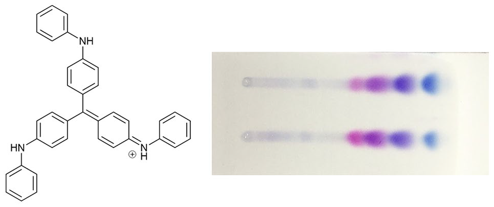
Liquid chromatography–mass spectrometry has been used to analyse a range of cationic aniline dyes from the 19th century. Mauveine from the Chandler museum is used as a standard for comparison. This consists of a typical W. H. Perkin mixture of mauveine A and B. Mauveine from a historic collection in Dresden is different and consists of mainly mauveine A and a monomethyl mauveine chromophore. Possible synthetic routes and its significance are discussed. Three samples of phenylated rosanilines have been analysed, and a list of 19 possible components compiled. An analysis by liquid chromatography–mass spectrometry works well on this complex mixture giving clear information on retention times and accurate mass molecular weights. Mono-, di- and triphenylrosanilines are present in two samples, and a third sample has mainly monophenylrosaniline. In each sample, a small amount of higher molecular weight homologues appear. The thin-layer chromatography plate, from left to right, has fuchsin or rosaniline then mono-, di- and triphenylrosaniline. The two spots on the right-hand side are blue, and the two spots on the left-hand side are red.
Liquid chromatography–mass spectrometry analysis of historical triphenylrosaniline (aniline blue) reveals 19 components.
Introduction
The Faculty of Chemistry and Food Chemistry of the Technical University of Dresden administers a Historical Collection of Dyes.
1
This collection is one of the oldest and largest collections of its kind and contains 8000 trade samples of synthetic dyes, coal-tar colours or aniline colours, in original bottles and cans made by approximately 80 manufacturers. It also houses more than 500 samples of natural dyes. The collection of dyes has nearly 300 historical triarylmethane dyes as well as a sample of mauveine given by W. H. Perkin. Mauve was discovered by Perkin2 in London in 1856 followed by Verguin’s
3
discovery of fuchsin or rosaniline in France in 1859 (Figure 1). This dye is sometimes called aniline red, rosaniline or magenta. Girard and de Laire
4
(Figure S1) discovered triphenylrosaniline by heating fuchsin or rosaniline with aniline in 1861. This insoluble blue dye was called bleu de Lyon or imperial violet. Hofmann played an important role in characterising rosaniline

The parent structures of mauveine, fuchsin and triphenylrosaniline.
Discussion
A sample of mauveine and three dyes from the Technical University of Dresden Historical Collection of Dyes were analysed by LC-MS. A standard mauveine from the Chandler museum in New York was analysed (see Figure 2 and Table 1). 13 A photograph is shown in the Supplemental Material (Figure S2). Perkin gave this mauveine to C. Chandler in 1906, and he was the recipient of the first American Chemical Society (ACS) Perkin Medal, which is still awarded today. 14 Perkin received a tea set and embossed silver tray from his American friends in commemoration of the 50th anniversary of his discovery of the dye mauve, New York, 6 October 1906 (Figure S3). 15 The chart has two scales on the vertical axis, one for the ultraviolet (UV) absorption at 550 nm in milli-Absorption Units (mAU) and one for the mass spectrometer in Electro Spray Mass Spectrometer (ES-MS) counts. The horizontal axis has the retention time, as the mauveine is eluted with gradient elution from a reverse phase silica column. This separates compounds by size and works well for cationic chromophores, which differ in size by one methyl group (CH2). 16 This sample is typical of the mauveine which W. H. Perkin produced by a unique manufacturing process and is rich in mauveine A (391, 37%) and B (405, 30%). 16 Figure 3 shows the structure of some typical mauveine chromophores. A previous analysis was done by nuclear magnetic resonance (NMR) 17 and high-performance liquid chromatography (HPLC).18,19 The composition is similar to that of the mauveine stored in London, Manchester, Bradford and Sudbury.20–24 This is different to the mauveine which is obtained by Perkins patented method, which has become the source of much research suggesting that Perkin carried out research and development to improve the method for making mauveine but kept it secret from potential competitors. 25

A reference sample of mauveine.
Relative peak areas for Figure 2.


The structures of some mauveine chromophores.
Figure 4 and Table 2 show data from the LC-MS analysis of mauveine from the Technical University of Dresden. W. H. Perkin visited here in the 19th century. A picture of the bottle (Figure S4), a card for it (Figure S5), with the names ‘violet au bichromate’ and ‘violet pate’, and some skeins from Perkin (Figure S6) are shown in the Supplemental Material. This mauveine is dominated by a monomethyl mauveine (377, 13%) and mauveine A (393, 28%) with very little mauveine B present. The monomethyl mauveine chromophores have been suggested as mauveine C25a or C25b. 19 This mauveine analysis came as a surprise because all previous samples of Perkin’s mauveine are rich in mauveine A and B, and were made by a unique manufacturing process. 16 This sample could be the first example of Perkin’s 2 mauveine which was made by his patented method of 1856 in which the toluidine content has been increased above 10%. The percentage of toluidine in the aniline which is oxidised controls the products which form. Oxidation of aniline containing 10% toluidine gives pseudomauveine and a monomethyl mauveine, but as the percentage of toluidine increases, more methylated mauveine chromophores form. 15 However, this particular composition containing a monomethyl mauveine, mauveine A and little mauveine B was not observed. Mauveine B forms efficiently like mauveine A, and we would have expected more mauveine B to form and others. 19 A similar match to this sample was only observed when p-toluidine was replaced with N-tert-butyl-p-toluidine in the synthesis, and the front fraction from a column was collected. 26 Figure 5 and Table 3 show data from the analysis of a rare plate 6 Victorian stamp. 27 This mauveine may have been made from oxidising aniline containing a low percentage of toluidine (10%–15%). Since it is different from Perkin’s mauveine, and more similar to Schunck’s 28 or Caro’s mauveine, 29 it has been suggested that Caro would have made it. 30 However, the discovery of mauveine from Perkin at the Technical University of Dresden, which might have come from oxidising aniline low in toluidines, suggests that the mauveine on the stamp could have been made by Perkin. Perkin has stated, though, that he made mauveine from toluidine-rich mixtures of aniline as the yield is higher.
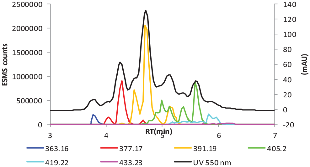
LC-MS chart of mauveine from the Technical University of Dresden.
Relative peak areas for Figure 4.

LC-MS chart of mauveine from a plate 6 Victorian 6d stamp. 27
Relative peak areas for Figure 5.

Triphenylrosaniline: Girard and de Laire’s bleu de Lyon or imperial violet
Three samples of historic dye from the Dresden archive were provided to analyse and characterise by LC-MS.4,5 Samples 1 and 2 were blue, and sample 3 was red. A thin-layer chromatography (TLC) plate of samples 1 and 2 showed two weaker red spots and higher up the plate two blue spots (Figure 6). These are identified shortly by LC-MS.

TLC analysis of sample 1 (top) and sample 2 (bottom) of triphenylrosaniline eluted with secBuOH/EtOAc/H2O/HOAc (60:30:9.5:0.5).
The dyes were analysed by LC-MS under conditions of gradient elution that are used for the analysis of mauveine samples. The separation and analysis work very well. The results from the analysis of sample 1 are shown in Figure 7, with Tables 4–7; for sample 2, Figure 8 with Tables 8–11; and for sample 3, Figure 9 with Tables 12–15. Table 16 is a list of the proposed dyes, which are present. The chart in Figures 7–9 shows four groups of dyes which have been separated by LC-MS counted from left to right. The mass spectral data for each group are shown as Groups 1–4 under each chart and are colour-coded. The same colours are repeated for each group, representing different molecular weights, because not enough different colours were available. The relative peak area for each group is shown in the tables. The four groups of compounds are mainly rosaniline, mono-, di- and triphenylrosaniline. Each of these is a mixture because the rosaniline starting material is made from aniline rich in toluidine and has four possible homologues. 32

Sample 1 LC-MS chart of the historic dyes formed from the condensation of rosaniline with aniline.
Group 1 in Figure 7.

Group 2 in Figure 7.

Group 3 in Figure 7.

Group 4 in Figure 7.


Sample 2 LC-MS chart of the historic dyes formed from the condensation of rosaniline with aniline.
Group 1 in Figure 8.

Group 2 in Figure 8.

Group 3 in Figure 8.

Group 4 in Figure 8.


Sample 3 LC-MS chart of the historic dyes formed from the condensation of rosaniline with aniline.
Group 1 in Figure 9.

Group 2 in Figure 9.
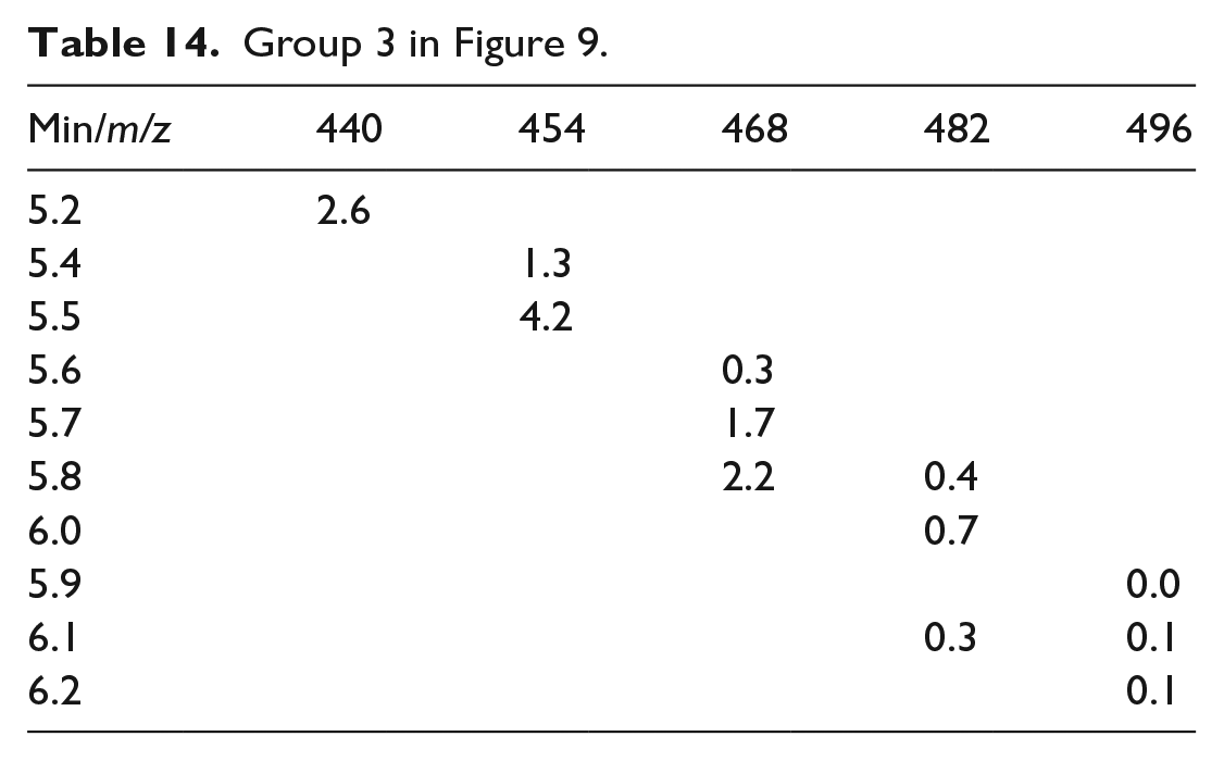
Group 3 in Figure 9.
Group 4 in Figure 9.

Four groups of dyes made from rosaniline with either none, one, two or three arylamines showing the proposed structures, formulae, and measured and calculated accurate masses{ }. 31 .

rdb: rings plus double bonds. Δppm refers to the accuracy of the accurate mass measurement.
Samples 1 and 2
In Figure 7 or 8, Group 1, there are four compounds of molecular weights 288–302–316–330. Each differs by a mass of 14 (CH2) corresponding to one methyl group. The proposed structures of these compounds
Sample 3
The same four groups of peaks appear here in sample 3, which were present in samples 1 and 2. In Figure 9, Group 1, there are four compounds of molecular weights 288–302–316–330. The proposed structures of these compounds
Interpretation
Table 16 lists suggested structures for the products based on their accurate mass data. Most of the compounds differ in molecular structure by one methyl group, which is sufficient for them to be separated on a reverse phase silica column and characterised by ES-MS counts. Elution on a TLC plate with the Meth-Cohn and Smith
17
eluent gives, from left to right, the four groups of spots, which correspond to the groups of compounds seen in the LC-MS (Figure 6). These are rosaniline (red), monophenylrosanilines (red/purple), diphenylrosanilines (dark blue) and triphenylrosanilines (light blue). The order of elution of the spots is reversed on a reverse phase silica column from that on a TLC plate. On reverse phase silica, the separation is based on size, so a difference in size of one methyl group is sufficient for the compounds to separate from a large mixture. The diphenylrosanilines are a darker blue colour, and the triphenylrosanilines are a lighter turquoise blue colour. These studies show that this reaction on an industrial manufacturing scale did not go to completion, and the two different blue compounds would give different shades of blue as was reported.
5
The synthesis involves a condensation of aniline with rosaniline. The central positive charge must activate each arylamine to nucleophilic attack, or the resonance form of an imine shown in Table 16 condenses with aniline. Either mechanism is likely to be similar in energy. It is not an oxidation reaction, so toluidines should have no preference to react over aniline as they do in oxidations to form mauveine or rosaniline. Hence, the main products, which are still mixtures, have a phenylamine as the outer unit since aniline low in toluidine was used.
5
The thermal condensation of aniline hydrochloride in boiling aniline to make diphenylamine shows that the method can work even on unactivated anilines.34–36 Additional isomers not drawn here must still be possible, such as the condensation of compounds
Conclusion
LC-MS analysis of the mauveine sample in the historical archive of the Technical University of Dresden shows that it contains mainly mauveine A and a monomethyl mauveine chromophore. According to the records, this mauveine was given to the collection by W. H. Perkin in the 19th century. The synthesis might be following Perkin’s
2
patented procedure with an increased toluidine content in the aniline, or by modifying a unique process we introduced to explain the composition of Perkin’s mauveine stored in museums which is rich in mauveine A and B.16,26 Perkin’s original 1856 method would be expected to give both mauveine A and B and other mauveine chromophores.17–19 This is the first sample of mauveine attributed to Perkin which is not a characteristic composition of mauveine A and B found in a number of museums.13,20–24 It suggests the possibility that mauveine used for some of the rare plate 6 Victorian 6d stamps might have come from Perkin’s, and not just Caro’s, factory.27,30 Three samples of phenylated rosanilines have been analysed by LC-MS. The method works well for cationic aniline dyes. Samples 1 and 2 are similar and show a complex mixture of products consisting of some starting material (rosaniline or fuchsin), monophenylated, diphenylated and triphenylated rosanilines. Each group is a mixture because the rosaniline starting material, which aniline reacted with, is four homologues
Experimental
For analytical separation, an Agilent 1290 Infinity HPLC system consisting of a quaternary HPLC pump, cooled auto sampler compartment, column compartment and diode-array UV-Vis detector was used. A Gold C-18 column (2.1 mm × 150 mm, Thermo Scientific, UK) was used for separation with a water/methanol gradient (both 0.1% v/v formic acid) from 40% to 100% MeOH in 7 min. The flow rate was 0.5 mL min−1, column temperature 40 °C and sample volume 5 µL. The mass spectrometer (ES-MS) used was a MAXIS II Ultra-High Resolution Time-Of- Flight (UHR-TOF) LC-MS System (Bruker UK Ltd, England) with Electro Spray Ionisation (ESI) source connected to the UV-Vis detector by a short length of Poly Ether Ether Ketone (PEEK) tubing. The ES-MS was operated in positive ion mode with a capillary voltage of 4.5 kV using sodium formate clusters for calibration and methyl stearate as lock mass. Mass spectra were recorded automatically.
Sample preparation
Samples of four dyes were provided in small plastic bags. These were cut open with scissors, and a grain or two of dye was removed with a spatula and placed in a small sample vial (2 cm × 4 cm). The spatula was cleaned each time to avoid cross-contamination. The grains were dissolved in MeOH and sent for analysis in a sealed sample vial. Mauveine was supplied in a sample vial.
Supplemental Material
JCR_TUD_Supplemental_1_Literature – Supplemental material for Liquid chromatography–mass spectrometry analysis of cationic aniline dyes from the Technical University of Dresden Historical Collection of Dyes
Supplemental material, JCR_TUD_Supplemental_1_Literature for Liquid chromatography–mass spectrometry analysis of cationic aniline dyes from the Technical University of Dresden Historical Collection of Dyes by Michael John Plater, Andrea Raab and Horst Hartmann in Journal of Chemical Research
Supplemental Material
JCR_TUD_Supplemental_2_Photographs – Supplemental material for Liquid chromatography–mass spectrometry analysis of cationic aniline dyes from the Technical University of Dresden Historical Collection of Dyes
Supplemental material, JCR_TUD_Supplemental_2_Photographs for Liquid chromatography–mass spectrometry analysis of cationic aniline dyes from the Technical University of Dresden Historical Collection of Dyes by Michael John Plater, Andrea Raab and Horst Hartmann in Journal of Chemical Research
Footnotes
Acknowledgements
We are grateful to Prof. Dr. Horst Hartmann of the Historische Farbstoffsammlung, Technische Universität Dresden and to the Chandler museum, New York, for samples to analyse.
Declaration of conflicting interests
The author(s) declared no potential conflicts of interest with respect to the research, authorship, and/or publication of this article.
Funding
The author(s) received no financial support for the research, authorship, and/or publication of this article.
Supplemental Material
Supplemental material for this article is available online.
References
Supplementary Material
Please find the following supplemental material available below.
For Open Access articles published under a Creative Commons License, all supplemental material carries the same license as the article it is associated with.
For non-Open Access articles published, all supplemental material carries a non-exclusive license, and permission requests for re-use of supplemental material or any part of supplemental material shall be sent directly to the copyright owner as specified in the copyright notice associated with the article.