A new 3-hydroxyphthalimide-based turn-on fluorescent probe is designed and synthesized. This probe can be used to determine the presence of Hg2+ ions by fluorescence spectroscope with high selectivity over other metal ions in aqueous solution. The analytical detection limit for Hg2+ is as low as 6.5 × 10⁻⁷ M. The recognition mechanism is attributed to Hg2+-promoted carbonothioate group cleavage and a subsequent excited-state intramolecular proton transfer mechanisms.
New 3-hydroxyphthalimide-based fluorescence probe 1 selectively determines Hg2+ ion in aqueous solution.
Introduction
Recently, the selective signaling of environmentally hazardous metal ions and anions has attracted growing research interest.1–4 Hg2+ is one of the most toxic and dangerous species in nature, which can result in prenatal brain damage, cognitive and motion disorders, vision and hearing loss, and even death.5,6 Therefore, it is very important to monitor its concentration in environmental and biological samples. Among the various detection techniques developed to date, fluorescence signaling is especially attractive due to its simplicity, specificity, and low detection limits.7 Numerous fluorescent probes for Hg2+ have been reported so far.8–15 However, many of them suffer from fluorescence quenching or poor water solubility. Therefore, developing water-soluble and turn-on fluorescent probes for the detection of Hg2+ is still a challenging task.
3-Hydroxyphthalimide and its derivatives possess a large Stokes shift upon excitation because of an excited-state intramolecular proton transfer (ESIPT) process in the excited state. In addition, 3-hydroxyphthalimide possesses many favorable optical properties, such as emission in the green region, good photostability, and a high fluorescent quantum yield. Therefore, 3-hydroxyphthalimide is an ideal candidate for the design of fluorescent probes.16,17 However, so far, 3-hydroxyphthalimide-based urn-on fluorescence probes for Hg2+ have still not been reported. Here, we reported a new 3-hydroxyphthalimide derivative, 2-butyl-1,3-dioxoisoindolin-4-yl O-phenyl carbonothioate (1), which can be used as a highly selective and sensitive fluorescent probe for detection of Hg2+ in aqueous solution through Hg2+-promoted carbonothioate group cleavage reaction and a subsequent ESIPT process.
Results and discussion
The synthetic route toward probe 1 is outlined in Scheme 1. First, 2-butyl-4-nitroisoindoline-1,3-dione (2) was prepared from the reaction of 4-nitroisobenzofuran-1,3-dione with butan-1-amine. Second, reduction of 2 with SnCl2 in acid solution gave 4-amino-2-butylisoindoline-1,3-dione (3). Next, compound 3 was transformed into 4-hydroxy-2-butylisoindoline-1,3-dione (4) by diazotization and hydrolysis. Finally, probe 1 was synthesized by the esterification of 4 using phenyl chloromethanethioate and N-ethyldiisopropylamine in CH2Cl2 at room temperature. The structure of the probe 1 was identified by infrared (IR), nuclear magnetic resonance (NMR), and ESI-High Resolution Mass Spectrometry (ESI-HRMS) (See Figures S1–S3 in the Supporting Information).
Synthetic route toward probe 1
The sensing behavior of probe 1 toward various metal ions, including Hg2+, K+, Na+, Mg2+, Hg2+, Cd2+, Fe3+, Zn2+, Co2+, Ni2+, Cu2+, Pb2+, Cr3+, and Mn2+, was first investigated in 4-(2-hydroxyethyl)-1-piperazineethanesulfonic acid (HEPES) buffer solution (pH = 7.0) by fluorescence measurements. As shown in Figure 1, probe 1 did not display any appreciable emission when excited at 400 nm. However, a significant increase in the fluorescence intensity at 515 nm was observed after addition of 2.0 equiv. of Hg2+ ions, the other tested metals did not cause such significant fluorescence changes under the same condition. So it can be concluded that probe 1 has higher selectivity for recognition of Hg2+ than for the other metal ions tested.
Fluorescence emission changes of 1 (10 μM) in HEPES buffer (pH = 7.0) in the presence of 20 μM of various metal ions. Inset: fluorescent color changes of 1 upon addition of 20-μM Hg2+ (λex = 400 nm).
Next, the fluorescence response of probe 1 to Hg2+ over time was studied. It can be seen from Figure 2 that the fluorescence intensity at 515 nm increased gradually with increasing reaction time when 2.0 equiv. of Hg2+ was added to a solution of probe 1 in HEPES buffer solution. In addition, the fluorescence intensity remained when the reaction time exceeded 10 min. Therefore, probe 1 provides a rapid analytical method for Hg2+ detection.
The fluorescence spectra of 1 (10 μM) incubated with Hg2+ (20 μM) in HEPES buffer solution (pH = 7.0) at different reaction times (0–16 min). Inset: Time-dependent fluorescence intensity (515 nm) changes of probe 1 (10 μM) upon addition of 2.0 equiv. of Hg2+ in HEPES solution (pH = 7.0) at room temperature.
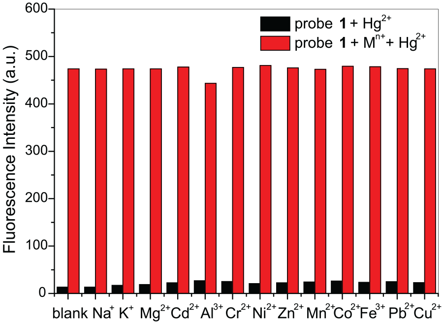
To further exploit the utility of probe 1 as an ion-selective probe for Hg2+, competitive experiments were carried out. As shown in Figure 3, the fluorescence intensity of 1 (10 μM) in the presence of 2.0 equiv. of Hg2+ ion was almost unaffected by the addition of 5 equiv. of other metal ions. These results also suggested that compound 1 could be used as a potential fluorescent probe for Hg2+.
Fluorescence responses of probe 1 (10 μM) in the presence of different metal ions (50 μM) (black bars), followed by the addition of Hg2+ (20 μM) (red bars) (λex at 400 nm). Each spectrum was acquired 10 min after Hg2+ addition.
To conduct a quantitative analysis of probe 1, fluorescence spectra titrations of probe 1 for different concentrations of Hg2+ were carried out. As shown in Figure 4, free probe 1 (10 μM) had no fluorescence. However, the fluorescence intensity at 515 nm gradually increased with the addition of Hg2+ (0–1.5 equiv.). When the amount of added Hg2+was about 1.2 equiv., the fluorescence intensity reached a maximum. Moreover, an excellent linear relationship of emission intensity versus Hg2+ concentration (0–1.0 equiv.) was observed (R = 0.9923, y = 451.32x − 0.845) (Figure 5). The detection limit of probe 1 for Hg2+ is 6.5 × 10⁻⁷ M based on the International Union of Pure and Applied Chemistry (IUPAC) definition of CDL = 3Sb/m.18 These results show that probe 1 could be used to detect Hg2+ quantitatively using the fluorescence spectroscopy method
Fluorescence spectra of probe 1 (10 μM) in the presence of increasing concentrations of Hg2+. Each spectrum was acquired 10 min after Hg2+ addition. Inset: fluorescence intensity changes of 1 upon addition of Hg2+ (λex = 400 nm).
The fluorescence intensities of probe 1 (10.0 µM) were linearly related to the concentration of Hg2+ (λex at 400 nm).
Considering the effect of H+ both for probe 1 and 1 + Hg2+, pH from 2.0 to 11.0 was tested (Figure 6). The emission of probe 1 is very weak in the pH 2.0–9.0 and in the pH 10.0–11.0, a fluorescence enhancement appears. The hydrolysis of the carbonothioate group is the most possible reason for this phenomenon. The response of probe 1 toward Hg2+ was pH dependent. With increasing pH value from 7.0 to 9.0, the probe becomes more sensitive to Hg2+. At pH 8.0, the fluorescence signal reaches the maximum value. It turned out that the probe could function over a pH range of 7.0–9.0 and react selectively toward Hg2+, which meets the demand to detect Hg2+ in natural sources of water.
Fluorescence intensity changes of 1 (10 μM) and Hg2+ (20 μM) with respect to different pH levels in HEPES buffer solution (λex = 400 nm, λem = 515 nm).
To investigate the practical applications of the probe 1, water samples (tap water, drinking water sample) were spiked with 2, 4, and 8 μM of Hg2+ and analyzed by the proposed method.19 A good agreement was obtained between the added and measured mercury amounts. The percentage recovery was found to be in the range of 97.8%–116% (Table 1). The result shows satisfactory recovery for the samples and further demonstrated the utility of our proposed method for effective and fast Hg2+ detection.
Analysis results of Hg2+ ion in two water samples.
Water samples
LOQ (μM)
Found (Hg2+)
Added (μM)
Found (μM)
Recovery (%)
Tap water
0.04
0
2
2.32
116
4
3.97
99.3
8
8.16
102
Drinking water
0.04
0
2
2.01
101
4
3.91
97.8
8
7.86
98.3
LOQ: limit of quantification.
To explore the sensing mechanism of probe 1 for Hg2+, the reaction products of 1-Hg2+ were separated. The fluorescence product was characterized as 4-hydroxyl-2- butylisoindoline-1,3-dione (4) by 1H NMR, 13C NMR, and ESI-MS (see Figures S4–S6 in the Supporting Information), which is in agreement to the previous Hg2+-promoted cleavage reaction of a carbonothioate group20 and an ESIPT mechanism.16 Therefore, a reasonable sensing mechanism was proposed in Scheme 2.
The proposed sensing mechanism of probe 1 for Hg2+.
Conclusion
In summary, a new 3-hydroxyphthalimide-based Hg2+ fluorescent probe has been designed and synthesized. The probe 1 shows no fluorescence in HEPES buffer (10 mM, pH 7.0) but displays an obvious fluorescence enhancement after addition of Hg2+ ions over other metal ions. The recognition mechanism is attributed to Hg2+-promoted carbonothioate group cleavage and a subsequent ESIPT mechanism.
Experimental
All reagents were obtained from commercial sources and were of AR grade. Melting points were determined with an XT4A micromelting point apparatus and are uncorrected. NMR spectra were measured on a Varian Mercury 300 spectrometer. ESI-MS spectra were obtained on a Finnigan Trace MS spectrometer. Fluorescence spectra measurements were performed on a Fluoro-Max-P spectrofluorimeter. 4-Hydroxyl-2-butylisoindoline-1,3-dione (4) was synthesized according to a previously reported procedure.16
Synthesis of 2-butyl-1,3-dioxoisoindolin-4-yl O-phenyl carbonothioate (1)
In a 50-mL flask, 4-hydroxyl-2-butylisoindoline-1,3-dione (4) (0.44 g, 2 mmol) and phenyl chlorothionocarbonate (0.34 g, 3 mmol) were dissolved in dry CH2Cl2 (14 mL) and N-ethyldiisopropylamine (350 μL) was added. The resulting mixture was stirred in room temperature for 10 h. After evaporation of the solvent, the product was purified by silica column chromatography using petroleum ether/ethyl acetate (v/v, 8:1) as eluent to afford probe 1 (0.54 g, 76% yield) as a pale color solid; m.p.: 95−96 °C; IR (νmax, KBr, cm−1): 3464, 2952, 1715, 1603, 1395, 1277, 1065, 865, 694; 1H NMR (300 MHz, CDCl3), δ 7.78–7.80 (m, 2H), 7.45–7.50 (m, 3H), 7.30–7.36 (m, 3H), 3.68 (t, J = 7.2 Hz, 2H), 1.63–1.70 (m, 2H), 1.32–1.42 (m, 2H), 0.94 (t, J = 7.2 Hz, 3H). 13C NMR (75 MHz, CDCl3), δ 13.6, 20.0, 30.4, 37.9, 121.6, 121.7, 123.3, 126.9, 128.1, 129.7, 134.1, 135.7, 148.5, 153.6, 165.3, 167.3, 193.2. ESI-HRMS: m/z [M + H]+calcd for C19H18NO4S: 356.0912; found: 356.0970.
Fluorescence spectroscopy analysis
A stock solution of compound 1 in HEPES buffer solution was diluted and the pH was adjusted to 7.0 to deliver the final concentration of the probe (10 μM, pH = 7.0).
Solutions of metal ions were prepared from Pb(NO3)2, Al2(SO4)3, and the chlorides of Na+, K+, Mg2+, Cd2+, Cr3+, Ni2+, Zn2+, Mn2+, Co2+, Fe3+, and Cu2+, respectively, and were dissolved in water (3.0 × 10⁻³ M). Fluorescence titration was performed on 3-mL solution of probe 1 in a quartz cell of 1 cm optical path length, by adding different stock solutions of cations into the quartz cell portionwise using a microsyringe. The fluorescence spectra were recorded using a fluorescence spectrophotometer (λex = 400 nm, λem = 515 nm) after 10 min with the addition of different analytes at room temperature.
Supplemental Material
revised_Support_Information – Supplemental material for A new 3-hydroxyphthalimide-based turn-on fluorescent probe for Hg2+ detection in aqueous solution
Supplemental material, revised_Support_Information for A new 3-hydroxyphthalimide-based turn-on fluorescent probe for Hg2+ detection in aqueous solution by Xue-Tao Pan, Qiao Li, Yuan-Yuan Xu and Sheng-Li Hu in Journal of Chemical Research
Footnotes
Declaration of conflicting interests
The author(s) declared no potential conflicts of interest with respect to the research, authorship, and/or publication of this article.
Funding
The author(s) disclosed receipt of the following financial support for the research, authorship, and/or publication of this article: The study was funded by the 2018 Open Foundation of the Hubei Key Laboratory of Pollutant Analysis & Reuse Technology (No. PA180205).
ORCID iD
Sheng-Li Hu
Supplemental material
Supplemental material for this article is available online.
References
1.
AragayGPonsJMerkoçiA. Chem Rev2011; 111: 3433.
2.
WangWNTaoHRPiHM, et al. J Chem Res2018; 42: 112.
3.
QiaoRXiongWZBaiCB, et al. J Chem Res2018; 42: 194.
4.
LvYJWeiW. J Che Res2018; 42: 611.
5.
HaradaMC. Rev Toxicol1995; 25: 1.
6.
BenoitJMFitzgeraldWFDammanAW. Environ Res1998; 78: 118.
7.
LakowiczJR. Principles of fluorescence spectroscopy. New York: Springer, 2006.
8.
NolanEMLippardSJ. Chem Rev2008; 108: 3443.
9.
KimHNRenWXKimJS, et al. Chem Soc Rev2012,41: 3210.
10.
HuJHuZLiuS, et al. Sens Actuators B2016; 230: 639.
SunYZhaoDFanS, et al. Sens Actuators B2015; 208: 512.
20.
ShuWWangYWuL, et al. Ind Eng Chem Res2016; 55: 8713.
Supplementary Material
Please find the following supplemental material available below.
For Open Access articles published under a Creative Commons License, all supplemental material carries the same license as the article it is associated with.
For non-Open Access articles published, all supplemental material carries a non-exclusive license, and permission requests for re-use of supplemental material or any part of supplemental material shall be sent directly to the copyright owner as specified in the copyright notice associated with the article.