Different drugs containing a basic nitrogen atom were crystallised with 2,4-dinitrophenol to study the mode of complexation in search of an antidote to 2,4-dinitrophenol poisoning. The protonated forms of quininium, quinidinium and trazodonium form N–H···O hydrogen bonds to the deprotonated O atom of the 2,4-dinitrophenolate anion, whereas haloperidolium forms a bifurcated N–H···(O,O) hydrogen bond to the deprotonated O atom of 2,4-dinitrophenol and an O atom of the adjacent nitro group. Hydrogen-bonded chains occur in the quininium, quinidinium and haloperidolium crystal structures, whereas the trazodonium structure consists of ion pairs. These results are discussed with a view to lowering the toxicity of 2,4-dinitrophenol in the body in the case of an overdose.
Fundamental research to lower the toxicity of 2,4-dinitrophenol in the body in the case of an overdose. A 2,4-dinitrophenol–haloperidolium complex or salt is shown below.
Introduction
2,4-Dinitrophenol (DNP) is available over the Internet as a recreational drug for weight loss.1 Its side effects were discovered by the poisoning of French munition workers in explosive production factories.2 It acts as a metabolic stimulant, but it is far too toxic for human consumption3,4 and a number of deaths1 have been reported since its evaluation in clinical trials5 in the 1930s as a weight loss treatment.6–11 It is believed to function by the uncoupling of oxidative phosphorylation.12–16 The clinical trials reported some euphoria and for other psychological reasons overdose is easy, but a small overdose of the recommended 200–300 mg per day can be fatal after a few days.9–11 Death occurs from overheating or hyperthermia and other very unpleasant side effects such as tachycardia, diaphoresis and tachypnoea.1 Currently, there is no antidote for DNP poisoning8 apart from using ice packs3 or a cold bath1 to lower the body temperature. There appears to be little guidance about its use on the Internet, and it does not have the classification of a recreational drug. Chemical companies sell it moistened with water as a desensitised explosive, but this requirement does not apply to the capsules sold for human consumption.
Discussion
Antidote theory for DNP
This paper reports our initial studies aimed at finding an antidote for DNP poisoning. Only a small number of compounds were evaluated previously, which included quininium because of its antipyretic properties.8 None were successful although cooling water was beneficial. New drugs are likely to be expensive and may not be pursued by pharma because the number of deaths is low compared to other fatalities. Ideally, an existing drug might be applied which will complex to DNP lowering its availability and hence toxicity in the body. Either DNP or the DNP–drug complex should be excreted from the body. Therapies based on supramolecular complex chemistry are known such as the Akzo Nobel (Schering–Plough) functionalised cyclodextrins, which reduce the availability of neuromuscular blocking agents after an anaesthetic and enhance their rate of elimination.17,18 DNP is quite acidic with a pKa of 4.0,19–21 so it is approximately 8–10 times more acidic than acetic acid. Many drugs are basic, especially central nervous system (CNS) active drugs,22 so they might form an acid–base complex with DNP in the body as the mode of supramolecular complexation (acid + base = salt) (Figure 1). Precedent for this scheme is provided by the protonation of the base 1,4-diazabicyclo[2.2.2]octane (DABCO) and its crystallisation with DNP.23 In blood, DNP turns yellow with an icteric tint6 showing that DNP is deprotonated to the 2,4-dinitrophenolate anion 3. Dilute hydrochloric acid decolourises the dye. This may be due to the buffering capacity of the blood or because of other basic components. The DNP anion might complex with a basic drug that is injected in the protonated form to make it water soluble. Figure 2 shows their possible mode of complexation. Many of the basic drugs studied here, which can be injected,24–26 are administered in the protonated form, which is water soluble. The binding constants K1 and K2 for the equation in Figure 127 are likely to be influenced by interfering acids, bases or ions. However, rational design of the basic drug or host may be pursued to favour further electrostatic, hydrophobic and van der Waals forces between the drug or host and DNP to give a stronger complex.
Scheme proposing the binding of DNP 1 with a basic drug 2 to give solvated ions in solution then a complex or salt [3 + 4].
Left: molecular structure of the quininium–DNP complex 14 showing 50% displacement ellipsoids. Right: molecular structure of the quinidinium–DNP complex 15 showing 50% displacement ellipsoids.
Initial studies have focussed on the crystallisation of various basic drugs with DNP to develop an understanding of the mode of binding. All the drugs studied, in their neutral form, gave yellow solutions with DNP in dichloromethane (DCM), indicating that the drug is deprotonating DNP. Some formed only oils but some gave crystalline products (Table 1).
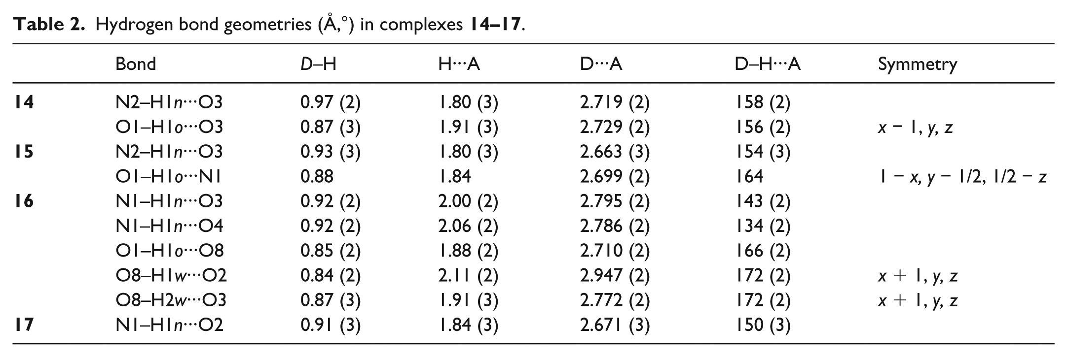
Hydrogen bond geometries (Å,°) in complexes 14–17.
Molecular structure of the drug
Colour of the solution of DNP with the drug in dichloromethane
Morphology of the DNP–drug complex
5
Quininium
Bright yellow
Crystalline
6
Quinidinium
Bright yellow
Crystalline
7
Chlorpromazine
Bright yellow
Oil
8
Haloperidolium
Bright yellow
Crystalline
9
Loperamide
Bright yellow
Oil
10
Hydroxyzine
Bright yellow
Oil
11
Trifluoroperazine
Bright yellow
Oil
12
Trazodonium
Bright yellow
Crystalline
13
Diphenhydramine
Bright yellow
Oil
DNP: 2,4-dinitrophenol.
Apart from haloperidolium all were purchased as their hydrochloride salts from Sigma-Aldrich.
All crystallisations were performed by treating a yellow solution of the drug 5–13 and DNP in DCM with light petroleum ether and allowing the solvent to partially evaporate over 3 days. DNP 1 is purchased as a moist solid, a so-called desensitised explosive, and was weighed out dampened with water as accurately as possible. The drugs were purchased as hydrochloride or double hydrochloride salts and were easily converted to the neutral base for crystallisation experiments. The formation of oils in these experiments was noted but they were not studied any further as the mode of binding illustrated by the crystal structures was investigated. The drugs are frequently designed with asymmetry, which will lower their crystallinity with DNP 1. An oil might also indicate a mixture of salt, DNP and unprotonated amine, which is less desirable as the complexation between the drug and DNP is weaker. The yellow colour is a characteristic of deprotonated DNP.
Crystal structures
The molecular structure of complex 14 is shown in Figure 2 and confirms that proton transfer from DNP (C6H4N2O5) to quininium (C20H24N2O2) and complexation have occurred to yield a C20H25N2O2+ quininium cation protonated at the bridgehead N2 atom and a C6H3N2O5− anion linked by an N2–H···O3 hydrogen bond (Table 2). The absolute structure is well defined (see section ‘Experimental’) and the quininium stereogenic centres are C10 R, C11 S, C13 S and C17 R, which is consistent with previous results.28 The N3 and N4 nitro groups are twisted from the C21–C26 plane by 39.28 (11)° and 7.12 (4)°, respectively. The quininium hydroxyl group forms an O1–H···O3 hydrogen bond to the deprotonated phenolic oxygen atom in an adjacent anion. Altogether, the N–H···O and O–H···O hydrogen bonds generate [100] chains in the extended structure.
Hydrogen bond geometries (Å,°) in complexes 14–17.
Bond
D–H
H···A
D···A
D–H···A
Symmetry
14
N2–H1n···O3
0.97 (2)
1.80 (3)
2.719 (2)
158 (2)
O1–H1o···O3
0.87 (3)
1.91 (3)
2.729 (2)
156 (2)
x − 1, y, z
15
N2–H1n···O3
0.93 (3)
1.80 (3)
2.663 (3)
154 (3)
O1–H1o···N1
0.88
1.84
2.699 (2)
164
1 − x, y − 1/2, 1/2 − z
16
N1–H1n···O3
0.92 (2)
2.00 (2)
2.795 (2)
143 (2)
N1–H1n···O4
0.92 (2)
2.06 (2)
2.786 (2)
134 (2)
O1–H1o···O8
0.85 (2)
1.88 (2)
2.710 (2)
166 (2)
O8–H1w···O2
0.84 (2)
2.11 (2)
2.947 (2)
172 (2)
x + 1, y, z
O8–H2w···O3
0.87 (3)
1.91 (3)
2.772 (2)
172 (2)
x + 1, y, z
17
N1–H1n···O2
0.91 (3)
1.84 (3)
2.671 (3)
150 (3)
The molecular structure of complex 15 as shown in Figure 2 confirms that the same protonation reaction has occurred to form a C20H25N2O2+ quinidinium cation protonated at the bridgehead N2 atom and a 2,4-dinitrophenolate anion linked by an N2–H···O3 hydrogen bond (Table 2). As is well known, quininium and quinidinium are diastereoisomers,28 and the stereogenic centres in the cation in complex 15 are C10 S, C11 R, C13 S and C17 R (i.e. C10 and C11 in complex 15 have the opposite configurations to the equivalent atoms in complex 14). In the anion, the nitro groups containing N3 and N4 are twisted from their attached ring by 21.2 (2)° and 3.1 (4)°, respectively. The hydrogen bonding behaviour of the hydroxyl group in complex 15 is different from that in complex 14. In complex 15, it forms an O–H···N link to the quinolone-ring N1 atom of an adjacent cation. Altogether, the N–H···O and O–H···N hydrogen bonds in complex 15 generate [010] chains in the extended structure.
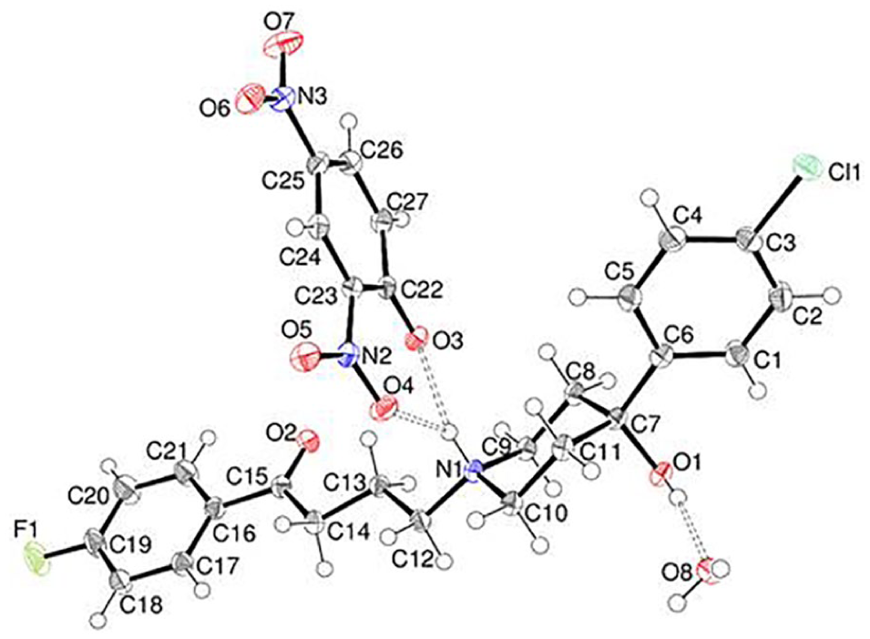
The asymmetric unit of complex 16 (Figure 3) shows that proton transfer has occurred to form a haloperidoliumium (C21H24ClFNO2+) cation and a DNP anion accompanied by a water molecule of crystallisation. The cation is protonated at N1 in the piperidine ring, which adopts a typical chair conformation: the exocyclic N1–C12 and C7–C6 bonds take on equatorial orientations and the N1–H1 and C7–O1 bonds are in axial positions. The N1–C12–C13–C14 torsion angle is −162.68 (13)° and the dihedral angle between the terminal halogenated rings is 75.14 (7)°. In the anion, the N2 and N3 nitro groups are twisted from the C22–C27 ring by 11.0 (3)° and 0.9 (2)°, respectively. The N1–H1n moiety forms a bifurcated hydrogen bond to O3 and O4 (Table 2), which may correlate with the smaller degree of twist of the N2 nitro group in complex 16 compared to the equivalent species in complexes 14 and 15. It may also be seen that the H1n···O2 separation is noticeably longer in complex 16 compared to complexes 14 and 15. The hydroxyl group in complex 16 forms an O–H···O hydrogen bond to the water molecule of crystallisation (O8) and O8 itself forms two O–H···O links to atoms O2 and O3 in adjacent molecules. Taken together, the classical hydrogen bonds in complex 16 generate [100] chains in the crystal.
The molecular structure of the haloperidolium–DNP complex 16 showing 50% displacement ellipsoids.
As with the other compounds described here, complex 17 is a molecular salt arising from the predicted proton transfer reaction between DNP and trazodonium to form a C19H23ClN5O+ trazodonium cation and a DNP anion linked by an N–H···O hydrogen bond (Figure 4). The piperazine ring adopts its usual chair conformation and atom N4 adjacent to the propyl chain is protonated, with the N–H moiety lying in an axial orientation. The N2–C7–C8–C9 and C7–C8–C9–N4 torsion angles are 69.4 (3)° and 171.9 (2)°, respectively, and the dihedral angle between the C1–C6/N1–N3 ring system and the chlorophenyl ring is 77.47 (7)°. In the anion, the dihedral angles subtended by the N6 and N7 nitro groups with respect to their attached ring are 14.9 (3)° and 4.4 (3)°, respectively. The only classical hydrogen bond in complex 17 is N4–H1n···O2, thus isolated ion pairs occur in the crystal.
The molecular structure of the trazodonium–DNP complex 17 showing 50% displacement ellipsoids.
Conclusion
The crystallisation of DNP with some basic drugs was studied. The compounds quininium 5, quinidinium 6, haloperidolium 8 and trazodonium 12 gave crystalline adducts or complexes 14–17. The hydrogen atom of the protonated nitrogen forms a hydrogen bond to the oxygen anion of the 2,4-dinitrophenolate anion 3. In the haloperidolium complex 16, the delocalised anion of 2,4-dinitrophenolate 3 forms a bifurcated hydrogen bond to the hydrogen atom of the protonated amine. These modes of crystallisation help to understand how binding occurs between DNP and basic drugs and may serve to help develop models for new hosts which will efficiently bind to DNP and reduce its toxicity in the body. Currently, nothing is understood about drug–DNP interactions with the compounds studied here. The development of efficient antidotes based on the strategy described here may be easier than developing competitive inhibitors of DNP in enzymic pathways.
Experimental
DNP was purchased from Sigma-Aldrich. It comes as a moistened solid which accounts for the water of crystallisation present in the haloperidolium–DNP complex 16. The commercial drugs (500 mg), which are typically hydrochloride salts, were dissolved in water (100 mL) then treated with dilute KOH (2 M) until precipitation was complete. The white precipitate was dissolved in DCM (100 mL), and the layers were separated in a separating funnel. The lower DCM layer was dried over MgSO4, filtered and then evaporated to dryness. The yields are nearly quantitative. A portion of the neutral solid (100 mg) and an equimolar amount of moistened DNP was dissolved in DCM (50 mL) and mixed with light petroleum ether (50 mL). The solution was left to partially evaporate for 3 days covered with aluminium foil with tiny holes in it. It is best to harvest crystals before all the solvent has evaporated as in some cases, as with the crystallisation of trazodonium, excess drug can precipitate.
Intensity data for 14–17 were collected using a Rigaku AFC11 CCD diffractometer at T = 100 K with Cu Kα radiation (λ = 1.54184 Å), and the structures were solved by direct methods and completed and optimised by least-squares refinement against ǀFǀ2 using SHELXL-2014.29 The N and O-bond H atoms were located in difference maps and their positions were freely refined (atom H1o attached to O1 in 15 was refined as riding in its as-found relative position). The C-bound H atoms were geometrically placed (C–H = 0.95–1.00 Å) and refined as riding atoms. The methyl groups in structures 14 and 15 were allowed to rotate, but not to tip, to best fit the electron density. The constraint Uiso(H) = 1.2Ueq(C) or 1.5Ueq(methyl C) was applied in all cases. Full details including weak C–H···O and C–H···Cl interactions are available in the deposited CIFs.
14 C26H28N4O7 (C20H25N2O2·C6H3N2O5), Mr = 508.52, intense yellow plate, 0.21 mm × 0.14 mm × 0.02 mm, orthorhombic, space group P212121 (no. 19), Z = 4, a = 6.60730 (10) Å, b = 17.69540 (10) Å, c = 20.0701 (2) Å, V = 2346.57 (4) Å3. Number of measured and unique reflections = 29,209 and 4282, respectively (−7 ⩽ h ⩽ 7, −19 ⩽ k ⩽ 21, −24 ⩽ l ⩽ 19; 2θmax = 136.5°°; RInt = 0.057). Final R(F) = 0.029, wR(F2) = 0.076 for 342 parameters and 4175 reflections with I > 2σ(I) (corresponding R-values based on all 4282 reflections = 0.030 and 0.077, respectively), Flack absolute structure parameter = −0.03 (9), CCDC deposition number 1895848.
15 C26H28N4O7 (C20H25N2O2·C6H3N2O5), Mr = 508.52, yellow plate, 0.10 mm × 0.06 mm × 0.01 mm, orthorhombic, space group P212121 (no. 19), Z = 4, a = 8.36879 (8) Å, b = 14.35646 (14) Å, c = 20.6601 (2) Å, V = 2482.23 (4) Å3. Number of measured and unique reflections = 39,895 and 4680, respectively (−10 ⩽ h ⩽ 10, −16 ⩽ k ⩽ 16, −25 ⩽ l ⩽ 25; 2θmax = 140.7°; RInt = 0.058). Final R(F) = 0.037, wR(F2) = 0.093 for 338 parameters and 4454 reflections with I > 2σ(I) (corresponding R-values based on all 4680 reflections = 0.039 and 0.095, respectively), Flack absolute structure parameter = −0.04 (8), CCDC deposition number 1895849.
16 C27H29ClFN3O8 (C21H24ClFNO4·C6H3N2O5·H2O) Mr = 577.98, green block, 0.10 mm × 0.06 mm × 0.05 mm, triclinic, space group P (no. 2), Z = 2, a = 8.8721 (5) Å, b = 13.0756 (4) Å, c = 13.2152 (7) Å, α = 65.161 (4)°, β = 78.463 (5)°, γ = 89.576 (4)°, V = 1358.09 (12) Å3. Number of measured and unique reflections = 23,251 and 4940, respectively (−10 ⩽ h ⩽ 10, −15 ⩽ k ⩽ 15, −15 ⩽ l ⩽ 15; 2θmax = 136.5°; RInt = 0.055). Final R(F) = 0.043, wR(F2) = 0.130 for 374 parameters and 4603 reflections with I > 2σ(I) (corresponding R-values based on all 4940 reflections = 0.045 and 0.132, respectively), CCDC deposition number 1895850.
17 C25H26ClN7O6 (C19H23ClN5O·C6H3N2O5) Mr = 555.98, intense orange plate, 0.06 mm × 0.05 mm × 0.01 mm, triclinic, space group P (no. 2), Z = 2, a = 10.0521 (4) Å, b = 10.3245 (4) Å, c = 12.8000 (5) Å, α = 76.793 (3)°, β = 85.914 (3)°, γ = 74.963 (3)°, V = 1248.90 (9) Å3. Number of measured and unique reflections = 45,012 and 4551, respectively (−12 ⩽ h ⩽ 12, −12 ⩽ k ⩽ 12, −15 ⩽ l ⩽ 15; 2θmax = 136.5°; RInt = 0.099). Final R(F) = 0.057, wR(F2) = 0.133 for 355 parameters and 3769 reflections with I > 2σ(I) (corresponding R-values based on all 4551 reflections = 0.072 and 0.141, respectively), CCDC deposition number 1895851.
Footnotes
Acknowledgements
We thank the National Mass Spectrometry Service Centre (University of Swansea) for mass spectrometry data and the UK National Crystallographic Service (University of Southampton) for intensity data collections.
Declaration of conflicting interests
The author(s) declared no potential conflicts of interest with respect to the research, authorship and/or publication of this article.
Funding
The author(s) received no financial support for the research, authorship and/or publication of this article.
ORCID iD
M John Plater
References
1.
GrundlinghJDarganPIEl-ZanfalyM, et al. J Med Toxicol2011; 7: 205.