
Abstract
Neuropathic pain (NP) is a refractory chronic pain disorder with a complex pathogenesis and limited effective treatment options. In recent years, exosomes derived from mesenchymal stem cells (MSC-Exos) have attracted attention as promising therapeutic agents due to their anti-inflammatory, neuroregenerative, and immunomodulatory properties. This study aimed to investigate the therapeutic effects and underlying mechanisms of human umbilical cord mesenchymal stem cell-derived exosomes (hUC-MSC-Exos) in a rat model of neuropathic pain induced by chronic constriction injury (CCI) of the sciatic nerve. hUC-MSC-Exos were isolated via ultracentrifugation and administered to CCI rats. Behavioral tests demonstrated that hUC-MSC-Exos significantly ameliorated both mechanical allodynia and thermal hyperalgesia in CCI rats. Further mechanistic investigations indicated that hUC-MSC-Exos downregulated the expression of pro-inflammatory cytokines (NF-κB, TNF-α, IL-6) and modulated astrocyte activation and polarization, thereby contributing to neuropathic pain relief. These findings elucidate the potential mechanisms through which hUC-MSC-Exos alleviate NP and provide an experimental foundation for their future clinical application.
Keywords
Introduction
Neuropathic pain (NP) is a refractory chronic pain condition resulting from damage or disease affecting the somatosensory nervous system. Its pathogenesis involves complex mechanisms, including peripheral and central sensitization, inflammatory responses, and neuroplastic alterations. 1 Clinical manifestations of NP encompass spontaneous pain, allodynia, and hyperalgesia, which significantly impair patients’ quality of life. 2 Current therapeutic options for NP remain limited; conventional pharmacotherapies such as opioids and antidepressants often yield suboptimal outcomes and are associated with considerable adverse effects. 3 Consequently, the development of novel treatment strategies represents a major focus of contemporary research.
In recent years, mesenchymal stem cells (MSCs) have demonstrated considerable therapeutic potential in various diseases owing to their anti-inflammatory, neuroregenerative, and immunomodulatory properties. 4 Notably, exosomes derived from MSCs (MSC-Exos) serve as key mediators of intercellular communication by delivering diverse bioactive molecules, such as proteins, mRNAs, miRNAs. These vesicles retain the therapeutic benefits of their parent cells while avoiding the risks associated with direct cell transplantation. 5 Previous studies have shown that exosomes derived from human umbilical cord MSCs (hUC-MSC-Exos) exert neuroprotective and reparative effects in contexts including neurodegenerative disorders, ischemic brain injury, and spinal cord injury.6,7 However, their role in neuropathic pain and the underlying mechanisms are not yet fully elucidated.
In neuropathic pain (NP), aberrant activation of spinal dorsal horn astrocytes—particularly their polarization toward a pro-inflammatory A1 phenotype—constitutes a critical mechanism underlying central sensitization and pain persistence. 8 Emerging evidence suggests that MSC-derived exosomes (MSC-Exos) can suppress microglial and astrocytic activation and alleviate neuroinflammation via the delivery of anti-inflammatory factors such as miR-124 and miR-21. 9 Nonetheless, it remains unclear whether hUC-MSC-Exos can mitigate neuropathic pain by modulating the A1/A2 phenotypic transition of astrocytes, and the specific molecular pathways involved require further elucidation.
The chronic constriction injury (CCI) model is a well-established rodent model that recapitulates key pathological features of human neuropathic pain (NP), including mechanical allodynia and thermal hyperalgesia. 10 Studies have demonstrated that astrocyte activation in the spinal dorsal horn and subsequent release of pro-inflammatory cytokines (e.g., TNF-α, IL-1β, IL-6) play pivotal roles in the initiation and maintenance of neuropathic pain in CCI models. 11 Moreover, the NF-κB signaling pathway has been implicated in regulating of inflammatory responses and neuronal sensitization. 12 Thus, investigating whether human umbilical cord mesenchymal stem cell-derived exosomes (hUC-MSC-Exos) alleviate NP by modulating astrocyte activation, pro-inflammatory cytokine production, and the NF-κB pathway holds substantial scientific and clinical relevance.
This study aims to investigate the therapeutic effects and underlying mechanisms of human umbilical cord mesenchymal stem cell-derived exosomes (hUC-MSC-Exos) on neuropathic pain using a rat chronic constriction injury (CCI) model. We will focus on the regulatory effects of hUC-MSC-Exos on the phenotypic polarization of spinal dorsal horn astrocytes, and employ proteomic and molecular biological approaches to identify involvement of key signaling pathways, including NF-κB and STAT3. This study is the first to systematically link hUC-MSC-Exos, the A1/A2 polarization switch of astrocytes, and the NF-κB inflammatory pathway in a CCI model. The findings are expected to provide experimental evidence to support a cell-free, exosome-based therapeutic strategy for neuropathic pain and to inform clinical translation.
Materials and methods
Culture and identification of mesenchymal stem cells
Following informed consent from the parturient and her family, umbilical cords were aseptically collected from healthy full-term cesarean section infants delivered by mothers who tested negative for four major infectious diseases. The cords were rinsed in phosphate-buffered saline (PBS) containing penicillin-streptomycin. Wharton’s jelly tissue was isolated and placed in T75 flasks containing primary stem cell culture medium (Youkang Primary Stem Cell Culture Medium) for incubation. After 7–12 days, cells were observed under an inverted light microscope. When cell proliferation reached approximately 80% confluence (characterized by a swirl-like morphology), the cells were digested with a gentle stem cell enzyme, centrifuged, and harvested as primary mesenchymal stem cells (MSCs) for continued expansion. Cells from passages 3 to 6 (P3–P6) were used for subsequent differentiation assays, identification, and exosome extraction. MSCs were identified via flow cytometry using positive markers (CD73, CD90, CD105) and negative markers (CD34, CD45, HLA-DR). Adipogenic differentiation was induced using an adipogenic induction medium (ADP) and confirmed by Oil Red O staining. Osteogenic differentiation was induced using an osteogenic induction medium and verified by Alizarin Red staining.
Isolation and identification of exosomes
The cell culture medium was replaced with exosome-depleted fetal bovine serum and incubated for 48 h. The supernatant was collected and centrifuged at 2000g and 4°C for 30 min. The resulting supernatant was further centrifuged at 10,000g and 4°C for 45 min to remove larger vesicles. The supernatant was then filtered through a 0.45 μm membrane, and the filtrate was ultracentrifuged at 100,000g and 4°C for 70 min. The pellet was resuspended in 10 mL of pre-cooled PBS and subjected to another round of ultracentrifugation under the same conditions. The final pellet was resuspended in 100 μL of pre-cooled PBS for storage and subsequent characterization. Exosome structure was examined using transmission electron microscopy (TEM). Particle size distribution was analyzed via nanoparticle tracking analysis (NTA). Specific surface markers (CD9, CD63, CD81) were detected using nanoflow cytometry.
Experimental animals
Specific pathogen-free (SPF) healthy male Sprague-Dawley rats, aged 6–8 weeks, were obtained from Beijing Sipeifu (SBF) Biotechnology Co., Ltd. All animals were housed in the Animal Management Center of Taiyuan Central Hospital under controlled conditions: a 12-h light/dark cycle, room temperature maintained at 22°C–24°C, relative humidity at 50%–60%, and ad libitum access to food and water. Bedding, cages, and feed were replaced regularly. Animal Ethics All experimental procedures were approved by the Animal Ethics Committee of Taiyuan Central Hospital (Approval No. HLK-20240221-001) and were in accordance with the NIH Guide for the Care and Use of Laboratory Animals.
Experimental grouping and establishment of the CCI rat model
A total of 36 Sprague-Dawley rats were randomly assigned to three groups (n = 12 per group) using a random number table: sham, CCI, and Exo groups. After 1 week of acclimatization, rats were anesthetized with sevoflurane. The left hind limb was shaved and disinfected. An incision was made to expose and bluntly dissect the sciatic nerve. Using 4-0 Mersilk sutures, the sciatic nerve was ligated just proximal to its trifurcation. The ligation tightness was adjusted to elicit a slight muscle twitch in the hind limb. Four ligatures were placed at intervals of approximately 1 mm. Muscle and skin were then sutured layer by layer. In the sham group, the sciatic nerve was exposed without ligation, while all other procedures remained identical to those in the CCI group. Within 30 min after surgery, rats in the Exo group received a tail vein injection of 100 μg exosomes (dissolved in 100 μL saline). 9 Rats in the other two groups received an equal volume of saline. The Exo group was injected with 100 μg of exosomes at the same time each day for three consecutive days. The other two groups were injected with the same volume of normal saline at the same time for three consecutive days. Nociceptive behavioral tests, including the paw withdrawal threshold (PWT) to mechanical stimulation and the paw withdrawal latency (PWL) to thermal stimulation, were conducted on postoperative days 3, 7, and 14 to evaluate the therapeutic effect of exosomes on neuropathic pain in CCI rats.
Measurement of paw withdrawal threshold (PWT) and paw withdrawal latency (PWL)
For mechanical sensitivity testing, rats were placed individually in an elevated wire mesh enclosure and allowed to acclimate for 30 min. Von Frey filaments were applied to the mid-plantar surface of the hind paw using the “up-and-down” method, starting with a 2.0 g filament. Each filament was applied vertically until bent into a “C” or “S” shape and held for 6–8 s. A positive response (rapid paw withdrawal, shaking, or licking) was recorded as “X,” and a negative response (no withdrawal) as “O.” The 50% mechanical paw withdrawal threshold (in grams) was calculated using the formula: 50% threshold (g) = (10[Xf + kδ])/10,000,where Xf is the log value of the final filament used, k is a value based on the response pattern, and δ is the mean difference between log stimulus intensities. Measurements were taken at 10-min intervals, and the average of three consecutive tests was defined as the PWT. For thermal sensitivity testing, rats were placed individually in transparent plexiglass chambers on a glass plate and allowed to acclimate for 30 min. A plantar analgesia tester (e.g., IITC Life Science or Ugo Basile) was used to apply a radiant heat source to the plantar surface of the left hind paw. The device automatically recorded the time from heat onset to paw withdrawal, defined as the PWL. A cutoff time of 20–25 s was set to prevent tissue damage. Measurements were taken at 10-min intervals, and the average of three tests was recorded as the thermal PWL. 11
Spinal cord tissue collection
Following anesthesia with sevoflurane, the dorsal fur of the rats was shaved. The thoracic wall and diaphragm were incised along the costal margin to expose the heart. A perfusion needle was inserted into the left ventricle at the apex, and 50 mL of saline was rapidly infused. The right auricle was incised to allow outflow. Ice-cold saline was perfused initially at a rapid rate, then slowly, until the liver became pale. The spinal column was then exposed, and the L4–L5 spinal segment was identified and carefully excised. The lumbar enlargement was collected, snap-frozen, and stored at −80°C for subsequent analysis.
Western blot analysis
Frozen spinal cord tissue was homogenized in ice-cold RIPA lysis buffer supplemented with protease inhibitors. The lysate was centrifuged, and the supernatant was collected. Protein concentration was determined using a bicinchoninic acid (BCA) assay. Equal amounts of protein were separated by SDS-PAGE and transferred to a PVDF membrane. The membrane was washed three times (10 min each) with TBST and blocked with 5% BSA for 2 h at room temperature with shaking. Subsequently, the membrane was incubated overnight at 4°C with primary antibodies (diluted 1:1000), followed by incubation with horseradish peroxidase (HRP)-conjugated secondary antibodies for 1 h at room temperature. Protein bands were visualized using an ECL detection reagent (Beyotime Biotechnology, Shanghai, China), and band densities were semi-quantified using ImageJ software. The primary antibodies used were: rabbit polyclonal anti-C3, rabbit polyclonal anti-S100A10, and mouse monoclonal anti-GAPDH (all purchased from ImmunoWay Biotechnology Company). The secondary antibodies used were: HRP-conjugated goat anti-rabbit and HRP-conjugated goat anti-mouse (both purchased from Wuhan Boster Biological Technology Co., Ltd.).
ELISA for spinal cord inflammatory cytokines
Protein extracts from spinal cord tissue, prepared as described for Western blot analysis, were used for cytokine quantification. The total protein concentration of each sample was measured and normalized using a BCA protein assay kit (Beijing Lamblade Biotechnology Co., Ltd.). According to the manufacturer’s instructions, commercial ELISA kits were employed to determine the concentrations of IL-6, NF-κB, and TNF-α in the spinal cord tissues. IL-6 was determined by an ELISA kit from Beijing BioLegend Biotechnology Co., Ltd. NF-κB and TNF-α were measured using ELISA kits from Elabscience Biotechnology Co., Ltd.
RNA extraction and quantitative reverse transcription polymerase chain reaction (qRT-PCR)
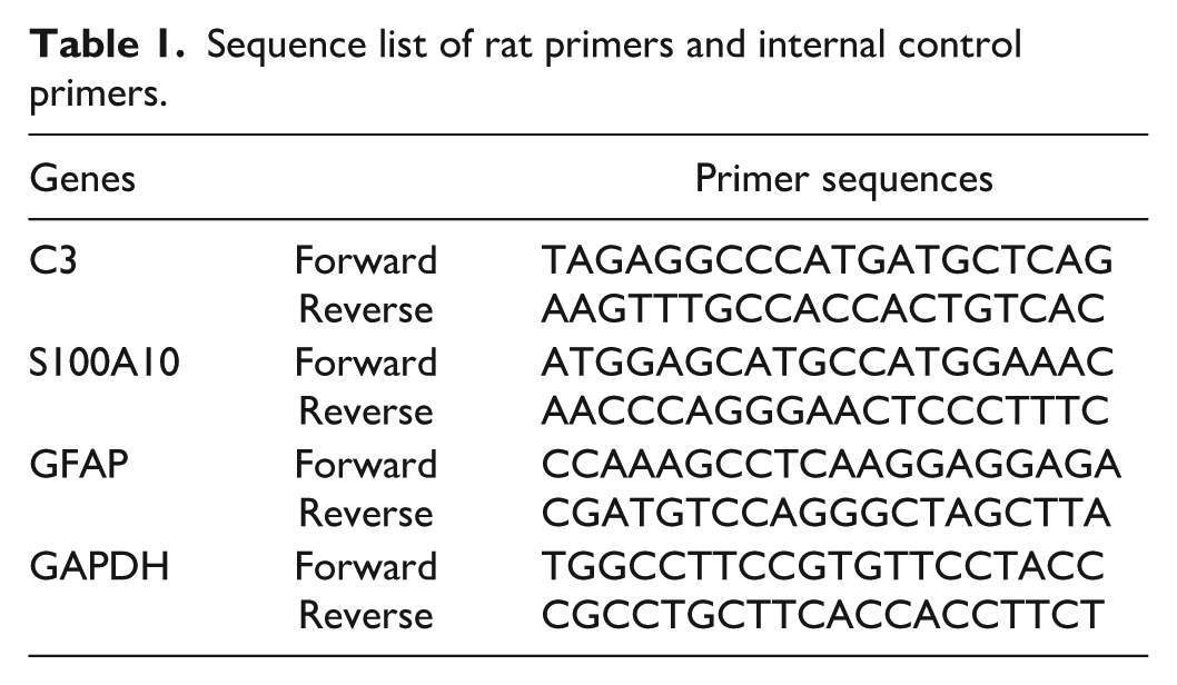
Total RNA was extracted from frozen spinal cord tissue using an Ultrapure RNA Kit (CWBIO, Beijing, China) and reverse-transcribed into cDNA using a Reverse Transcription Kit. Experiments were performed using the CFX96 Real-Time PCR Detection System according to the instructions of the ChamQ Universal SYBR qPCR Master Mix kit. The primer and internal reference sequences are listed in Table 1.
Sequence list of rat primers and internal control primers.
Statistical analysis
All experiments were performed with at least three independent biological replicates. GraphPad Prism software (version 9.0) was used for graphical presentation and statistical analysis. Data from multiple groups across different time points were analyzed using two-way analysis of variance (ANOVA) followed by Bonferroni’s post hoc test. A value of p < 0.05 was considered statistically significant.
Results
Phenotypic characterization of mesenchymal stem cell-derived exosomes
Microscopic examination revealed cell migration from tissue explants by day 3 (Figure 1(a)). By day 7, cell density reached approximately 80% confluence in certain regions, indicating suitability for passaging. After subculturing, the cells displayed a characteristic spindle-shaped, fibroblast-like morphology with strong adherence and robust growth 48 h post-seeding (Figure 1(b)).

(a) Human umbilical cord mesenchymal stem cells crawling out of the tissue block on day 3. (b) Growth status of human umbilical cord mesenchymal stem cells after 48 h of P1 adherent culture.
Flow cytometric analysis confirmed the phenotypic identity of human umbilical cord MSCs (hUC-MSCs), showing high expression of positive surface markers (CD73, CD90, CD105) and absence of hematopoietic markers (CD34, CD45, HLA-DR; Figure 2). Following approximately 3 weeks of culture in adipogenic induction medium (ADP1 and ADP2), lipid droplet formation was observed in hUC-MSCs. Fixed with 4% paraformaldehyde and stained with Oil Red O, orange-red lipid droplets were visible under an inverted microscope (Figure 3). After about 4 weeks in osteogenic induction medium, noticeable changes in cell morphology occurred. Subsequent fixation and staining with Alizarin Red S revealed extensive red mineralized nodules, indicating calcium deposition (Figure 4). Transmission electron microscopy (TEM) images showed that hUC-MSC-derived exosomes exhibited a typical cup-shaped or saucer-like morphology surrounded by a lipid bilayer, consistent with exosomal ultrastructural characteristics (Figure 5(a)). Nanoparticle tracking analysis (NTA) revealed a unimodal size distribution with a mean particle diameter of 77 nm (Figure 5(b)). Furthermore, nano-flow cytometry confirmed the presence of exosome-specific surface markers CD9, CD63, and CD81 (Figure 5(c)). These results collectively demonstrate the successful isolation and identification of exosomes derived from hUC-MSCs, which exhibit both the morphological hallmarks and molecular signature typical of exosomes.

Nanoflow detection results of human umbilical cord mesenchymal stem cells.

Adipogenic induction and differentiation of hUC-MSCs.

Osteogenic induction and differentiation of hUC-MSCs.

(a) Exosomes possess a lipid bilayer and exhibit a vesicle-like morphology. (b) Exosome particle size is 77 nm. (c) Expression of specific markers of hUC-MSCs.
Exosome injection alleviated neuropathic pain in CCI rats
Behavioral assessments conducted on postoperative day 3 (D3) revealed that, compared to the Sham group, both the paw withdrawal threshold (PWT) and paw withdrawal latency (PWL) were significantly reduced in the CCI group (p < 0.001). Relative to the CCI group, exosome-treated rats (Exo group) showed no statistically significant difference in PWT (p > 0.05), but exhibited a marked increase in PWL (p < 0.001). On postoperative days 7 and 14 (D7, D14), the CCI group continued to display significantly lower PWT and PWL values compared to the Sham group (p < 0.001). In contrast, the Exo group demonstrated a significant increase in both PWT and PWL relative to the CCI group at these time points (p < 0.001). The sustained reduction in nociceptive thresholds in the CCI group confirmed successful establishment of the neuropathic pain model. The significant recovery of mechanical and thermal pain sensitivity in the Exo group at D7 and D14 indicates that exosome treatment effectively alleviated neuropathic pain in CCI rats (Figure 6).

The impact of exosomes on pain behavior in CCI rats. (a) Mechanical paw withdrawal threshold (PWT) of rats in Sham group, CCI group, and Exo group. (b) Thermal withdrawal latency (PWL) of rats in Sham group, CCI group, and Exo group ns indicates p > 0.05; * denotes the difference between the Exo group and the CCI group; # denotes the difference between the Sham group and the CCI group; ****p < 0.001; ####p < 0.001.
Exosomes attenuated neuroinflammatory rResponses in the spinal cord
On the seventh post-operative day (POD 7), 50% of the rats in each group were humanely sacrificed (n = 6), and the other 50% were euthanized on POD 14 (n = 6). Spinal cord tissues at the lumbosacral enlargement were collected immediately after euthanasia, and the concentrations of NF-κB, TNF–α, and IL-6 were quantified via ELISA (Figure 7). On both D7 and D14, the CCI group showed significantly elevated expression of NF-κB (p < 0.001), TNF-α (p < 0.001), and IL-6 (D7: p < 0.01; D14: p < 0.001) compared to the Sham group. Following intravenous administration of exosomes, the Exo group exhibited a significant reduction in the concentrations of NF-κB (p < 0.001), TNF-α (p < 0.001), and IL-6 (D7: p < 0.01; D14: p < 0.001) relative to the CCI group at both time points. No statistically significant differences were observed between D7 and D14 within the Exo group (p > 0.05). These results demonstrate that exosome treatment effectively suppressed spinal neuroinflammation, with a significant anti-inflammatory effect already evident by D7.

The effect of exosomes on inflammatory factors in the spinal dorsal horn of CCI rats. (a–c) represent the intra-group and inter-group comparison results of NF-κB, TNF-α, and IL-6 at D7 and D14 in the spinal cord Sham group, CCI group, and Exo group, respectively. Here, nsp > 0.05; *p < 0.05; **p < 0.01; ****p < 0.001.
Exosomes promoted the expression of the A2 phenotype in spinal cord astrocytes in rats
Astrocytes exhibit two distinct phenotypes. The authors hypothesized that UC-MSC-Exos promoted the expression of the A2 phenotype in astrocytes in CCI rats. The expression of A1 (GFAP mRNA, C3) and A2 (GFAP mRNA, S100A10 mRNA) genes was analyzed via qRT-PCR. As shown in Figure 8, on D14, the gene expression of the A2 phenotype in the UC-MSC-Exos group was significantly higher than that in both the Sham and CCI groups, while the gene expression of the A1 phenotype in the CCI group was significantly elevated. The qRT-PCR results were further confirmed by Western blot analysis (Figure 9).

Changes in mRNA content of A1, A2, and GFAP in spinal cord astrocytes of CCI rats treated with exosomes. (a–c) Represent the intra-group and inter-group comparison results of GFAP, S100A10 mRNA, and C3 mRNA in the spinal dorsal horn at D7 and D14 for the Sham group, CCI group, and Exo group, respectively.

Changes in A1 and A2 protein content in spinal cord astrocytes of CCI rats treated with exosomes. (a–d) Represent the intra-group and inter-group comparison results of C3 and S100A10 in the spinal dorsal horn at D7 and D14 for the Sham group, CCI group, and Exo group, respectively. Here, nsp > 0.05; *p < 0.05; **p < 0.01; ***p < 0.001; ****p < 0.0001.
Discussion
The alleviative effect of hUC-MSC-Exos on neuropathic pain
This study demonstrated that administration of human umbilical cord mesenchymal stem cell-derived exosomes (hUC-MSC-Exos) significantly alleviates pain-related behaviors in a rat model of chronic constriction injury (CCI), corroborating previous findings that mesenchymal stem cell-derived exosomes mediate anti-inflammatory and neuroprotective effects via their bioactive molecular cargo (e.g., miRNAs, proteins). 5 Importantly, exosome-based therapy retains the therapeutic benefits of parent cells while circumventing risks associated with whole-cell transplantation, offering a promising novel strategy for the treatment of neuropathic pain (NP).
Neuropathic pain pathogenesis involves the upregulation of pro-inflammatory factors, which are primarily released by immune cells (e.g., microglia, macrophages) and glial cells (e.g., astrocytes). These factors contribute to pain sensitization through complex signaling cascades. For example, TNF-α activates neuronal TNF receptors (TNFR1/2), upregulates sodium channels (e.g., Nav1.3, Nav1.8), enhances neuronal excitability, and promotes microglial activation and subsequent release of IL-1β and IL-6. 13 Elevated spinal TNF-α levels in CCI rats are correlated with hyperalgesia, and TNF-α neutralization alleviates pain. 14 Similarly, IL-6 activates astrocytes via the gp130/JAK-STAT3 pathway, amplifies neuroinflammation, and upregulates TRPV1 expression, thereby enhancing nociception. 15 IL-6 knockout mice exhibit markedly reduced NP symptoms. 16
Consistent with these mechanisms, hUC-MSC-Exos treatment significantly increased the mechanical paw withdrawal threshold (PWT) and thermal paw withdrawal latency (PWL) on days 7 and 14 post-CCI. Concurrently, ELISA revealed pronounced reductions in spinal levels of NF-κB, TNF-α, and IL-6. The lack of significant differences in inflammatory mediator levels between day 7 (D7) and day 14 (D14) in the Exo group indicates that exosome treatment effectively suppressed neuroinflammation as early as D7. These findings support the conclusion that hUC-MSC-Exos alleviate NP, at least in part, through downregulation of pro-inflammatory factors. NF-κB serves as a master transcriptional regulator of neuroinflammation and pain sensitization. It activates pro-inflammatory signaling in microglia, astrocytes, and neurons, promotes the expression of TNF-α, IL-1β, and IL-6, 12 and contributes to chronic pain maintenance. 17 In early NP stages, NF-κB activation drives microglial release of TNF-α and IL-1β, amplifying inflammatory responses. 18 At later stages, it upregulates NMDA receptors and Nav1.3 channels in spinal dorsal horn neurons, facilitating neuronal hyperexcitability and central sensitization, 19 and promotes astrocyte-derived IL-6 and chemokine release, perpetuating chronic pain. 20 In this study, hUC-MSC-Exos treatment induced synchronous reductions in NF-κB, TNF-α, and IL-6 levels by D7, suggesting that exosomes may inhibit NF-κB nuclear translocation and downstream inflammatory gene expression. The early and sustained control of neuroinflammation underscores the therapeutic potential of hUC-MSC-Exos for NP and provides an experimental foundation for optimizing treatment windows in future clinical applications.
The role of astrocytes in neuropathic pain
Astrocytes, the most abundant glial cells in the central nervous system (CNS), play a critical role in the initiation, maintenance, and chronicity of neuropathic pain (NP). Through their highly branched processes, astrocytes form specialized “microdomains” that allow precise regulation of individual synapses and vascular segments.
21
They contribute to neurovascular coupling, modulate local synaptic plasticity, and supply energy substrates to neurons via the astrocyte-neuron lactate shuttle (ANLS).
22
As integral components of the tripartite synapse,
23
astrocytes clear synaptic glutamate through the glutamate-glutamine cycle—a process dependent on GLT-1 and GLAST transporters—and release gliotransmitters such as
UC-MSC-Exos promote A2 phenotype polarization of astrocytes
Astrocyte polarization plays an important role in NP. This study observed that on D14, GFAP mRNA expression in the CCI group was significantly higher than that in the Sham and Exo groups, indicating that NP can activate astrocytes. The expression in the Exo group was significantly lower than that in the CCI group, suggesting that exosome treatment may inhibit the reactivity of astrocytes. Further experiments are needed to confirm this. On D14, S100a10 mRNA levels were significantly increased in the Exo group, while C3 mRNA levels were significantly elevated in the CCI group. This result was further confirmed by subsequent protein expression detection. Collectively, these data indicate that after UC-MSC-Exos treatment, spinal astrocytes transform toward the A2 phenotype (neuroprotective phenotype), characterized by upregulation of A2 markers (e.g., S100a10). Thus, our findings reveal a novel mechanism through which UC‑MSC‑Exos alleviate NP—namely, by promoting the phenotypic transition of astrocytes from the pro‑inflammatory (A1) to the anti‑inflammatory (A2) state.
In recent years, exosomes, as a novel type of nanoscale extracellular vesicles, have demonstrated great potential in the treatment of neuropathic pain. miR-133b-modified mesenchymal stem cell (MSC)-derived exosomes can significantly improve motor function and promote axonal regeneration by delivering miR-133b, thus offering providing a new strategy for spinal cord injury and potential neuropathic pain treatment. 31 Neuron-derived exosomes (NDEs) can inhibit the overactivation of microglia and astrocytes by delivering miR-124-3p,thereby reducing neuroinflammation and secondary injury, indicating their potential in neuroprotection. 32 Pericyte-derived exosomes (PC-Exos) can reduce vascular leakage, inhibit inflammatory responses, and promote angiogenesis after spinal cord injury, which in turn improving neurological recovery, potentially by maintaining blood-nerve barrier integrity to alleviate neuropathic pain. 33 This experiment used human umbilical cord-derived mesenchymal stem cell exosomes, which offer significant biosafety advantages. Studies have shown that hucMSC-Exos express low levels of MHC molecules, low immunogenicity, and are less likely to cause immune rejection. 34 Compared with embryonic stem cells, the umbilical cord is readily available as medical waste and raises no ethical concerns. 35 HucMSC-Exos exhibit good tissue compatibility, and multiple animal experiments have confirmed the absence of significant toxic reactions after intravenous injection. 36 HucMSC-Exos deliver miRNAs such as miR-124 and miR-21 to inhibit M1 polarization of microglia and downregulate the expression of pro-inflammatory factors such as TNF-α and IL-1β 9 (this study investigated their effect on astrocyte polarization). They carry neurotrophic factors such as BDNF and NGF to promote neuronal survival and axonal regeneration 37 and regulate long-term potentiation (LTP) of synaptic transmission in the spinal dorsal horn. 38 The nanoscale size of hucMSC-Exos (30–150 nm) enables them to effectively cross the blood-brain/blood-nerve barrier, 39 leading to better biological effects. Wharton’s jelly of the umbilical cord contains a large number of MSCs, with a single umbilical cord yielding 1–5 × 10⁶ MSCs, 40 effectively overcoming source limitations. Moreover, hucMSCs can be stably passaged for over 15 generations while maintaining differentiation potential. 41 Compared with bone marrow MSCs, hucMSCs secrete 30%–50% more exosomes per cell, 42 providing stronger support for experiments. Although exosomes show optimistic prospects for intervention in neuropathic pain, challenges remain, such as poor tissue-specific targeting ability that impairs therapeutic efficacy, 43 and after systemic administration, 60%–70% of exosomes are captured by the liver and spleen, with only 5%–10% reaching the nerve injury site. 44 Additionally, animal models (e.g., CCI, SNI) differ somewhat from human chronic pain pathology to some extent, and long-term safety data (e.g., tumorigenicity, immune tolerance) lack systematic evaluation. 45
Effective treatment of neuropathic pain is a major challenge for healthcare professionals. Current treatment regimens struggle to achieve low-dose efficacy with minimal side effects. This study is the first to demonstrate that human umbilical cord-derived mesenchymal stem cell exosomes (UC-MSC-Exos) effectively alleviate neuropathic pain in chronic constriction injury (CCI) rats by inhibiting neuroinflammation mediated by the NF-κB signaling pathway and promoting A2 phenotype polarization of astrocytes. This finding provides an important experimental basis for developing exosome-based NP treatment strategies. Future research should further explore the clinical application of exosomes.
The author acknowledges the limitations of this experiment. Additional research is needed to determine whether the specific effects exerted by UC-MSC-Exos are attributed to microRNAs (miRNAs; e.g., miR-124-3p, miR-21-5p), proteins, or lipids. It remains to be verified whether there is a causal relationship between the downregulation of the NF-κB pathway and astrocyte A2 polarization, as well as the specific factors influencing astrocyte polarization.
Conclusion
This study utilized a chronic constriction injury (CCI) rat model to evaluate the therapeutic potential of umbilical cord mesenchymal stem cell-derived exosomes (UC-MSC-Exos) in alleviating neuropathic pain (NP) and to elucidate the underlying mechanisms. The results indicate that UC-MSC-Exos significantly attenuated mechanical allodynia and thermal hyperalgesia in CCI rats. Furthermore, exosome treatment downregulated the expression of key pro-inflammatory mediators—NF-κB, TNF-α, and IL-6—in spinal cord tissues and promoted a shift in astrocyte polarization toward the neuroprotective A2 phenotype.
Footnotes
Declaration of conflicting interests
The authors declared no potential conflicts of interest with respect to the research, authorship, and/or publication of this article.
Funding
The authors disclosed receipt of the following financial support for the research, authorship, and/or publication of this article: This study was funded by the Science and Technology Bureau of Taiyuan City (202268 and 202232).