
Abstract
With the increasing use of surgical robots, robust and accurate segmentation techniques for brain tissue in the brain magnetic resonance image are needed to be embedded in the robot vision module. However, the brain magnetic resonance image segmentation results are often unsatisfactory because of noise and intensity inhomogeneity. To obtain accurate segmentation of brain tissue, one new multiphase active contour model, which is based on multiple descriptors mean, variance, and the local entropy, is proposed in this study. The model can bring about a more full description of local intensity distribution. Also, the entropy is introduced to improve the performance of robustness to noise of the algorithm. The segmentation and bias correction for brain magnetic resonance image can be simultaneously incorporated by introducing the bias factor in the proposed approach. At last, three experiments are carried out to test the performance of the method. The results in the experiments show that method proposed in this study performed better than most current methods in regard to accuracy and robustness. In addition, the bias-corrected images obtained by proposed method have better visual effect.
Introduction
With the development of pattern recognition, artificial intelligence, and image processing technology, various kinds of robotics have been widely used. Robots mainly include the following three kinds: first, industrial robotics applied to manufacturing, logistics, and agriculture. Second, special robotics used in medical and other fields. Third, service robotics applied to customer service, escort, and design. Medical robotics is the robotics that has been applied in many medical fields, such as diagnosis, treatment, rehabilitation, nursing, and functional assistance. Among them, surgical robots are the most widely used and promising. The powerful functions they provide overcome the poor accuracy, long operation time, fatigue, and lack of vision in traditional surgery.
Currently, encephalopathy is one of the serious diseases that threaten people’s health. With the development of medical technology and robotics, brain surgery robots have been increasingly used in the planning, navigation, and orientation of cerebral surgery. And the computer vision has been applied in neurosurgery 1,2 and robotic brain surgery. 3 –5 Brain surgery robot uses embedded computer vision module to collect brain image data, and then accurately segment, detect, and locate brain tissue. Due to the accurate segmentation algorithm, precise location information of brain tissue can be used during brain surgery. Image segmentation is the foundation of computer vision applications. In the computer vision module of the brain surgery robot, the segmentation effect of the brain image directly determines the positioning accuracy and the success rate of the operation. However, the brain magnetic resonance (MR) image segmentation is difficult because of the complicated brain structure and the inferior quality image. The brain MR image is affected by acquisition sequences and radio frequency coils. So the intensity variation across the image reflects the intensity inhomogeneity. Also, image is usually vulnerable to noise that causes unexpected results. Therefore, more accurate and robust segmentation algorithms for brain image with intensity inhomogeneity and noise are very important and necessary. And the algorithm should meet the following four conditions. The first one refers to the full description of gray value distribution, which can overcome the intensity inhomogeneity and ensure segmentation accuracy. The second one is that it should be robust to serious noise. Third, it can implement the multiphase segmentation. Finally, the bias field can be estimated.
In the past decades, many active contour models (ACMs) that are considered as a well-performed class of segmentation algorithms with promising result have been proposed. 6 For the sake of images with intensity, some ACMs introduce the local grayscale statistics. 7 –10 Vese and Chan 7 provided a piecewise model, which can solve the problem in some case. The method is computationally inefficient, which limits its application. Li et al. 8 presented an ACM with local binary fitting (LBF) energy to obtain the correct object boundaries in segmentation images with inhomogeneous intensity. Subsequently, a number of methods were proposed, whose effects were similar to LBF. 11,12 However, the LBF method fails to achieve accurate results sometimes because the local intensity information is only represented by means. It is not enough to describe intensity distribution in a local domain with same mean and different texture. To solve the problem, Wang et al. presented a new ACM based on local Gaussian distribution fitting energy. 9 In the local Gaussian distribution fitting (LGDF) model, the local grayscale statistics are described not only with means but also with variances, which enable LGDF with better robustness on the intensity inhomogeneity. However, the above methods could not work very well for segmenting the brain MR image.
Recently, some novel ACM-based brain MR image segmentation methods have been proposed in literature. 13 –21 However, these methods have different drawbacks and cannot provide the desirable results. The method proposed by Akram et al. 13 is not able to carry out the bias field estimation. The methods proposed by Li et al., 14,15 Gharge and Bhatia, 16 and Konduri and Thirupathaiah 17 cannot accurately describe the local intensity distribution of brain MR image. The methods proposed by Gharge and Bhatia, 16 Konduri and Thirupathaiah, 17 and Zhang 18 cannot be used to implement multiphase segmentation for brain tissue. The methods proposed by Zhang et al. 19,20 and Chen et al. 21 are sensitive to serious noise.
In this article, one ACM based on multiple descriptors (MDACM) is presented. And the proposed method is capable of effectively dealing with the intensity inhomogeneity and serious noised image. Moreover, the proposed algorithm can complete segmentation and bias correction at the same time. In our method, local entropy is introduced to describe the intensity distribution combined with the means and the variance, which can yield more accuracy segmentation. Besides, the MDACM is more robust to noise because we adopt the entropy for the local gray statistic. Two level set functions are utilized to segment white matter, gray matter, cerebrospinal fluid, and background. Additionally, by introducing bias field factor, the MDACM can complete bias correction at the same time of segmentation.
The proposed segmentation model can be embedded into the visual module of brain surgery robots and help to obtain the accurate segmentation of brain tissue. The segmentation results can provide important information for the diagnosis and treatment of brain diseases. These preliminary processing can not only reduce the burden of doctors but also improve the success rate of operation.
The remaining parts are arranged as follows: “Background” section gives reviews of some typical ACM. “Proposed method” section introduces the new MDACM model. “Experimental results” section gives the results of brain MR image segmentation. The final section gives the conclusion.
Background
The LBF model
The LBF model takes full advantage of the local intensity information to achieve the segmentation for images with inhomogeneous intensity. 8,18 The LBF energy can be expressed as following
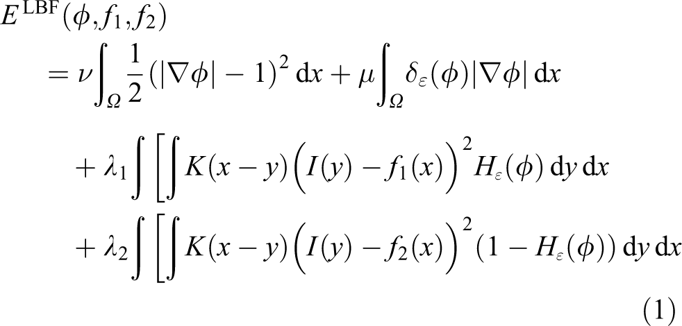
where υ, μ,
As the LBF uses local means
The LGDF model
In the study, 9 the local gray statistics are described with both means and variances, which enable LGDF with better robustness on the intensity inhomogeneity. In Bayesian framework, Wang et al. proposed the following energy functional

where
Because the LGDF model utilizes two descriptors, both mean and variance, it is more robust to images with inhomogeneous intensity. But the segmentation accurate is not good enough for the images with complex brain tissue structure. And it is two-phase segmentation and no bias field estimation.
The local intensity clustering model
In the study, 14 an ACM based on local intensity clustering (LIC) is proposed. And a defined bias field is used to represent the intensity inhomogeneity. The LIC energy functional can be expressed as follows

where
The LIC can achieve segmentation as well as bias field correction. And it can realize multiphase segmentation and better results in accuracy, efficiency, and robustness. Nevertheless, it is not able to deal with the images at the presence of serious noise.
Proposed method
Local entropy
In Shannon’s information theory,
22
a probabilistic system includes n events, and if the occurrence probability of an event i is

And the information quantity of the whole probabilistic system with n events is defined as

SHIOZAKI proposed to use the definition of SHANNON entropy to represent the image entropy.
23
The entropy of

where

where
The different parts of images have different information. To describe the local image, we define the entropy of a continuous subarea
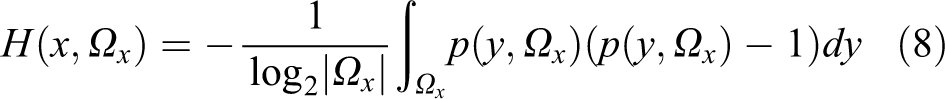
where
The entropy can reflect the richness of image information. And it can be used as the measurement standard of the homogeneity of the partition. The partition is more homogeneous, and the entropy is higher.
In the proposed method, we take the local entropy together with mean and variance as the local image descriptors to obtain higher fitting degree for image intensity distribution. The value of local entropy is not sensitive to a single noise pixel because it is determined by several pixels in a continuous domain. And therefore the local entropy has a certain filtering effect. In addition, the normalization of intensity probability can also smooth the noise, which can guarantee the robustness to noise of our MDACM method, especially for segmentation brain MR image with serious noises.
Image model
The proposed algorithm adopts the following common model to describe the images in the presence of inhomogeneous intensity

where I is the observed image, b is the inhomogeneous intensity component, J standards for the true image, and n is additive noise. The whole image domain is represented with Ω. The following part is the more detailed statement of J and b. 14,22
Assumption 1: The inhomogeneous intensity component b changes slowly, so the value of b in the neighborhood of point x is approximately a constant, that is,
Assumption 2: Image J includes N disjoint regions, that is,
Therefore, we have the value of subneighborhood

MDACM fitting energy
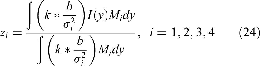
We take the mean, variance, and entropy of image as descriptors to fit intensity distribution. The data fitting term of the model is defined in the Bayesian framework. So the fitting energy of every point can be written as

where
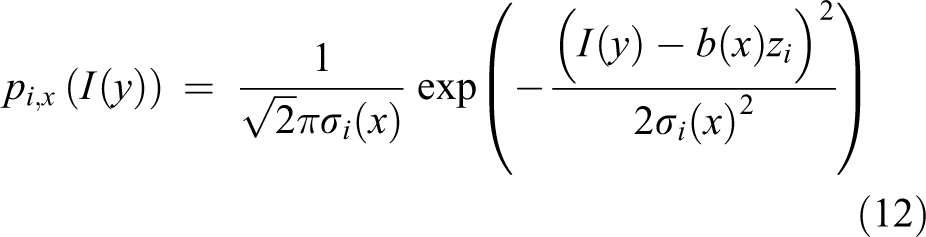
where
Then we introduce a non-negative window function
where a is a positive constant and
The above

where the local entropy
MDACM
It is difficult to get the solution of the minimization of
In traditional two-phase segmentation, only one level set function is enough to divide the image into foreground and background. In multi-phase segmentation

where
The differential of H is denoted as δ, which is computed by

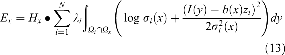
So the final data fitting term of the four-phase model for brain MR image is rewritten in the following form

For convenience, where we represent the constants
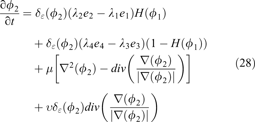
The energy

where μ and υ are positive coefficients. The second term is the regularizing term, which can ensure stable evolution of ϕ. The deviation of ϕ from the signed distance function is defined as following

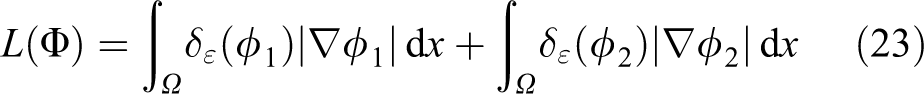
The third term is the length regularization term. And whose definition formula is
To get the correct boundary, we minimize the energy functional.
Energy minimization
The gradient descent method is used to complete the minimization of the total energy formula (21) for image segmentation. Image segmentation is achieved by iterative process, taking one variable in turn to iterate.
Each variable and the level set evolution formula are given as follows.
By fixing the variables Φ, b, Θ and minimizing E relate to
By fixing the variables Φ, b, Z and minimizing

By fixing the variables Φ, Θ, Z and minimizing

Keeping b, Z, and Θ fixed and minimizing

where

Implementation
The implementation of our algorithm includes the following seven steps:
Step 1: Compute the local entropy
Step 2: Initialize the level set functions
Step 3: Calculate the means
Step 4: Calculate the variances
Step 5: Update the level set functions
Step 6: Update the bias factor b.
Step 7: Check whether meet the convergence criteria. If not, rerun Step3.
Experimental results
To evaluate the performance of the MDACM, we apply it to some brain MR image. The experiments are implemented in Matlab 2013 in PC with Inter CPU 3.20 and 4 GB RAM and Windows 7 operating system. In this section, the parameters are as follows:
The validity of MDACM
The first experiment is designed to demonstrate the validity of the MDACM model. We apply it to segment brain MR image and correct the bias field.
The experiment results are shown in Figure 1. Figure 1(a), (b), (c), and (d), respectively, shows the original images, segmentation result images, bias field images, and images after correction. It is obvious that the intensity distribution of the same tissue is more homogeneous in the images after correction than in the original images. Figure 1(e) displays the histograms of original images. Figure 1(f) displays the histograms of the images after correction. It can be seen that the histogram of original image has no well-separated peaks because of the inhomogeneous intensity. However, the histogram shown in Figure 1(f) has three well-separated peaks. The inhomogenous intensity in the images has been obviously improved.

Applications of MDACM to brain MR image with inhomogeneous intensity. (a) Original images; (b) segmentation results; (c) bias fields; (d) bias-corrected images; (e) histograms of the original images; (f) histograms of the bias corrected images. MR: magnetic resonance.
The experiment results in this subsection show that the MDACM has the ability of segmenting the brain MR image with inhomogeneous intensity as well as correcting bias field.
Comparison with multiphase LBF model and LIC model
The second experiment is designed to show the comparison of the segmentation accuracy. The three models of MDACM, multiphase LBF, and LIC are employed in segmenting the brain MR image as shown in Figure 2.

Comparison of the MDACM with the multiphase LBF and the LIC models for brain MR image. (a) Original images; (b) results of the multiphase LBF; (c) results of the LIC; (d) results of the MDACM. MR: magnetic resonance; LBF: local binary fitting; LIC: local intensity clustering.
The original images with intensity inhomogeneity are shown in Figure 2(a). The results of the multiphase LBF, the LIC, and the MDACM are shown in Figure 2(b) to (d). In Figure 2(b) to 2(d), these three models segment the brain MR image with different results. As shown in Figure 2(b), there are lots of errors in sedimentation of cerebrospinal fluid as gray matter. Compared with the multiphase LBF model, the LIC has better segmentation results as shown in Figure 2(c). However, there are still some false segmentation regions in brain tissue. In contrast, the proposed method has the best segmentation results that are most similar to the true segmentation.
In order to give a quantitative comparison of our MDACM model with the other two models, we select the dice similarity coefficient (DSC), the false negative ratio (FNR), the false positive ratio (FPR), and the ratio of segmentation error (RSE) to descript the segmentation accuracy. 24 Figure 3 is the histogram of the DSC value. And Table 1 lists the data statistics for the other three indexes. The ideal segmentation results should be DSC→1, FPR→0, FNR→0, RSE→0.

Accuracy comparison of the three models for brain MR image. MR: magnetic resonance.
FNR, FPR, and RSE values.

FNR: false negative ratio; FPR: false positive ratio; RSE: ratio of segmentation error; LBF: local binary fitting; LIC: local intensity clustering.
As can be seen from Figure 3, for the left image, the DSC value of the proposed algorithm MDACM is close to 0.9, the LIC has a DSC value of 0.85, while the DSC value of the multiphase LBF is 0.78. And for the right image, the DSC value of the MDACM is 0.88, the LIC has a DSC value of 0.83, while the DSC value of the multiphase LBF is 0.67. Obviously, the proposed algorithm has the highest DSC value and the best segmentation effect.
In Table 1, the value of each index is the average number of two images. And the values of the MDACM are the lowest, with the FNR 0.0307, the FPR 0.0152, and the RSE 0.0081.The values of the multiphase LBF are the highest, with the FNR 0.0307, the FPR 0.0152, and the RSE 0.0081. And the values of the LIC are between the above two methods. The data show that the proposed MDACM algorithm has the best segmentation effect.
The MDACM is obviously superior to the multiphase LBF method. On the one hand, the two descriptors, such as variance and local entropy, are added, so the intensity description is more accurate. On the other hand, the algorithm realizes the bias field correction, so the effect of intensity inhomogeneity on segmentation is weakened. The LIC algorithm takes into account the bias field compared with the multiphase LBF, so the performance is better than the multiphase LBF. While compared with the proposed MDACM, the LIC lacks two descriptors of variance and local entropy, so the accuracy is lower than the MDACM.
Robustness to noise
The proposed method is robust, which is validated in the third experiment. The segmentation results of noisy images for the multiphase LBF, the LIC, and the MDACM are shown in Figure 4. The same images corrupted with Gaussian white noise with different standard deviations are shown in Figure 4(a). The standard deviations of Gaussian noise are 0.1, 0.15, 0.2, and 0.5 from left to right. Figure 4(b) and (c) shows the segmentation images of the multiphase LBF and the LIC. Figure 4(d) shows the segmentation of the MDACM. It is obvious that the segmentation results of the MDACM model are better than the other two methods.
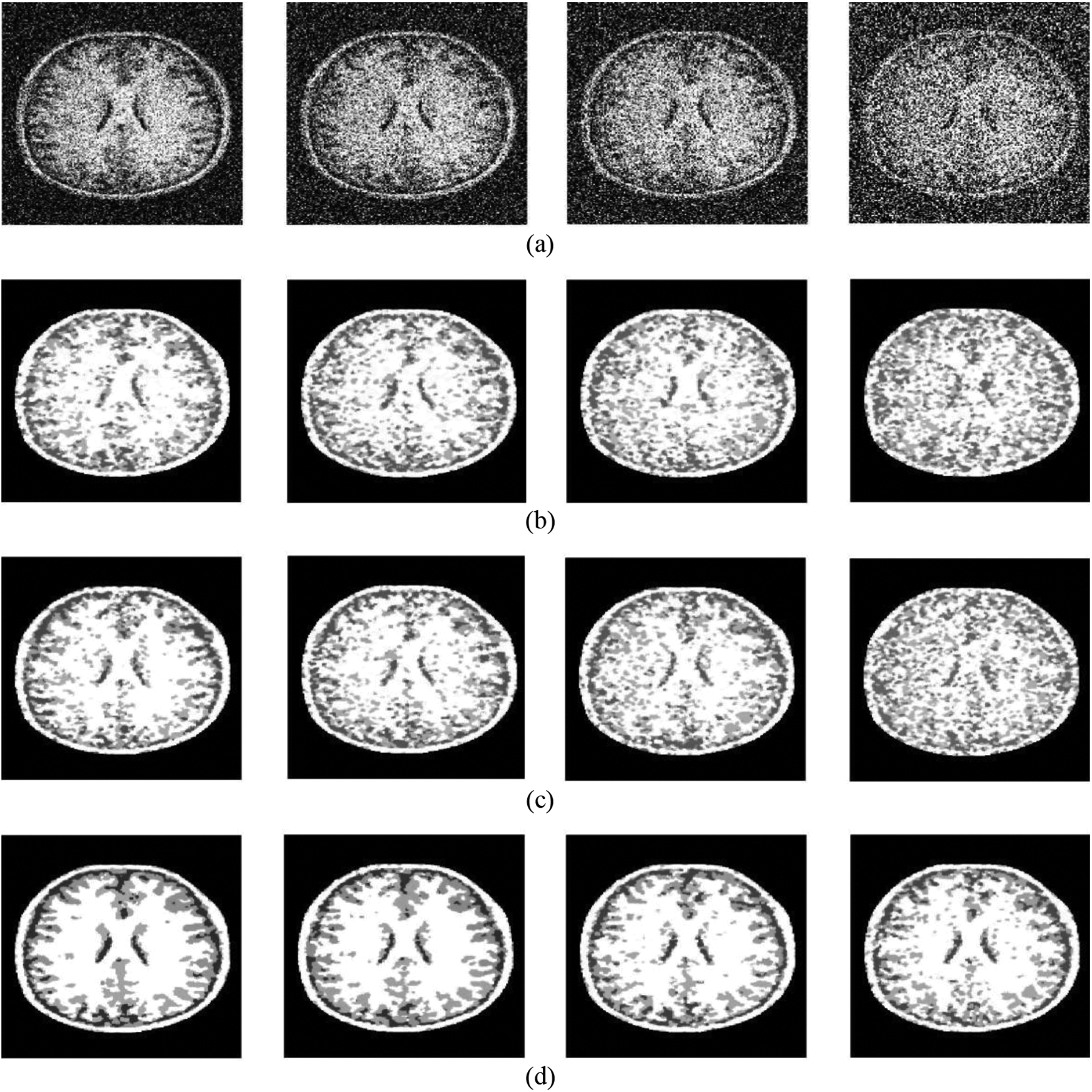
Segmentation results for the images with different Gaussian white noise. (a) Noisy brain MR image; (b) results of multiphase LBF; (c) results of LIC; (d) results of the proposed method. MR: magnetic resonance; LBF: local binary fitting; LIC: local intensity clustering.
To give a quantitative comparison, we also select the DSC, the FNR, the FPR, and the RSE to descript the segmentation accuracy. The corresponding DSC values of three models are shown in Figure 5. The values of FNR, FPR, and RSE are listed in Table 2.
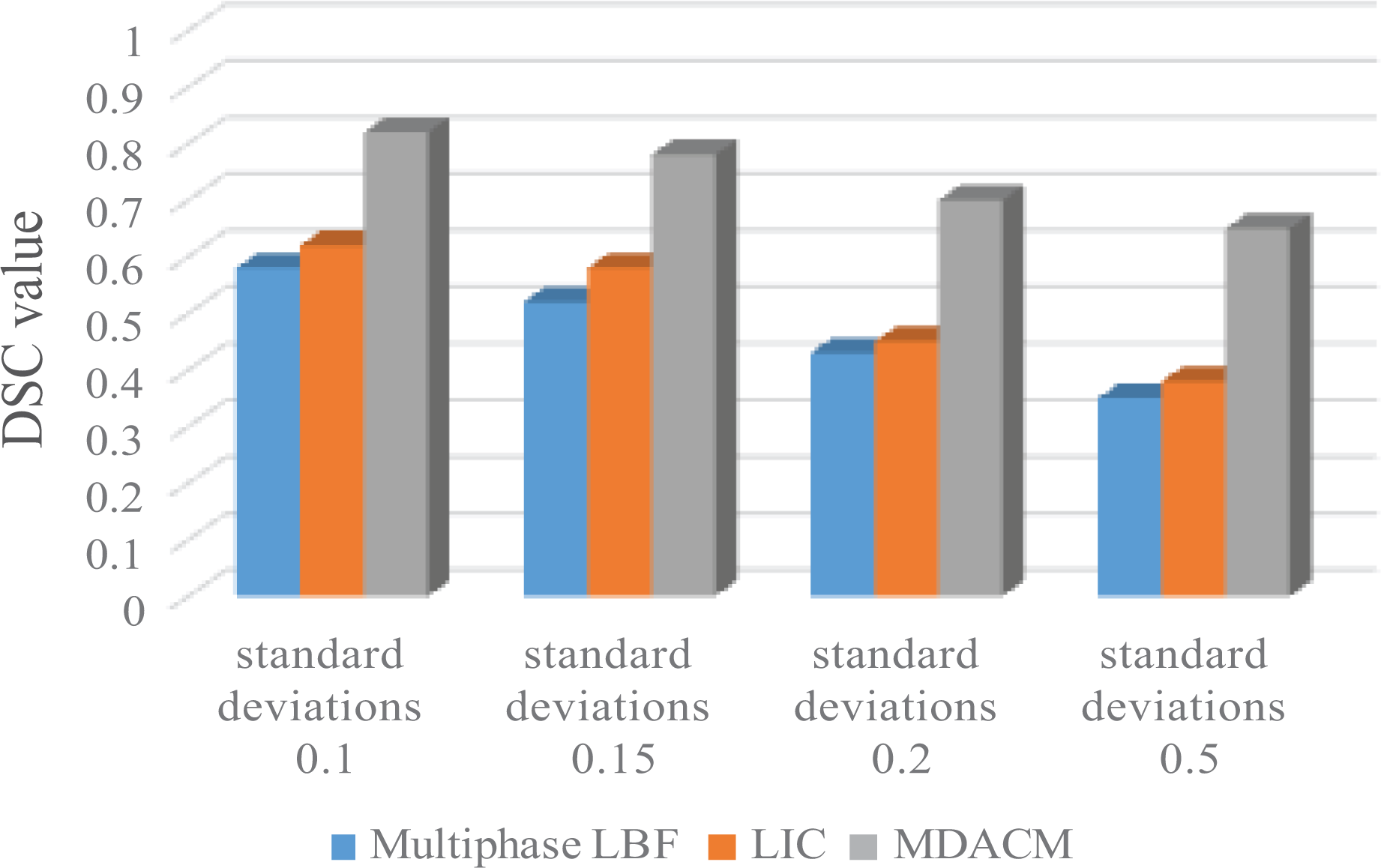
Robustness to noise comparison of the three models.
FNR, FPR, and RSE values.

FNR: false negative ratio; FPR: false positive ratio; RSE: ratio of segmentation error; LBF: local binary fitting; LIC: local intensity clustering.
As can be seen from Figure 5, in the case of the same noise, the DSC value of the proposed MDACM is the highest and the DSC value of the multiphase LBF is the lowest. For example, when the standard deviation of Gaussian noise is 0.1, the DSC of the MDACM is close to 0.8, the DSC value of the LIC is 0.61, and the DSC value of the multiphase LBF is only 0.53. The DSC value of the MDACM is nearly 60% higher than that of the multiphase LBF. And when the standard deviation of Gaussian noise is 0.5, the DSC of the MDACM is 0.63, the DSC value of the LIC is 0.37, and the DSC value of the multiphase LBF is only 0.32. The DSC value of the MDACM is nearly 100% higher than that of the multiphase LBF. It is worth noting that with the increase of noise level, the DSC values of various methods are decreased, and the decrease value of the MDACM is the least, which shows that the proposed MDACM has the best performance of robustness to noise.
As can be seen from Table 2, MDACM has the lowest FNR value of 0.1205, the lowest FPR value of 0.0801, and the lowest RSE value of 0.0627. While the multiphase LBF has the highest FNR value of 0.5125, the highest FPR value is 0.4768, the highest RSE value is 0.3532, and all above values are close to 5 times of those of MDACM.
The data in Figure 5 and Table 2 show that the proposed MDACM method has better robustness to noise than the multiphase LBF model and the LIC model. The main reason is that the algorithm introduces the local entropy as the descriptor on the basis of mean and variance, and the local entropy has the effect of smoothing noise. So the anti-noise performance of the algorithm is improved.
Conclusion
In this article, one multiphase ACM segmentation method named MDACM for brain MR image with inhomogeneous intensity and serious noise is proposed, which can be embedded into the visual module of brain surgery robots. Our model utilizes multiple descriptors, the mean, the variance, and the local entropy to guarantee the exhaustive expression of the local intensity distribution. The local entropy is successfully introduced to obtain desirable performance in the aspect of robustness to noise. The proposed method can complete segmentation and bias correction for brain MR image. Experimental results show our method meets all the four conditions of accurate segmentation of brain tissue: the full description of gray value distribution, robust to serious noise, multiphase segmentation, and bias field estimation. Comparisons with the LIC and the multiphase LBF models, experimental results demonstrate that the MDACM has better performance on the aspects of accuracy and robustness to serious noise. While achieving better accuracy, there are still some works to be improved. In the future, more efforts will be made to further improve the robustness to noise and real-time performance to adapt to the requirements of high precision and real-time of brain surgery robots. In addition, the MDACM model will be embedded into brain surgery robot for clinical tests to further improve the accuracy and applicability of the algorithm according to the doctor’s advises. It is believed that this will improve the localization of brain lesions and the success rate of surgery.
Footnotes
Declaration of conflicting interests
The authors declared no potential conflicts of interest with respect to research, authorship, and/or publication of this article.
Funding
The author(s) disclosed receipt of the following financial support for the research, authorship, and/or publication of this article: This work is supported by the National Science Foundation of China (61503274 and 61603080), and Fundamental Research Funds for the Central Universities (N16260004).