
Abstract
Introduction
Oral lichen planus (OLP) is a common oral mucosal disease frequently encountered in clinical practice affecting approximately 1–2% of the general population, with a higher prevalence in women and a potential for malignant transformation in 0.5–2% of cases.1,2 Its clinical manifestations typically include small papules that form dendritic, linear, or reticular white stripes, often accompanied by congestion, ulcers, blisters, atrophy, and erosion. Current treatments, such as corticosteroids, are limited by their symptomatic focus and inability to prevent recurrence or progression, underscoring the need for targeted therapies. 3 The onset of OLP is believed to be influenced by multiple factors, contributing to its chronic, refractory nature and recurrent flare-ups. 4 Prolonged erosion in affected areas not only restricts the patient’s ability to open their mouth but also leads to local fibrous hyperplasia and, in some cases, malignant transformation, significantly affecting their daily life and work. 2
Recent research indicates that OLP is an inflammatory condition mediated by T lymphocytes, with various cytokines playing key roles in lesion development. 5 IFN-γ and IL-4 were selected as representative cytokines of Th1 and Th2 cells, respectively, due to their well-established roles as key regulators of the Th1/Th2 balance, which is widely implicated in OLP pathogenesis.6,7 These cytokines are also frequently studied in OLP, providing a foundation for comparison with existing literature.8,9 While IFN-γ and IL-4 have been implicated in OLP, their differential expression across subtypes and their clinical implications remain underexplored. 10 The lamina propria of OLP mucosa contains a high density of lymphocytes, including Th0 precursor cells, which can differentiate into Th1 or Th2 cells depending on specific conditions. 6 Th1 cells predominantly produce cytokines such as IFN-γ, while Th2 cells mainly secrete IL-4. 7 The balance between Th1 and Th2 subsets is crucial for maintaining immune homeostasis and regulation. 11
This study aimed to examine the expression levels and significance of IFN-γ and IL-4 in the peripheral blood of OLP patients, with a focus on erosive and non-erosive subtypes.
Material and methods
General data
A total of 58 OLP patients, diagnosed by histopathology at our hospital between June 2020 and June 2022, were selected for the OLP group in this retrospective case-control study, comparing OLP patients with healthy controls. This study adhered to the STROBE guidelines for reporting observational studies. The sample size was determined by the availability of eligible patients diagnosed during this period; a priori power analysis was not conducted due to the retrospective design. This group consisted of 25 men and 33 women, aged 18 to 70 years, with a mean age of 52.35 ± 10.23 years. Additionally, 58 healthy volunteers who visited the hospital during the same period were included as the control group. No significant differences were observed in the general data between the two groups (P > 0.05). Furthermore, the 58 OLP patients were divided into two groups: 30 cases of the non-erosive type (including network, stripe, plaque, and papule types) and 28 cases of the erosive type (including erythema and atrophy types). No significant differences were found in age or gender between the two groups (p > .05).
Inclusion criteria: (1) OLP confirmed by pathological diagnosis; (2) No systemic treatment; (3) All subjects provided informed consent.
Exclusion criteria: (1) Presence of other oral mucosal lesions; (2) Presence of acute infections, systemic diseases, or tumors; (3) Use of antibiotics or immunosuppressants within the past 3 months; (4) Pregnancy or lactation.
Isolation of serum and peripheral blood mononuclear cells
Fasting peripheral venous blood was collected before 9 a.m. from both OLP patients and healthy controls. The blood was centrifuged, and the supernatant was carefully extracted. After sorting, the supernatant was immediately stored in a low-temperature refrigerator at −80°C for future use. Venous blood was anticoagulated with sodium citrate, diluted with an equal volume of D-Hank’s solution, and then separated by density gradient centrifugation. The mononuclear cells were subsequently collected.
Detection of IFN-γ and IL-4 mRNA levels by reverse transcription polymerase chain reaction (RT-PCR)
Total RNA was extracted from 1 × 10⁷ mononuclear cells using the Trizol Total RNA Extraction Kit, followed by reverse transcription to cDNA. PCR amplification was then performed using a PCR reagent kit. β-actin served as a reference primer, and the relative density values of IFN-γ, IL-4, and β-actin were compared for semi-quantitative analysis. The primer sequences for IFN-γ and IL-4 are as follows: • IFN-γ: Forward: 5′-CATTCAGATGTAGCGGATAA-3′, Reverse: 5′-TTTCGCTTCCCTGTTTTA-3’. • IL-4: Forward: 5′-TATGCTGAAACTTTGTAGT-3′, Reverse: 5′-TTTTGATGATCTCCTGTA-3’. • β-actin: Forward: 5′-CTCCATCCTGGCCTCGCTGT-3′, Reverse: 5′-GCTGTCACCTTCACCGTTCC-3’.
After electrophoresis on a 1.5% agarose gel, the relative density values of IFN-γ, IL-4, and β-actin were compared for semi-quantitative analysis.
Detection of IFN-γ and IL-4 protein levels through enzyme-linked immunosorbent assay (ELISA)
The protein levels of IFN-γ and IL-4 in serum were measured using ELISA, following the manufacturer’s instructions (Thermo Fisher Scientific, USA).
Statistical analysis
Statistical analysis was conducted using SPSS 17.0 software. Data from each group are presented as mean ± standard deviation, and comparisons were made using the t-test. A p-value of <.05 was considered statistically significant.
Results
IFN-γ and IL-4 mRNA levels in Both Groups
Compared to healthy controls, OLP patients exhibited significantly higher levels of IL-4 and IFN-γ mRNA (p < .05, Figure 1). IFN-γ and IL-4 mRNA Expression Levels in the Peripheral Blood of OLP Patients and Healthy Controls; The levels of IFN-γ and IL-4 were significantly higher in both the erosive and non-erosive OLP groups compared to the control group (*p < .05). Data are presented as mean ± standard deviation.
IFN-γ and IL-4 mRNA Levels in Different Clinical Types of OLP patients
Compared to healthy controls, both erosive and non-erosive OLP patients showed increased levels of IFN-γ and IL-4 mRNA (p < .05). However, the IFN-γ mRNA levels did not differ between the erosive and non-erosive OLP groups (p > .05). In contrast, IL-4 mRNA levels were significantly higher in erosive OLP patients than in non-erosive OLP patients (p < .05, Figure 2). IFN-γ and IL-4 mRNA Levels in Different Clinical Types of OLP Patients; *p < .05, compared with the control group; #p < .05, compared with the non-erosive OLP group. Both erosive and non-erosive OLP patients had significantly higher IFN-γ and IL-4 mRNA levels compared to controls (p < .05). IL-4 mRNA levels were significantly higher in erosive OLP patients than in non-erosive OLP patients (p < .05).
IFN-γ and IL-4 Protein Levels in Both Groups
Compared to healthy controls, OLP patients exhibited significantly elevated IFN-γ and IL-4 protein levels (p < .05, Figure 3). IFN-γ and IL-4 Protein Levels in Both Groups. *p < .05. OLP patients had significantly higher IFN-γ and IL-4 protein levels compared to healthy controls (P < 0.05).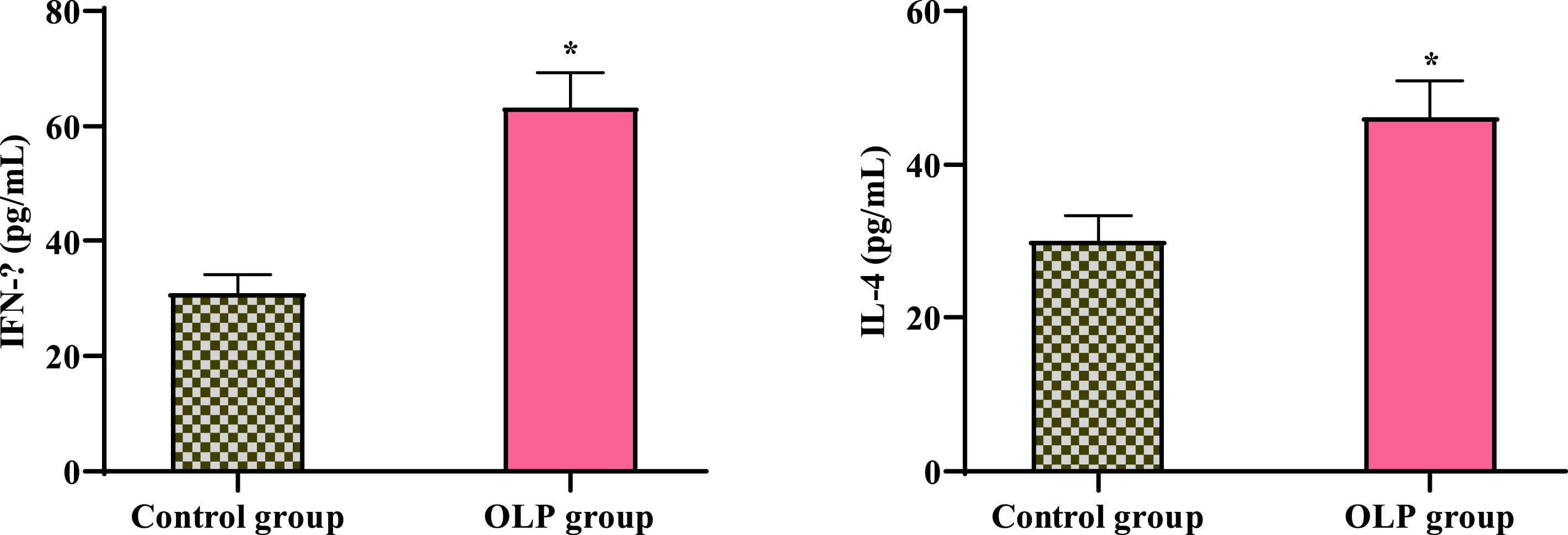
IFN-γ and IL-4 protein Levels in Different Clinical Types of OLP patients
Compared to healthy controls, both erosive and non-erosive OLP patients had increased IFN-γ and IL-4 protein levels (p < .05). However, the IFN-γ protein levels did not differ between the erosive and non-erosive OLP groups (P > 0.05). In contrast, IL-4 protein levels were significantly higher in erosive OLP patients than in non-erosive OLP patients (p < .05, Figure 4). IFN-γ and IL-4 Protein Levels in OLP Patients. *p < .05, compared with the control group; #p < .05, compared with the non-erosive OLP group. Both OLP subtypes showed increased IFN-γ and IL-4 protein levels compared to controls. Erosive OLP patients had higher IL-4 protein levels than non-erosive OLP patients.
Ratio of IFN-γ/IL-4 mRNA and protein expression
The ratio of IFN-γ/IL-4 mRNA expression was higher in both the erosive and non-erosive OLP groups compared to the control group (p < .05). The ratio of IFN-γ/IL-4 mRNA expression was lower in the erosive OLP group compared to the non-erosive OLP group (p < .05). A similar trend was observed for the ratio of IFN-γ/IL-4 protein expression (Figure 5). Ratio of IFN-γ/IL-4 mRNA and protein expression. *p < .05, compared with control group; #p < .05, compared with non-erosive OLP group. Both OLP groups showed a higher IFN-γ/IL-4 ratio compared to controls. The ratio was lower in the erosive OLP group than in the non-erosive OLP group. The same trend was observed for protein expression.
Correlation between IFN-γ and IL-4 expression in OLP patients
Correlation between IFN-γ and IL-4 at mRNA and protein levels.


Correlation Between IFN-γ and IL-4 at mRNA and Protein Levels; The scatter plots illustrate the correlation between IFN-γ and IL-4 at the mRNA level (left) and protein level (right). The red lines indicate the overall trend, showing a weak positive correlation between these two cytokines.
Discussion
This study demonstrated elevated IFN-γ and IL-4 expression in OLP patients, with higher IL-4 levels and a lower IFN-γ/IL-4 ratio in the erosive subtype compared to the non-erosive subtype. OLP is a common non-infectious chronic inflammatory condition of the oral mucosa, which occurs more frequently in women. 12 The primary lesion manifests as a white network pattern or plaque on the mucosal surface, with erosion developing in some patients within the affected area. 13 Clinically, based on the mucosal condition beneath the lesion, OLP is classified into hyperemic erosion type and non-erosive type. 14 In recent years, an immune imbalance has become increasingly recognized as a key factor in the pathogenesis of OLP. Specifically, the balance of T helper cells and their cytokine secretion in CD4 + T cells is believed to play a role in the development of OLP. 15 CD4 + T cells are thought to differentiate into both Th1 and Th2 cells. 16 Th1 cells primarily secrete cytokines such as IFN-γ, IL-2, IL-12, and TNF-α, which are involved in cellular immunity and delayed hypersensitivity reactions. Among these, IFN-γ is the classic cytokine secreted by Th1 cells. 17 In contrast, Th2 cells produce cytokines including IL-4, IL-6, and IL-10, which are associated with humoral immunity. IL-4 is a key cytokine secreted by Th2 cells. 18
In this experiment, it was found that relative to the control group, IFN-γ and IL-4 expression were high in peripheral blood of erosive and non-erosive OLP patients. The increased IFN-γ levels in OLP may reflect enhanced Th1-mediated cellular immunity and delayed hypersensitivity, consistent with reports of T-cell infiltration in OLP lesions. 5 Similarly, elevated IL-4 levels could indicate a compensatory Th2 response or local tissue repair processes, as suggested by studies linking IL-4 to humoral immunity and chronic inflammation. 18 The observed Th1/Th2 skewing may be driven by genetic polymorphisms affecting IFN-γ production, 19 environmental factors such as chronic stress or infections, or local immune microenvironment changes in OLP lesions. 5 Further mechanistic studies are needed to elucidate these contributors.
Meanwhile, IFN-γ mRNA and protein levels did not differ between the erosive and non-erosive OLP groups, but IL-4 mRNA and protein levels were higher in erosive OLP patients compared to non-erosive OLP patients, which is consistent with previous literature. 20 Previous studies have shown that patients with erosive and non-erosive OLP differ significantly in clinical presentation, malignant transformation potential, and cell proliferation. 21 This suggests that the expression of IL-4 may vary between different clinical types of OLP. Furthermore, it has been reported that both IFN-γ and IL-4 are key pathogenic factors in OLP. 9 However, other studies have indicated that OLP patients exhibit a low-level IFN-γ and high-level IL-4 expression profile in saliva. 10 It is also noteworthy that some studies suggest genetic polymorphisms may influence both IFN-γ levels and OLP development. 22 Our findings also showed a significantly elevated IFN-γ/IL-4 ratio compared to the control group. This result may indicate a greater dominance of Th1 cells over Th2 cells, which aligns with several reports.23,24
Taken together, the expression of IFN-γ and IL-4 plays a critical role in the pathogenesis of OLP. Further research into the balance of Th1/Th2 cytokines will enhance our understanding of OLP’s pathogenesis and potentially provide new insights for cellular-level treatments.
Limitations
This study has several limitations, including the retrospective design, lack of a priori sample size calculation, and absence of clinical severity correlations. These factors may limit the generalizability and clinical applicability of our findings. A limitation of this study is the lack of correlation between cytokine levels and clinical parameters such as erosion area, severity, or pain scores, which were not consistently documented in our retrospective cohort. Future prospective studies should explore these associations to better understand the clinical relevance of cytokine dysregulation.
Conclusion
Elevated IFN-γ and IL-4 expression, particularly the skewed IFN-γ/IL-4 ratio in erosive OLP, highlights the importance of Th1/Th2 imbalance in OLP pathogenesis. This warrants further investigation into cytokine-targeted therapies for this chronic condition.
Footnotes
Acknowledgement
The authors thank the staff of Beijing Stomatological Hospital for their support in data collection.
Ethical statement
Author contribution
Dr. Ge designed the study, performed data analysis, and drafted the manuscript. Co-authors contributed to data collection and manuscript revision.
Funding
The authors received no financial support for the research, authorship, and/or publication of this article.
Declaration of conflicting interests
The authors declared no potential conflicts of interest with respect to the research, authorship, and/or publication of this article.
Data Availability Statement
The data used in this study are available upon reasonable request from the corresponding author.