
Abstract
Cucurbitacins are triterpenoids commonly found in Cucurbitaceae and Cruciferae and have long been used in traditional medicine. Cucurbitacins demonstrate anti-inflammatory and anti-cancer activities. We investigated whether cucurbitacin D affects viability in breast cancer cells and its mechanism of action. An MTT assay was used to measure the viability of breast cancer cells. Western blot analysis was used to measure the expression of various modulators, such as p-p53, p-Stat3, p-Akt, and p-NF-κB. Doxorubicin and cucurbitacin D affected the viability of MCF7, MDA-MB-231, and SKBR3 cells. Cucurbitacin D and doxorubicin increased p-p53 expression in MCF7, SKBR3, and MDA-MB-231 cells. Cucurbitacin D suppressed p-Akt, p-NF-κB, and p-Stat3 expression in MCF7, MDA-MB-231, and SKBR3 cells. Doxorubicin alone did not decrease p-Akt and p-Stat3 levels. Cucurbitacin D decreased p-NF-κB and p-Stat3 levels. Doxorubicin in combination with cucurbitacin D increased p-p53 levels and suppressed Akt, NF-κB, Stat3, and Bcl-2 expression more than cucurbitacin D alone. Our results clearly demonstrate that cucurbitacin D could be a useful compound for treating human breast cancer.
Background
Breast cancer is one of the most common cancers in women and is the second leading cause of cancer-related mortality among women. Approximately 1.3 million people worldwide suffer from this disease.1,2 Many factors, such as genetic disorders, a western lifestyle, and dietary patterns, are considered as causes of breast cancer. Fortunately, there have been improvements in the survival period and quality of life with breast cancer therapy.3,4
Doxorubicin is an anthracycline antibiotic that is a US Food and Drug Administration (FDA)-approved chemotherapeutic drug. It has been widely used for the treatment of several cancers. Doxorubicin therapy has been recommended as the first-line treatment in the 2016 National Comprehensive Cancer Network’s breast cancer guidelines. 5 Therefore, doxorubicin is a very important agent used in breast cancer therapy. Doxorubicin induces DNA damage and leads to cancer cell apoptosis.6–9 However, resistance to doxorubicin develops in nearly 50% of treated patients. 10 Therefore, resistance to doxorubicin remains the major obstacle to successful treatment. The MCF7 cell line has an induced upregulation of activated Stat3 and shows resistance to doxorubicin.11–13
Signal transducers and activators of transcription (Stat) proteins are transcription factors. The Stat family is composed of Stats 1, 2, 3, 4, 5a, 5b, and 6. Stat3 activation is related to tumor development, for example, cell proliferation, survival, invasion, angiogenesis, and metastasis.14–18 Also, the Akt pathway has been shown to play a key role in several cancers. 19 Akt activation is associated with breast cancer and poor clinical outcomes. 20
Recently, interest in the anti-cancer effects of natural products is increasing, and research is actively underway.21–24 Cucurbitacins are triterpenoids commonly found in Cucurbitaceae and Cruciferae and have been used in traditional medicine for a long time. 25 Cucurbitacins demonstrate anti-inflammatory and anti-cancer activities. 26 Cucurbitacins have a high toxicity, but this is not a problem because the toxic dose is larger than the active dose. Cucurbitacins have potential as a new drug for inhibiting cancer progression. 27 There are several types of cucurbitacins. Cucurbitacin B induces cell cycle arrest by decreasing c-Myc in human breast cancer cells. 28 Cucurbitacin E inhibits proliferation in prostate cancer cells and causes disruption of the cytoskeleton structure. 29 Many studies have revealed that cucurbitacin D induces apoptosis by suppressing the activation of NF-κ B and Stat3.11,30 Also, it is known that cucurbitacin D induces apoptosis and autophagy in human T cell leukemia cells. 31
In this study, we investigated the anti-cancer effect of cucurbitacin D in several breast cancer cell lines. For this purpose, we examined whether cucurbitacin D affects viability in MCF7 (ER positive), SKBR3 (Her2 positive), and MDA-MB 231 (triple negative) cells and its mechanism of action.
Materials and methods
Reagents and antibodies
Cucurbitacin D was purchased from Extrasynthese (Genay, France). Dimethyl sulfoxide (DMSO) and 3-(4,5-dimethylthiazol-2-yl)-2,5-diphenyltetrazolium bromide (MTT) were purchased from Sigma–Aldrich (St. Louis, MO, USA). The antibodies against phospho-Akt, phospho-Stat3 (Tyr705), phospho-NF-κB p65 (Ser536), phospho-p53, total Akt, total Stat3, and total p53 were obtained from Cell Signaling Technology (Danvers, MA, USA). The antibodies against actin, Bcl-2, phospho-Erk, total Erk, and total NF-κB were obtained from Santa Cruz Biotechnology (Dallas, TX, USA). The tubulin antibody was obtained from Sigma–Aldrich.
Cell culture
MCF7, SKBR3, and MDA-MB 231 breast cancer cells obtained from the American Type Culture Collection (ATCC) were maintained in RPMI 1640 supplemented with 10% heat-inactivated fetal bovine serum (Invitrogen, Carlsbad, CA, USA) and 100 U/mL antibiotics-antimycotics (Invitrogen). Cells were maintained at 37°C in a humidified incubator with 5% CO2.
Cell viability assay
Cell viability was measured using the MTT assay. Cells were plated in 96-well flat bottom tissue culture plates at a density of 3 × 10³ cells/well and incubated for 24 h. Cells were cultured for an additional 24 h with cucurbitacin D (0.125–16 μg/mL) or doxorubicin (0.04–25 μM). After incubation, MTT reagents (0.5 mg/mL) were added to each well, and the plates were incubated in the dark at 37°C for another 2 h. The medium was removed, formazan was dissolved in DMSO, and the optical density was measured at 570 nm using an ELISA plate reader.
Western blot analysis
Cells were harvested, incubated in one volume of lysis buffer (50 mM Tris-Cl, pH 7.4, 1% NP-40, 0.25% sodium deoxycholate, 0.1% SDS, 150 mM NaCl, 1 mM EDTA, and protease inhibitor) for 20 min and centrifuged at 13,000 r/min and 4°C for 20 min. Aliquots containing 20 μg of protein were separated by SDS-polyacrylamide gel electrophoresis using 8%–12% gels and transferred to nitrocellulose membranes (Protran nitrocellulose membrane; Whatman, UK). Membranes were blocked with 5% nonfat milk and probed with specific primary antibodies. Membranes were then incubated with horseradish peroxidase-conjugated secondary IgG antibody (Calbiochem, San Diego, CA, USA) and visualized using an enhanced chemiluminescence detection system (Amersham ECL kit; Amersham Pharmacia Biotech, Inc., Piscataway, NJ, USA).
Statistical analysis
All experimental results are expressed as the mean ± standard deviation (SD) or mean ± SD of the mean of at least three separate tests. Student’s t-test was used for comparisons, and a P-value < 0.05 was considered statistically significant. All statistical analyses were performed using PRISM software (GraphPad Software, Inc., La Jolla, CA, USA).
Results
Effect of doxorubicin and cucurbitacin D on MCF7, SKBR3, and MDA-MB-231 cell viability
We investigated whether doxorubicin and cucurbitacin D affected the viability of MCF7, MDA-MB-231, and SKBR3 cells. For that purpose, all MCF7, MDA-MB-231, and SKBR3 cells were treated with different concentrations of doxorubicin (0.005, 0.05, 0.1, 0.25, 0.5, 1, 2.5, 5, and 10 μM) and cucurbitacin D (0.0025, 0.025, 0.05, 0.25, 0.5, and 2.5 μg/mL) for 24 h. Cell viability was then measured by MTT assay. We found that doxorubicin significantly suppressed growth in a dose-dependent manner in MCF7, MDA-MB-231, and SKBR3 cells (Figure 1(a)). We also found that cucurbitacin D significantly suppressed growth in all MCF7, MDA-MB-231, and SKBR3 cells in a dose-dependent manner (Figure 1(b)). Our results indicated that cucurbitacin D and doxorubicin significantly suppressed growth in MCF7, MDA-MB-231, and SKBR3 cells.
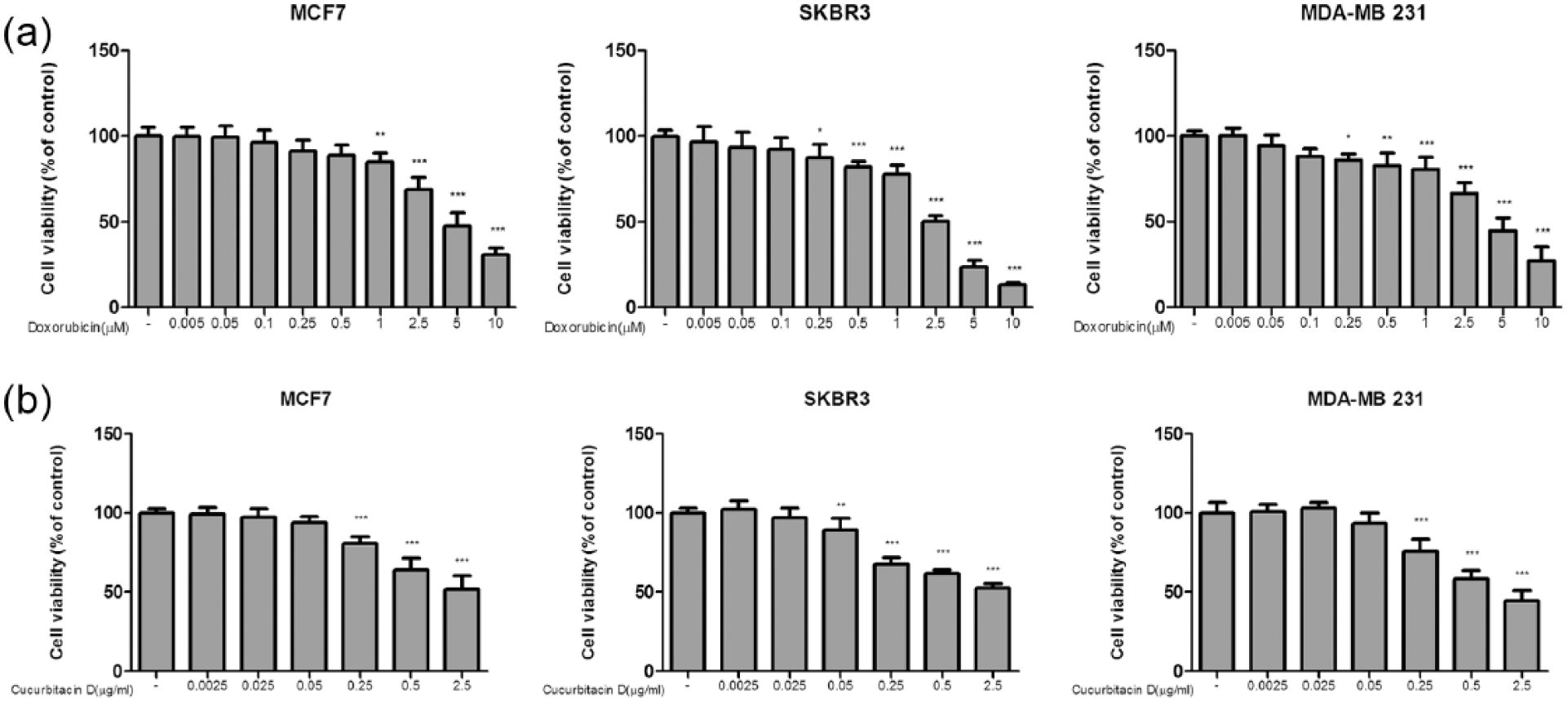
Effect of doxorubicin and cucurbitacin D on MCF7, SKBR3, and MDA-MB 231 cell viability. MCF7, SKBR3, and MDA-MB 231 cells were treated with different concentrations of doxorubicin and cucurbitacin D for 24 h. Cell viability was then measured using the MTT assay. Data are presented as the mean ± SD of three independent experiments. *P < 0.05; **P < 0.01; ***P < 0.001 by Student t-test.
Cucurbitacin D and doxorubicin increased p53 expression in MCF7, SKBR3, and MDA-MB-231 cells
We investigated whether doxorubicin and cucurbitacin D increased p-p53 levels in MCF7, SKBR3, and MDA-MB-231 cells. p53 has different multiple biological functions because it can regulate the expression of over 100 genes. p53 is a tumor suppressor gene that induces apoptosis, cell cycle arrest, and DNA repair. 32 For this experiment, we treated MCF7, SKBR3, and MDA-MB-231 cells with cucurbitacin D (0.5, 1, 2, and 4 μg/mL) or doxorubicin (0.25, 0.5, 1, and 2 μM) and performed western blot analyses. We found that cucurbitacin D and doxorubicin increased the levels of p-p53 in MCF7, SKBR3, and MDA-MB-231 cells (Figure 2(a)–(c)).

Cucurbitacin D and doxorubicin increased p-p53 levels in MCF7, SKBR3, and MDA-MB-231 cells. (a) MCF7, (b) SKBR3, and (c) MDA-MB-231 cells were treated with different concentrations of doxorubicin and cucurbitacin D for 24 h. Whole-cell lysates were analyzed by western blot to measure p-p53.
Cucurbitacin D suppressed Akt, NF-κB, and Stat3 expression in MCF7, SKBR3, and MDA-MB-231 cells
We investigated whether doxorubicin and cucurbitacin D inhibit Akt and Stat3 signaling in MCF7, MDA-MB-231, and SKBR3 cells. Activated-Akt, activated-NF-kB, and phospho-Stat3 play a critical role in cancer cells by increasing migration and invasion. Thus, Akt, Stat3, and NF-κB expression levels are very important in cancer therapy.33–35 For this experiment, we treated MCF7, MDA-MB-231, and SKBR3 cells with cucurbitacin D (0.5, 1, 2, and 4 μg/mL) or doxorubicin (0.25, 0.5, 1, and 2 μM) and performed western blot analyses. We found that cucurbitacin D decreased the levels of p-Akt and p-Stat3 in MCF7, MDA-MB-231, and SKBR3 cells. However, doxorubicin alone did not decrease p-Akt and p-Stat3 levels (Figure 3(a)). When we treated MCF7 cells with IL-6, we found that cucurbitacin D decreased p-NF-κB and p-Stat3 expression (Figure 3(b)).

Cucurbitacin D suppressed the expression of p-Akt, p-NF-κB, and p-Stat3 in human breast cancer cells. Constitutive activation of Akt and Stat3 was detected in MCF7, SKBR3, and MDA-MB 231 cells. Doxorubicin increased constitutive Stat3 phosphorylation in a dose-dependent manner in MCF7, SKBR3, and MDA-MB 231 cells. (a) Cucurbitacin D decreased p-Akt and p-Stat3 expression in MCF7, SKBR3, and MDA-MB 231 cells. MCF7 cells were treated with cucurbitacin D (0.5 μg/mL) and IL-6 (10 ng/mL). IL-6 increased constitutive NF-κB and Stat3 phosphorylation in MCF7 cells. (b) Cucurbitacin D suppressed p-NF-κB and p-Stat3 expression in MCF7 cells. Whole-cell lysates were analyzed by western blot with anti-pAkt, anti-Akt, anti-pNF-κB, anti-NF-κB, anti-pStat3, anti-Stat3, anti-actin, and anti-tubulin antibodies.
Combined effect of doxorubicin and cucurbitacin D on MCF7, SKBR3, and MDA-MB-231 cell viability
We investigated whether the combination of doxorubicin and cucurbitacin D affected the viability of MCF7, MDA-MB-231, and SKBR3 cells. For that purpose, we treated MCF7, MDA-MB-231, and SKBR3 cells with cucurbitacin D (0.5, 1, and 2 μg/mL) in the presence of doxorubicin (1 μM). Cell viability data from MTT assays were then analyzed by CompuSyn software to test a synergistic effect. 36 A combination of cucurbitacin D with doxorubicin showed the synergism. We found that cucurbitacin D combined with doxorubicin significantly suppressed cell growth more than cucurbitacin D alone (Figure 4(a)). Doxorubicin (1 μM) alone did not induce characteristic cell death morphology, but cucurbitacin D (2 μg/mL) alone induced characteristic cell death morphology in MCF7 and MDA-MB-231 cells. Additionally, doxorubicin combined with cucurbitacin D induced characteristic cell death morphology (Figure 4(b)).

Effects of combined treatment with cucurbitacin D and doxorubicin on morphology and viability in MCF7 and MDA-MB-231 cells. MCF7, SKBR3, and MDA-MB 231 cells were treated with different concentrations of doxorubicin, cucurbitacin D, or cucurbitacin D (0.5 and 2 μg/mL) and doxorubicin (1 μM) for 24 h. Combinational treatments of cucurbitacin D with doxorubicin. The effect is a cell viability and CI is combination index. (a) Synergistic effects were analyzed with CompuSyn software. Cell viability was then measured using the MTT assay. Indicated MCF7 and MDA-MB-231 cells were photographed using a phase-contrast microscope. (b) A change in cell morphology was observed in MCF7 and MDA-MB-231 cells (magnification ×20). Data are presented as the mean ± SD of three independent experiments. *P < 0.05; **P < 0.01; ***P < 0.001 by Student t-test.
Combination of cucurbitacin D with doxorubicin increased p-p53 levels and suppressed Akt, NF-κB, Stat3, and Bcl-2 expression in MCF7 cells
We investigated whether doxorubicin combined with cucurbitacin D increased p-p53 levels and suppressed Akt, NF-κB, Stat3, and Bcl-2 expression in MCF7 cells. We found that cucurbitacin D combined with doxorubicin significantly increased p-p53 levels and suppressed p-Akt, p-NF-κB, p-Stat3, and Bcl-2 levels more than doxorubicin or cucurbitacin D alone (Figure 5(a) and (b)).

Combined treatment with cucurbitacin D and doxorubicin suppressed p-Akt, p-Erk, p-NF-κB, and p-Stat3 expression in MCF7 cells. MCF7 cells were treated with cucurbitacin D (0.5 μg/mL) and doxorubicin (1 μM). (a) Cucurbitacin D (0.5 μg/mL) and doxorubicin (1 μM) decreased p-Akt, p-Erk, p-NFκB, and p-Stat3 expression in MCF7 cells. (b) Cucurbitacin D (0.5 μg/mL) and doxorubicin (1 μM) decreased Bcl-2 levels and increased p-p53 levels. Whole-cell lysates were analyzed by western blot with anti-pAkt, anti-Akt, anti-pErk, anti-Erk, anti-pNF-κB, anti-NF-κB, anti-pStat3, anti-Stat3, anti-Bcl-2, anti-p-p53, anti-p53, and anti-tubulin antibodies.
Discussion
In this study, we found that cucurbitacin D decreased cell proliferation by inhibiting Akt, NF-κB, and Stat3 signaling in the human breast cancer cells MCF7, MDA-MB-231, and SKBR3.
Trichosanthes kirilowii tuber extract is a traditional medicine used in East Asia for patients with diabetes symptoms. 37 Recently, it was reported that cucurbitacin D, isolated from Trichosanthes kirilowii tuber extract, induces apoptosis through inhibiting Stat3 activity in breast cancer cells. 30 In addition, cucurbitacin D is known to induce apoptosis and cell cycle arrest in ovarian, cervical, lung, and hepatocellular carcinoma.38–41 Therefore, we investigated whether doxorubicin and cucurbitacin D affected the viability of MCF7, MDA-MB-231, and SKBR3 cells. We found that cucurbitacin D and doxorubicin significantly suppressed cell growth in MCF7, MDA-MB-231, and SKBR3 cells.
Cucurbitacin B induces apoptosis in colon cancer cells expressing p53 42 and enhances the regulatory effect of p53-specific CTL in 16HBE/BPDE tumor cells by inhibiting JAK2/Stat3 activation. 43
Cucurbitacin E decreases the viability of pancreatic cancer cells by suppressing Stat3 phosphorylation and upregulating tumor suppressor p53. 44 We investigated whether doxorubicin and cucurbitacin D increased p-p53 levels in MCF7 and MDA-MB-231 cells. We found that cucurbitacin D and doxorubicin increased the levels of p-p53 in MCF7 and MDA-MB-231 cells.
Doxorubicin-treated MCF7 cells have shown to contain activated Stat3. Stat3 activation is a predictive marker of drug resistance because it is linked to the development of doxorubicin resistance in cancer cell lines.11,45,46 We found that cucurbitacin D decreased the levels of p-Akt and p-Stat3 in MCF7, MDA-MB-231, and SKBR3 cells. However, doxorubicin alone did not decrease p-Akt and p-Stat3 levels. We treated MCF7 cells with IL-6 to confirm whether cucurbitacin D inhibits the activity of NF-κB and Stat3. We found that cucurbitacin D decreased p-NF-κB and p-Stat3 levels. We investigated whether doxorubicin combined with cucurbitacin D affected the viability of MCF7, MDA-MB-231, and SKBR3 cells. We found that cucurbitacin D combined with doxorubicin significantly suppressed cell growth more than cucurbitacin D alone. Moreover, doxorubicin combined with cucurbitacin D changed the characteristic cell death morphology. Cucurbitacin D combined with doxorubicin significantly increased p-p53 levels and suppressed p-Akt, p-NF-κB, p-Stat3, and Bcl-2 levels more than doxorubicin or cucurbitacin D alone. Targeting Akt, NF-κB, and Stat3 may also be useful for treating breast cancer. Our study clearly demonstrates that cucurbitacin D inhibited Akt, NF-κB, and Stat3 activity in breast cancer cells and that cucurbitacin D combined with doxorubicin significantly suppressed cell growth more than cucurbitacin D alone.
Conclusion
Our study clearly demonstrates that cucurbitacin D inhibited Akt, NF-κB, and Stat3 activity in breast cancer cells and that doxorubicin combined with cucurbitacin D increased p-p53 levels and suppressed Akt, NF-κB, Stat3, and Bcl-2 expression more than cucurbitacin D alone. These results clearly demonstrate that cucurbitacin D could be used as a compound for treating human breast cancer.
Footnotes
Declaration of conflicting interests
The author(s) declared no potential conflicts of interest with respect to the research, authorship, and/or publication of this article.
Funding
This research was supported by a grant from the Korean Medicine R&D Project of the Ministry of Health and Welfare (B110043).