
Abstract
Background:
Gelsolin (GSN) is an actin-binding plasma protein with a pivotal role in the systemic response to acute tissue damage. The present study investigated GSN expression in the liver, spleen and blood serum in mice after burn.
Method:
A murine model of thermal injury was selected, and the animals were sacrificed at 8, 24, 48 and 72 h after injury. Real-time quantitative polymerase chain reaction (RT-PCR) was performed to determine the messenger RNA (mRNA) expression of GSN, and GSN protein expression was determined by enzyme-linked immunosorbent assay (ELISA).
Results:
We found that GSN mRNA and protein were expressed in the liver, spleen and blood serum of the mice. GSN expression in these tissues was the lowest among the tested time points at 8 h after burn injury. The mortality within 72 h among the mice subjected to burn injury was significantly lower in those treated with GSN than in those not treated with GSN. Treatment with GSN markedly increased the GSN levels in the liver, spleen and blood serum after injury.
Conclusion:
These results indicated that GSN treatment may affect the outcome of thermal injury via changes in the GSN content of multiple tissues.
Introduction
Severe burn injury can induce life-threatening systemic infections, also known as sepsis, as well as dysfunctions of multiple organs including the heart, lung, spleen, intestine, liver, brain and others. 1 The combination of systemic infection and visceral dysfunction can aggravate septic shock, leading to the formation of a vicious cycle that can result in death. Sepsis, as a leading cause of high morbidity and mortality among patients with critical illnesses, represents a complex clinical syndrome that results from serious infection followed by an amplified and dysregulated inflammatory response.2–4
To date, the progression of septic syndrome secondary to extensive burns is not well understood. Recently, gelsolin (GSN) levels have been reported to be altered in a number of disease conditions, and its depletion among tissues in burn cases and sepsis models has supported it to be a prognostic marker of general systemic health. 5 The administration of recombinant GSN to mice with cecal ligation and puncture-induced sepsis did not decrease the total amount of actin in plasma. However, the proportion of soluble to insoluble actin increased, indicating that the actin might have been cut and depolymerized by plasma GSN.6,7 Several lines of evidence have suggested that the administration of recombinant plasma-type GSN led to improvements in serious conditions including organ failure, lung injury, inflammatory response and microcirculation disorder caused by severe burns, trauma or hypoxia.8,9 However, it is not clear whether decreased GSN levels might be related to multiple organ failure and improve the outcome of sepsis.
Therefore, in the present study, we used a murine model of thermal injury and measured messenger RNA (mRNA) and protein expression of GSN in the liver, spleen and blood serum at different time points post injury in order to gain an improved understanding of the relationship between GSN and tissue damage after severe burns.
Material and methods
Experimental animals
All experimental manipulations were performed in accordance with the guidelines of Shandong University Animal Ethical Committee. Male C57BL/6 mice were purchased from Shanghai Laboratory Animal Center of the Chinese Academy of Science (Shanghai, PR China) and were aged 8–10 weeks at the time of entry into the study.
Animal thermal injury model
Male C57BL/6 mice used in our experiments (weight range, 18–22 g) were housed in separate cages in a temperature-controlled room. All animals had free access to water but were fasted overnight, and the hair on the animals’ backs was removed with 20% (w/v) sodium sulfide before the experiment. Mice were anesthetized and the dorsal and lateral surfaces of their bodies were shaved. Subsequently, the exposed back skin of the mice was immersed into water heated to 96°C for 8 s. The thermal injuries were confirmed by pathological examination. Sham-injured mice were subjected to all of the procedures except that the water used for immersion was at room temperature. 10 Lactated Ringer’s solution was administered intraperitoneally for initial resuscitation 6 h after injury, and then 1.0 mL of the same solution was administered at 12 and 24 h after thermal injury.
Experimental design
A total of 120 mice were used for the in vivo experiments and were randomly divided into three groups as follows: sham group (40 mice), burn group (40 mice), and burn with GSN (0.5 mg) treatment group (40 mice). All these groups were further divided into four subgroups of 10 mice each and animals of all groups were sacrificed at 8, 24, 48 and 72 h after thermal injury. GSN (0.5 mg) (Sigma-Aldrich, St. Louis, MO, USA) was injected into the tail veins of mice in the burn + GSN group. Spleen, liver and blood serum samples were harvested and stored immediately at −80°C for the measurement of GSN expression.
Extraction of total RNA and real-time quantitative polymerase chain reaction (RT-PCR)
Total RNA was extracted from liver and spleen using TRIzol® kits (Invitrogen, California, CA, USA).11,12 The concentration of purified total RNA was determined spectrophotometrically at 260 nm. GSN mRNA expression in liver and spleen was quantified by SYBR® Green two-step RT-PCR. After the removal of potentially contaminating DNA using DNase I, 1 μg of total RNA from each sample was used for reverse transcription with an oligo dT and SuperScript® II reverse transcriptase to generate first-strand complementary DNA, which was used as a template for quantitative PCR (qPCR). The mRNA level of β-actin was also measured as an internal control for each sample. Primers for GSN were forward 5’-GCC AGG ATC AGT CGA C-3’; reverse 5’-AAAGGC ACT GAT TGG TGA-3’ (Table 1). 13 The thermal cycling conditions were as follows: 94°C for 5 min, followed by 40 cycles of denaturation, annealing and amplification (94°C for 30 s, 57°C for 45s, and 60°C for 1 min), and a final extension period of 7 min at 72°C. Each sample was run in triplicate. A melting-curve analysis was performed to ensure the specificity of the products.
Primer sequences used for PCR.
GSN protein measurements using enzyme-linked immunosorbent assay (ELISA)
The level of GSN protein in each tissue was measured using commercially available ELISA kits according to the manufacturer’s instructions (BioSource, Worcester, MA, USA).
Statistical analysis
Data were presented as mean ± standard deviation. Datasets were examined by one-way analysis of variance, and mean values were compared between groups using Student’s paired t test. Simple linear regression was used to determine correlation coefficients. All statistical tests were two-sided and a P value of 0.05 or less was considered to indicate statistical significance.
Results
GSN expression in the liver and spleen tissue
To determine the gene expression of GSN, total RNA was extracted from liver tissue and analyzed using RT-qPCR. It was shown that normal control animals expressed GSN mRNA in the spleen and liver, and a significant difference was found between the sham injury and thermal injury groups. To evaluate the potential role of GSN, the expression levels of GSN mRNA and protein in the spleen and liver were measured at 8, 24, 48 and 72 h after thermal injury-induced sepsis. As shown in Figures 1 and 2, compared with the results for the sham-injured group, GSN mRNA and protein expression in the spleen and liver of the thermal injury group was markedly decreased at 8, 24, 48 and 72 h after thermal injury and was the lowest among the measured time points at 8 h (P <0.05). Treatment with GSN significantly elevated GSN mRNA and protein levels in the spleen and liver compared with those in the thermal injury group (P <0.05).

The mRNA and protein expression of GSN in the liver in C57BL/6 mice (n = 40 per group). RT-qPCR and ELISA were used to detect GSN mRNA and protein levels in mice. GSN mRNA and protein expression in the liver was markedly decreased at 8, 12, 48 and 72 h after thermal injury, compared with that in the sham-injured group. Treatment with GSN significantly increased GSN mRNA and protein expression in the liver, compared with that in the burn group. Statistical significance:*P <0.05 for burn injury group vs. sham injury group; #P <0.05 for the burn injury + GSN group vs. the burn group.
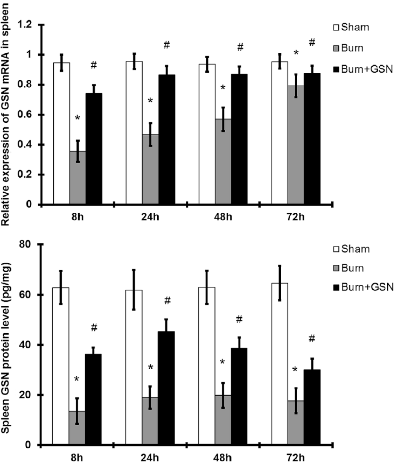
The mRNA and protein expression of GSN in the spleen in C57BL/6 mice (n = 40 per group). In vivo, GSN mRNA and protein expression shown by RT-PCR analysis and ELISA in the spleen were significantly decreased in thermal injury, compared with those in sham-injured mice. Treatment with GSN following thermal injury significantly increased the expression of GSN in the spleen, compared with that in the burn group. Statistical significance:*P <0.05 for the burn group vs. the sham injury group; #P <0.05 for the burn injury + GSN group vs. the burn group.
GSN expression in the blood serum
ELISA was used to detect GSN protein in the blood serum. As shown in Figure 3, the concentration of GSN protein in the blood serum was markedly decreased at 8, 24, 48 and 72 h after thermal injury compared with that in the sham-injured group at the same time points. The concentration of GSN in the blood serum was the lowest among the measured time points at 8 h after thermal injury (P <0.05). Treatment with GSN markedly increased the concentration of GSN protein in the blood serum at all measured time points after thermal injury (P <0.05).

The protein expression of GSN in the blood serum in C57BL/6 mice (n = 40 per group). The level of GSN protein was detected by ELISA. The expression of GSN in the blood sera of mice in the burn group was significantly lower than that in the sham-injured group. Treatment with GSN significantly increased the expression of GSN protein in the blood serum after thermal injury, compared with that in the burn group. Statistical significance:*P <0.05 for the burn group vs. the sham injury group; #P <0.05 for the burn injury + GSN group vs. the burn group.
Effect of GSN on the survival rate of the mice subjected to thermal injury
C57BL/6 mice were subjected to sepsis induced by thermal injury and GSN was administered at 8, 24, 48 and 72 h after burns. The mortality rates of the two groups were compared using the Kaplan–Meier method. As shown in Figure 4, the cumulative mortality was 80% at 72 h in the thermal injury group, whereas it was significantly decreased to70% in the thermal injury + GSN group (P <0.05). The administration of GSN (0.5 mg) improved the survival rate of mice after thermal injury.

Effect of GSN on the survival rate of the mice subjected to thermal injury.
Discussion
Recently, GSN has been found to be one of the most abundant actin-binding proteins; it controls actin dynamics and acts as a multifunctional regulator of cell metabolism and survival.14–16 The outcomes of sepsis and septic shock have not markedly improved in recent decades despite large amounts of research on the molecular pathways involved in these conditions. Recently, GSN has been found to be one of the most abundant actin-binding proteins; it controls actin dynamics and acts as a multifunctional regulator of cell metabolism and survival.15,16 A multitude of complex functions of GSN have been identified to be involved in various cellular signaling processes, including apoptosis, proliferation, differentiation, epithelial mesenchymal transition and carcinogenesis.17–19 GSN acts as an early biomarker and exogenous GSN can reduce morbidity from sepsis. 20 However, the temporal expression of GSN and the effect of exogenous GSN administration on the prognosis of burn-induced sepsis have not been fully clarified.
In this study, using RT-qPCR and ELISA, we found that GSN mRNA and protein were expressed in the liver, spleen and blood serum of mice. The current experiment was planned to examine GSN expression in different tissues after thermal injury and investigate the potential role of GSN during sepsis. Experiments in vivo showed that the expression of GSN was strongly decreased in the liver, spleen and blood serum in the burned mice, compared with that in sham-injured mice, and the lowest GSN level among all tested time points appeared at 8 h after thermal injury. Severe burn can cause low blood volume shock and, consistently with previous reports, the time period of the largest exudation amount during shock was within 8 h after burn injury. The results demonstrated that the levels of GSN present in multiple tissues might affect the outcome of thermal injury. GSN levels were found to increase in the burned mice upon treatment with GSN (0.5 mg) for 72 h. Since the levels of GSN were elevated after treatment, the increased GSN levels in the liver, spleen and blood serum might have been due to less depletion rather than increased biosynthesis of the protein. Further, the mortality rate in the thermal injury + GSN group was markedly decreased compared with that in the group subjected to thermal injury alone, suggesting that the administration of GSN (0.5 mg) could improve the survival rate of mice after thermal injury. These observations illustrated the effectiveness and importance of intervening after thermal injury with the administration of GSN. Furthermore, the results supported that the optimal time point for treatment with exogenous GSN is at 8 h after thermal injury. Further experiments are needed to provide a detailed insight into the mechanism(s) by which the levels of GSN in different tissues are related and whether and how monitoring GSN values would improve the outcome of sepsis. Nevertheless, this study has limitations. We only detected the protein expression of GSN in liver, spleen and blood serum, and we will detect the kidney, liver, even muscle and the plasma level of GSN in future. The free circulating actin as a score for the efficacy of plasma gelsolin would also be monitored. Further, the possible mechanism why increased GSN content could increase GSN mRNA expression as well as improving the survival rate of mice after thermal injury will be demonstrated in the further study.
Conclusions
Collectively, our data demonstrated that GSN was expressed in the liver, spleen and blood serum. Our results also showed that GSN levels were decreased in these tissues after thermal injury. Further, treatment with a high dosage of exogenous GSN increased GSN levels in the liver, spleen and blood serum in mice with burn-induced sepsis and improved their survival.
Footnotes
Declaration of conflicting interests
The authors declared no potential conflicts of interest with respect to the research, authorship, and/or publication of this article.
Funding
This article was supported by the Beijing Natural Science Foundation of China (Grant no.7163224) and the National Science Foundation for Postdoctoral Scientists of China (CPSF) (Grant no. 2015M572776).