
Abstract
Tea polyphenols (TP) are functional substances present in tea, which is one of the most promising preventive agents for cancer. This study was carried out to analyze the effects of TP on the ovarian cancer cells and possible mechanisms involved. TP led to inhibition of cell growth in a time- and dose-dependent manner, and promoted entry into the apoptosis-phase of the cell cycle. TP also decreased the invasion of ovarian cancer cells in vitro. In addition, TP treatment upregulated the mRNA expressions rate of Bax/Bcl-2 and downregulated Cyclin D and MMP2 mRNA expressions. Taken together, our data highlight that TP could be a potential therapeutic strategy for ovarian cancer. These findings also suggested that oncogens are involved in the anti-cancer effects of TP.
Introduction
Ovarian cancer, with the highest incidence of gynecological malignancies, is the leading cause of death of women worldwide. Despite considerable progress in recent decades in surgical oncology and the application of new radiation treatments and chemotherapy, mortality due to ovarian cancer remains high due to metastasis. Tea is consumed as a popular beverage worldwide. It is largely consumed in some Asian countries such as Japan, China, Korea, and parts of India, and a few countries in North Africa and the Middle East. 1 The consumption of green tea is also increasing in Western countries including the United States because of increasingly new investigations on its health benefits and anti-carcinogenic activities in various organs. Most of the medical activities of tea seem to be attributed to tea polyphenols (TPs). It is known to possess strong antioxidant and anti-carcinogenic activities.2–4 Numerous experimental studies demonstrate that the preventive mechanism of the TP during carcinogenesis has been suggested to be associated with inhibition of cellular proliferation and induction of apoptosis.5,6
In this study, we will investigate whether TP have the ability to regulate biological behavior of human ovarian cancer cells and whether these effects are associated with their effects on cell proliferation genes, tumor metastatic genes, and apoptosis regulation genes. Our studies will provide evidence that TP have the ability to inhibit the growth and invasion of ovarian cancer cells by targeting regulators.
Materials and methods
Materials
TP extract with a purity of 98% was obtained from Yuan Ye Bio-Technology (Shanghai, PR China). Cell Counting Kit-8 and Annexin V, FITC Apoptosis Detection Kit (Dojindo, Japan), Transwell chamber (Costar, USA), Matrigel (BD Biosciences, USA), RNA PCR Kit (AMV) Ver.3.0 and Trizol (Taraka Clontech, Japan), Thermo Scientific Maxima SYBR Green qPCR Master Mixes (Thermo Fisher Scientific, USA) were used for experiments.
Cell line
Ovarian cell lines (HO8910, SKOV3) were obtained from the Chinese Academy of Sciences, Shanghai Institutes for Biological Sciences. HO8910 and SKOV3 were cultured in RPMI-1640 supplemented with 10% fetal calf serum (FCS), 2 mM l-glutamine, 100 U/mL penicillin, 100 mg/mL streptomycin, and 50 mM 2-ME. The cell line was incubated at 37°C with 5% CO2. All mediums and supplements were obtained from Gibco BRL Life Technologies.
CCK-8 assay for cell growth inhibition
HO8910/SKOV3 cells were seeded at a density of 5×10³ cells in each well of the 96-well plates and treated with TP of different concentrations: HO8910 (10 μg/mL, 20 μg/mL, 40 μg/mL, 60 μg/mL, 80 μg/mL, 100 μg/mL), SKOV3 (100 μg/mL, 150 μg/mL, 200 μg/mL, 250 μg/mL, 300μg/mL). After 24 h and 48 h, CCK-8 (10 μL/well) was added to each well and incubated at 37°C for 4 h. Absorbance values at 450 nm were detected by the microplate reader.
Flow cytometric analysis
HO8910/SKOV3 cells were harvested directly or 24 h after TP treatment and washed with ice-cold phosphate-buffered saline (PBS). Annexin V, FITC Apoptosis Detection Kit was used to detect cell apoptosis in a FACScan instrument.
Migration assay
The migration assay was performed using Transwell chamber (pore size, 8 μm) in 24-well dishes. Approximately 2 × 10⁴ HO8910/SKOV3 cells in 100 μL of serum-free RPMI-1640 medium were placed in the upper chamber, and 500 μL of the same medium containing TP (SKOV3: 40 μg/mL, HO8910: 200 μg/mL) was placed in the lower chamber. The plates were incubated for 12 h at 37°C in 5% CO2, and then cells were fixed in methanol for 15 min and stained with 0.05% crystal violet in PBS for 15 min. Cells on the upper side of the filters were removed with cotton-tipped swabs and the filters were washed with PBS. Cells on the underside of the filters were examined under a microscope. Each clone was plated in triplicate in each experiment and each experiment was repeated at least three times.
mRNA expression measurement
Total RNA from HO8910/SKOV3 cells with TP (SKOV3 40 μg/mL, HO8910 200 μg/mL) was isolated using Trizol reagent according to the manufacturer’s recommendations. RNA PCR Kit (AMV) Ver.3.0 was used to synthesize cDNA as routine methods. Sequences of primers used to specifically amplify the genes of interest are as follows: Cyclin D: 5’-GCTGCGAAGTGGAAACCATC-3’ and 5’-CCTCCTTCTGCACACATTTGAA-3’, MMP2: 5’-TACAGGATCATTGG CTACACACC-3’ and 5’-GGTCACATCGCTCCAGACT-3’, Bax: 5’-CCCGAGVAG GTCTTTTTCCGAG-3’ and 5’-CCAGCCCATGATGGTTCTG AT-3’, Bcl-2: 5’-GGTGGGGTCATGTGTGTGG-3’ and 5’-CGGTTCAGGTA CTCAGTCATCC-3’. Amplifica-tion was performed in a thermal cycler.
Statistical analysis
All statistical analyses were performed using a statistical software package (SPSS, Inc., Version 10.0, Chicago, IL, USA). Statistical significance was defined as values of P <0.05. All P values were two-sided.
Results
Growth inhibitory effects of different concentrations of TP against ovarian cancer cells
First, we determined the in vitro growth of cancer cells upon treating with TP using CCK-8 assay. As shown in Figure 1, after 24 h, TP began to reduce the growth of HO8910 and SKOV3 cells at concentrations of 200 μg/mL and 40 μg/mL, respectively. After 48 h, an in vitro determination of the effective concentration needed for inhibition of HO8910 and SKOV3 cells was decreased. The data showed that TP inhibited growth of ovarian cancer cells in a concentration- and time-dependent manner. We found that the inhibition of 200 μg/mL TP-treated HO8910 and 40 μg/mL TP-treated SKOV3 cells were 22.36 ± 0.03% and 28.58 ± 0.03% (Figure 1). Then, the concentrations of 200 μg/mL and 40 μg/mL were chosen for following the assays of HO8910 and SKOV3 cells, respectively.
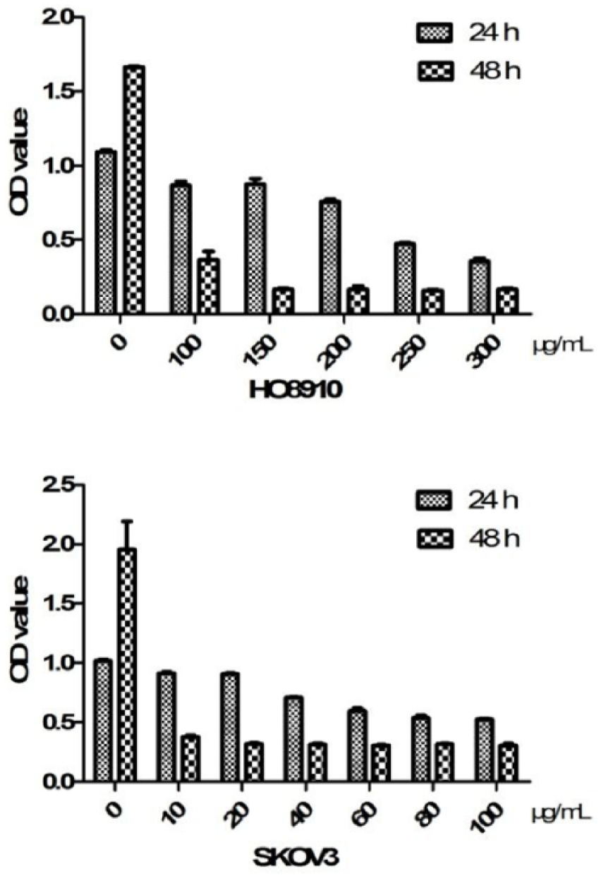
The inhibitory effect of TP on proliferation of HO8910 and SKOV3 cells. Cells were cultured in 96-well plates with TP. The CCK-8 test for cell proliferation was done at different concentrations and times of culture. The bar represents the median in each group. Results are representative of three independent experiments, P <0.05.
Induction of apoptosis by TP
We used flow cytometry to determine whether the inhibitory effect of TP on ovarian cancer cell proliferation was mediated, at least in part, through affecting cell apoptosis. We found that the apoptosis of TP-treated HO8910 and SKOV3 cells were up to 18.50 ± 1.40% and 32.00 ± 2.94%, respectively (Figure 2).

TP induced apoptosis of HO8910 and SKOV3 cells. Cells were recovered after 24 h of culture with TP and analyzed on FCM using Annexin V-PI assay. Scatter plots of the percentage of apoptotic cells were in the fourth quadrant. The bar represents the median in each group. Results are representative of three independent experiments, P <0.05.
TP affected the invasive ability of ovarian cancer cells
Then, we measured the migratory ability of TP-treated HO8910 and SKOV3 cells using the transwell assay with matrix. As shown in Figure 3, in the context of TP, HO8910 and SKOV3 showed a lower migratory ability at 24 h.

The effect of TP on the invasion of HO8910 and SKOV3 cells (100×). The cells treated with TP were in the upper chamber and the medium plus 10% FBS was added to the lower chamber. The cells on the underside of the filter were fixed.
TP regulated the gene expressions of cell proliferation, apoptosis, and invasion of ovarian cancer cells
To investigate the mechanism by which TP induces apoptosis and migration inhibition in HO8910 and SKOV3 cells, we detected expression levels of several molecules by semi-quantitative RT-PCR. As shown in Figure 4, analysis showed that TP resulted in a reduction in Cyclin D, MMP2 gene expression, and the ratio of Bax/Bcl-2 gene expression in HO8910 and SKOV3 cells.
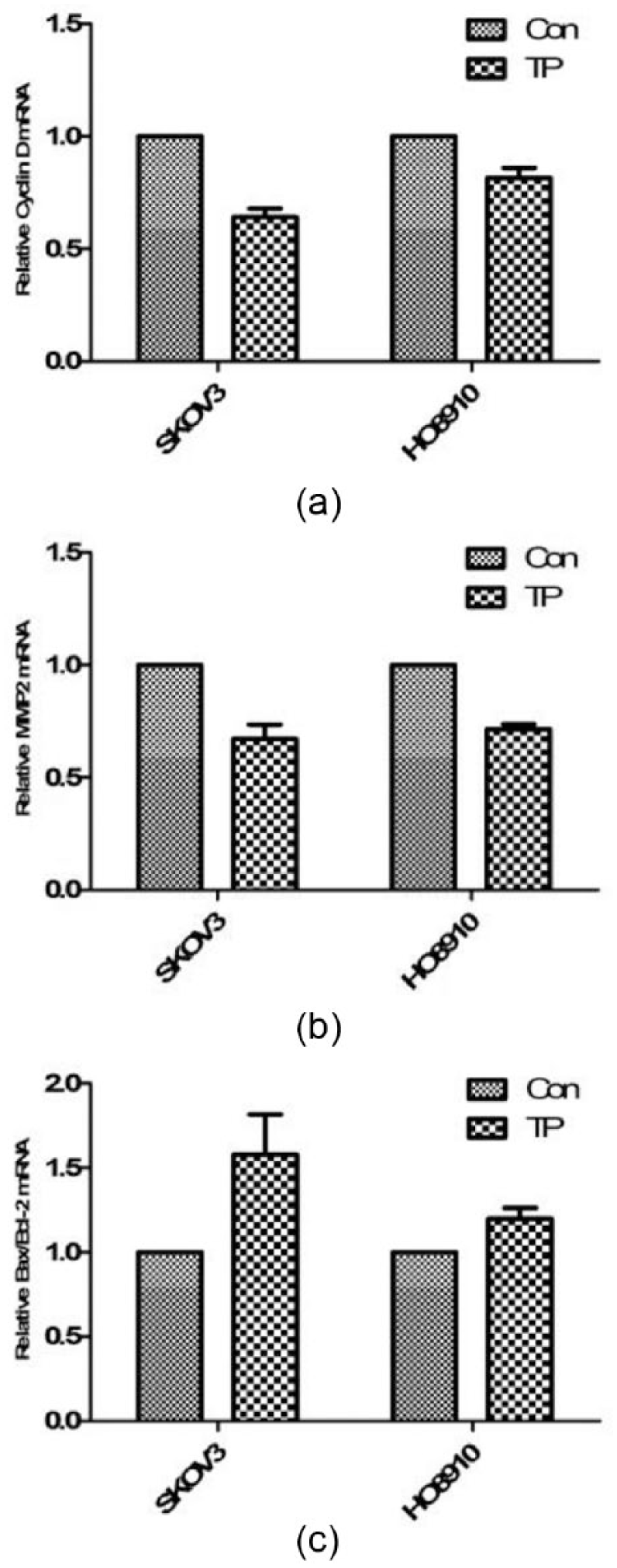
Expression of Cyclin D (a), MMP2 (b), and Bax/Bcl-2 mRNA (c) in HO8910 and SKOV3 cells after TP treatment detected by real-time PCR. Data analysis was done using the 2–∆∆CT method for relative quantification and all samples were normalized to β-actin. Results are representative of three independent experiments, P <0.05.
Discussion
Cancer constitutes one of the greatest challenges in terms of developing preventive, therapeutic, and diagnosis methods. Cancer cells have the characteristics of unlimited proliferation, immortality, and invasion, and they normally display several differences in metabolism, gene expression, and survival mechanisms. Tea is one of the most widely consumed beverages in the world. It contains biochemically active components that have been hypothesized to prevent cancer risk. Song et al. have shown that those women who drank at least one cup of green tea a day may reduce the risk of ovarian cancer. 7 As expected, tea and its components can inhibit carcinogenesis through a wide variety of mechanisms. These compounds may induce apoptosis, deregulate cell cycle, inhibit growth, proliferation, angiogenesis, enzyme activities, and gene transcription, among many other mechanisms.8–10
In this study, we investigated the function and mechanism of TP on the biological behavior of ovarian cancer cells HO8910 and SKOV3. TP inhibited proliferation and promoted the apoptosis of HO8910 and SKOV3 cells in a significant dose-dependent manner and decreased the invasion in both two cell lines. Cyclin D is a cell cycle regulator gene and it plays an important role in the proliferation of cancer cells. TP could downregulate the Cyclin D mRNA expression. Our findings showed that TP prevents ovarian cancer cells from entering the S phase, thus inhibiting obvious proliferation.
MMP2 is a member of the family of matrix metalloproteinases (MMPs) and the main enzyme of degradation of extracellular matrix and basement membrane. It plays a key role in the invasion and metastasis of cancer cells. 11 Recent studies show that overexpression of MMP2 in malignant tumor tissue is correlated with prognosis.12,13 Our findings showed that the numbers of ovarian cancer cells treated with TP penetrating the matrix are significantly decreased. Real-time PCR results also showed decreased expression of MMP2. The results suggested that TP inhibit the invasion of ovarian cancer cells by downregulating MMP2 expression.
Bcl-2 family genes are important apoptosis-related genes. Bcl-2 is an inhibitor of apoptosis proteins. However, Bax is a pro-apoptotic protein and the main apoptosis molecule in the Bcl-2 family. When Bax is combined with Bcl-2, it inactivates Bcl-2. Once Bcl-2 is neutralized and weakened, active Bax will transpose to mitochondria and stimulate mitochondrial outer membrane permeabilization (MOMP) to release pro-apoptotic molecules into proteins, thus inducing apoptosis of cancer cells.14,15 Our findings showed that TP is able to increase the rate of Bax/ Bcl-2 mRNA expression, thus inducing apoptosis.
All in all, this study demonstrated that TP can induce apoptosis and inhibit proliferation and invasion of ovarian cancer cells through regulating the expression of Cyclin D, MMP2, Bax, and Bcl-2. Thus, we can conclude that TP with general consumption, low toxicity, and anti-tumor effect will be a potentially promising therapeutic agent.
Footnotes
Declaration of conflicting interests
The author(s) declared no potential conflicts of interest with respect to the research, authorship, and/or publication of this article.
Funding
This research was supported by the Suzhou social development project (SYS201605 and SYS201338).