
Abstract
We investigated the effect of the probiotic Lactobacillus acidophilus on acute phase parameters in infected animals and to evaluate its possible use as alternative to replace the classical anti-inflammatory drugs as a trial to avoid the side effect of these drugs and its disadvantages. Forty albino rats were divided into four groups, group A was given saline orally and kept as normal-control rats, group B was orally given Lactobacillus acidophilus at a dose regimen of 108 CFU/day and kept as normal-treated rats for 6 weeks, group C was experimentally infected with Salmonella typhimurium (0.2 mL of 1.5 × 10⁸ CFU/mL) and received saline orally to be kept as diseased-control rats, while group D was orally given Lactobacillus acidophilus (108 CFU/day) for 6 weeks and experimentally infected with Salmonella typhimurium and kept as diseased-treated rats. Results of group D revealed significant decrease in ESR, fibrinogen, TIBC, UIBC, and ceruloplasmin, especially on the 34th day post infection. On the other hand, significant increase in total proteins, albumin, total iron, and transferrin saturation percentage was revealed, when compared with group C. These data indicate that the probiotic Lactobacillus acidophilus may alter acute phase proteins after infection and significantly reduce the degree of inflammation.
Introduction
Currently, the general trend in the world is the use of natural products to attain wanted therapeutic effects and to avoid unwanted adverse effects. These products have minimal side effects that are usually associated with the use of classical chemical drugs, at the same time they can eliminate diseases, reduce inflammation, and various infections of humans and animals. For example, and not as a limitation, the current treatment of inflammation with steroidal and non-steroidal anti-inflammatory drugs (SAIDs and NSAIDs) have various adverse side effects that necessitate their use with care, such as gastric ulceration, induction of asthma, bleeding disorders and renal effects, 1 immunosuppression, 2 increased intraocular pressure, cataract formation, 3 and delayed wound healing and osteoporosis, 4 Therefore, scientists must search for alternative means to these drugs which are less dangerous and still effective.
Probiotics are one of the most important of these natural products which are friendly microbiota, can boost immunity, reduce inflammation and offset side effects of antibiotics and anti-inflammatory drugs, and modulate pharmacokinetics of a drug 5 that received a lot of attention and consideration recently. A number of new studies showed that the administration of probiotics such as Lactobacillus acidophilus and Bifidobacteria spp. can prevent infection by improving the immune response and reducing excess inflammation due to the regulation of cytokine function 6 and may ameliorate the associated tissue breakdown. 7 Accordingly, probiotics may act as microbial ingredients or healthy foods which have a role in the management of inflammatory disorders. 8 Recent studies concluded that some probiotics strains, including Lactobacillus strain Lp91 has a potent anti-inflammatory property. 9
The definition of probiotics has evolved over the years, but the consensus designates probiotics as ‘nonpathogenic, live microbial, mono- or mixed-culture preparations’, which, when applied to humans or animals in high enough doses, beneficially affect the host by improving the intestinal microbial balance and its properties. 10
It is worth mentioning that scientists have observed that during bacterial or parasitic infections, physical and chemical trauma, malignant tumors, and immunologic disorders lead to a series of local and systemic reactions of the organism, the so-called acute-phase response, 11 which is the sum of the systemic and metabolic changes occurred by release of acute phase proteins (APPs) in response to inflammatory stimulus. 12 APPs are a group of blood proteins that change in concentration in animals subjected to external or internal challenges, such as infection, inflammation, surgical trauma, or stress. One of the characteristic changes of this phase is the intensification or retardation of synthesis of APPs. APPs are blood proteins primarily synthesized by hepatocytes as part of the acute phase response. 13 It was shown that in mammals, higher acute phase response is induced more by bacteria and their components than by viruses. 14 Proteins with a transient increase synthesis and plasma concentration are called positive APPs, whereas proteins whose synthesis decreases are referred to as negative APP. 11 Most of the positive APPs have important host protective anti-inflammatory or antimicrobial functions; 15 whereas the function of negative acute phase proteins is associated with maintenance of homeostasis. 16 APPs have been studied widely in human medicine, especially as biomarkers of diseases, inflammatory processes, and various infections, to diagnose and monitor the success of diseases in clinical practice as reported by Endre and Westhuyzen, 17 where the practical uses and advantages of APPs assays have been reported and mentioned in various scientific reports and papers which published in the last few years.
Salmonella infection is considered one of the most dangerous infections in both human and animal fields. It causes severe illnesses in human that may be life-threatening resulting in thousands of deaths in humans worldwide. 18 Human Salmonellosis is related with society costs like medical costs, the value of time lost from work, the value of premature death, etc. It also causes large economic losses in animals. So controlling Salmonella infection by safe yet effective means is, therefore, a must.
Therefore, the present study was conducted to investigate the possible effect of probiotics on APP parameters in rats experimentally infected with Salmonella typhimurium and to evaluate their ability to reduce the degree of inflammation associated with infection indicated by decrement of the markers of inflammation.
Experimental
Animals
Forty albino rats weighing in the range of 200–250 g were used in this study. The rats were cared for in accordance with the guidelines for the care and use of laboratory animals stated by The European Commission Directive 86/609/EEC for ethics of animal experiments. 19 The rats were maintained in standardized conditions in terms of food, water, light, and temperature. All the animals were fed on normal balanced diets and water ad libitum, and were kept in suitable cages at room temperature. Excreta of the rats were cleaned regularly and hygienic and sanitary measures were adopted.
Diets and experimental design
Animal basal diets were formulated according to NRC 1992 20 (Tables 1 and 2). Animals were housed in individual suitable cages; and then after 1 week of the adaptation period, the rats were divided into four groups (of 10 rats each) as follows: Group A included 10 rats given saline orally and kept as a normal-control group; group B included 10 rats orally given Lactobacillus acidophilus by gavage at a dose regimen of 108 CFU/day, from the first day till the end of experiment (6 weeks) and kept as a normal-treated group; Group C included 10 rats experimentally infected with 0.2 mL (1.5 × 10⁸ CFU/mL) of Salmonella typhimurium on the 7th day of the experiment and received saline orally to be kept as diseased-control group; and Group D included 10 rats orally given Lactobacillus acidophilus (108 CFU/ day) by gavage from the first day till the end of experiment (6 weeks) and experimentally infected with 0.2 mL (1.5 × 10⁸ CFU/mL) Salmonella typhimurium on the 7th day of the experiment and kept as diseased-treated group.
Ingredient composition (%) of the basal diet.

The calculated chemical analysis (%) of the basal diet.

Probiotic bacteria
Freeze-dried Lactobacillus acidophilus bacterial cell culture as an example of probiotics was obtained from a food additives company and was given orally to the rats in groups B and D after the first day of the experiment till the end of experiment. The preparation of bacteria was according to the method described by Heon et al. 21 Briefly, in a sterile Erlenmyr’s flask, 5 gm of freeze-dried Lactobacillus acidophilus culture was mixed with 45 mL of sterile broth and incubated anaerobically at 37°C for 48–72 h. Then counting of the colony forming unit per mL of the incubated broth was performed using MRS agar which was incubated at 37°C for 48–72 h; then dilution was made in 1 L of brain heart infusion broth to give a final concentration of 108 CFU/mL. Prepared bacterial suspensions were stored at 4°C till the beginning of the experiment.
Experimental infection by microorganism and induction of acute inflammation
Inflammation was induced in each rat of groups C and D by oral gavage of a single dose of the prepared Salmonella typhimurium ATCC14028 suspension (0.2 mL of 1.5 × 10⁸ CFU/mL) on the 7th day of the experiment period.
Blood sampling
Two blood samples were collected from the retro-orbital venous plexus located at the medial canthus of the eye by means of capillary tubes. Blood samples were collected from all rats of all groups on the 14th, 24th, and 34th days post infection and divided as follows:
- First part: 0.5 mL of blood was received on dipotassium EDTA and used for measuring ESR and for separation of plasma to measure the total protein and fibrinogen.
- Second part: 0.5 mL of blood was left for 1 h to clot and then centrifuged at 3000 rpm for 15 min for separation of clear serum that was stored at −20°C until use for estimation of biochemical parameters.
Clinicopathological assays
Hematological assay
ESR estimation
ESR was determined by filling a Wintrobe hematocrit tube as if for packed cell volume determination, and allowed to stand for 1 h. The level of the top of the erythrocytes column was recorded and read from the scale on the left side. The measurement is recorded as number of millimeters according to Thrall. 22
Biochemical assays
Acute phase reactants assays
Albumin, total proteins, ceruloplasmin, and iron were assayed by using commercial kits (Stanbio-laboratory, USA), which were purchased from Gama Trade Company for chemicals. Fibrinogen concentration was estimated by heat precipitation test according to the methods described by Thrall, 22 and then the fibrinogen concentration is calculated by subtracting the total proteins value of the plasma in heated tube at 56°C for 10 min from that of unheated normal plasma. Plasma total proteins were determined by the reaction described by Weichselbaum. 23 In this method, copper ions in alkaline media react with peptide bonds of proteins producing violet color, which is proportional to the amount of protein present in the plasma sample when was measured colorimetrically at 550 nm. Albumin was determined by colorimetric method according to Doumas. 24 Serum total iron concentration was determined by the methods described by Tobacco et al. 25 TIBC was determined by the method described by Tietz. 26 UIBC was measured by the traditional calculation method described by Siek et al., 27 using the unsaturated iron-binding capacity (UIBC) and serum iron by the following equation: (TIBC μg/dl = UIBC μg/dl + serum iron μg/dl), so (UBIC μg/dl = TIBC in μg/dl - Iron in μg/dl). Concerning ceruloplasmin, the method employed in this work was based on that was described by Sunderman and Nomoto 28 with the modifications of Lewis et al., 29 in which, ceruloplasmin catalyses the oxidation of p-phenylenediamine (PPD) to yield a purple colored product. The color intensity is proportional to the concentration of ceruloplasmin in the sample. 28 Transferrin saturation % was determined by calculating from serum iron levels and serum total iron binding capacity levels with the following formula that was reported by Voyvoda et al.: 30 Transferrin saturation (%) = (total iron / TIBC) × 100.
Histopathological examination
Autopsy samples were taken from the liver and intestine of rats in different groups at the end of experimental period and fixed in 10% formol saline for 24 h. Washing was done under tap water and then samples were subjected to serial dilutions of alcohol (methyl, ethyl, and absolute ethyl alcohols) for dehydration. Specimens were cleared in xylene and embedded in paraffin at 56°C in hot air oven for 24 h. Paraffin tissue blocks were prepared for sectioning at 4 microns thickness by rotary microtome. The obtained tissue sections were collected on glass slides, deparaffinized, stained by hematoxylin and eosin, and examination was performed by the light electric microscope. 31
Statistical analysis
Data were expressed as mean ± SE of the mean. Data analyses were performed using SPSS statistical software program (SPSS Inc., SPSS v20 for Windows, Chicago, IL, USA, 2011). 32 One way ANOVA was used to calculate whether there was a difference between treatments. LSD post-hoc multiple-comparison test was used to evaluate the statistical difference among experimental groups. Differences between means at P <0.05 were considered significant.
Results
Hematological study
The data of ESR of different experimental groups are demonstrated in Table 3. As shown, there was a significant increase in ESR in rats of group C during the period of the experiment, compared to rats in group A, while oral supplementation of Lactobacillus acidophilus probiotics to the rats in group D produced a significant decrease in ESR values at the end of experimental period on the 34th day post infection, when compared with group C.
Effect of Lactobacillus acidophilus probiotic supplementation on ESR and fibrinogen concentration (means ± SE).

Significantly different from group A.
Significantly different from group C.
Biochemical study
Results of fibrinogen in different experimental groups are mentioned in Table 3. As shown, there was a significant increase in plasma fibrinogen concentration in rats of group C during the period of the experiment, compared to rats kept as control in group A, while oral supplementation of Lactobacillus acidophilus probiotics to rats in group D caused significant decrease in plasma fibrinogen concentration on the 24th and the 34th days post infection, when compared to group C.
Data of total plasma proteins of different experimental groups are demonstrated in Table 4. As shown, there was a significant decrease in plasma total protein concentration in rats in group C on the 24th and 34th days post infection, compared to rats in group A. Oral supplementation of Lactobacillus acidophilus probiotics to rats in group D caused significant increase in plasma total protein concentration on the 24th and 34th days post infection compared to group C. Serum albumin levels of different experimental groups are listed in Table 4. As shown, there was a significant decrease in serum albumin concentration in rats in group C, during the experimental period, compared to rats in group A. Meanwhile, oral supplementation of Lactobacillus acidophilus probiotics to rats in group D produced a significant increase in serum albumin concentrations at the end of experiment on the 34th day post infection.
Effect of Lactobacillus acidophilus probiotic supplementation on total proteins and albumin concentrations (means ± SE).

Significantly different from group A.
Significantly different from group C.
The data of total serum iron in different groups are illustrated in Table 5. As shown, there was a significant decrease in serum iron concentration in rats in group C during the experimental period on the 14th, 24th, and 34th days post infection, compared to rats kept as controls in group A. Meanwhile, oral supplementation of Lactobacillus acidophilus probiotics to rats in group D revealed significant increase in serum iron concentration at the end of the experimental period on the 34th day post infection, when compared with group C.The results of TIBC of different groups are illustrated in Table 4. As shown, there was a significant increase in TIBC in the rats in group C during the experimental period, compared to rats kept as a control. However, oral supplementation of Lactobacillus acidophilus probiotics to rats in group D resulted in a significant decrease in serum TIBC at the end of the experimental period on the 34th day post infection, when compared with group C. Data of UIBC of different experimental groups are shown in Table 5. As shown, there was a significant increase in UIBC in the rats in group C during the experimental period, compared to rats in group A. Meanwhile, oral supplementation of Lactobacillus acidophilus probiotics to rats in group D caused a significant decrease in UIBC on the 24th and 34th days post infection, compared to rats in group C.
Effect of Lactobacillus acidophilus probiotic supplementation on total iron, total iron binding capacity, and unsaturated iron binding capacity (means ± SE).

Significantly different from group A.
Significantly different from group C.
Changes of plasma transferrin saturation % of different experimental groups are listed in Table 6. It is shown that there was a significant decrease in serum transferrin saturation % in rats in group C during the experimental period, compared to rats in group A, while oral supplementation of Lactobacillus acidophilus probiotics to rats in group D produced a significant increase in serum transferrin saturation % at the end of experimental period on the 34th day post infection, compared to group C.
Effect of Lactobacillus acidophilus probiotic supplementation on transferring saturation % and ceruloplasmin (means ± SE).

Significantly different from group A.
Significantly different from group C.
The data of ceruloplasmin of different experimental groups are demonstrated in Table 6. It is clear that there was a significant increase in ceruloplasmin in rats in group C during the experimental period, compared to rats in control group A. Yet, oral supplementation of Lactobacillus acidophilus probiotics to rats in group D resulted in a significant decrease in plasma ceruloplasmin concentration at the end of experimental period on the 34th day post infection, when compared with group C.
Histopathological study
The histopathological examination of the liver and intestines of rats in control group A and rats in orally supplemented Lactobacillus acidophilus probiotics group B revealed no abnormal changes. Meanwhile, the examined organs of rats in groups C and D showed histopathological alterations which were more severe in group C, when compared with those observed in group D. In groups A and B, no obvious histopathological lesions were recorded in both livers and intestine (Figures 1a, b, 2a, b); while in group C, the liver exhibited severe inflammatory reaction with degenerative changes involved the most hepatic cells (Figure 3a) and the intestine exhibited mononuclear cellular infiltration of the intestinal wall mainly lymphocytes (Figure 4a). Meanwhile, the liver of group D demonstrated less hepatic degenerative changes, just congestion was observed in the central veins and sinusoids (Figure 3b) and no histopathological alteration observed in the intestine as recorded in Figure 4b.

The histopathological examination of liver (a) in rats of control group and the effect of Lactobacillus acidophilus probiotic supplementation (b) on the histological structure of the liver of albino rats. (n = 10).
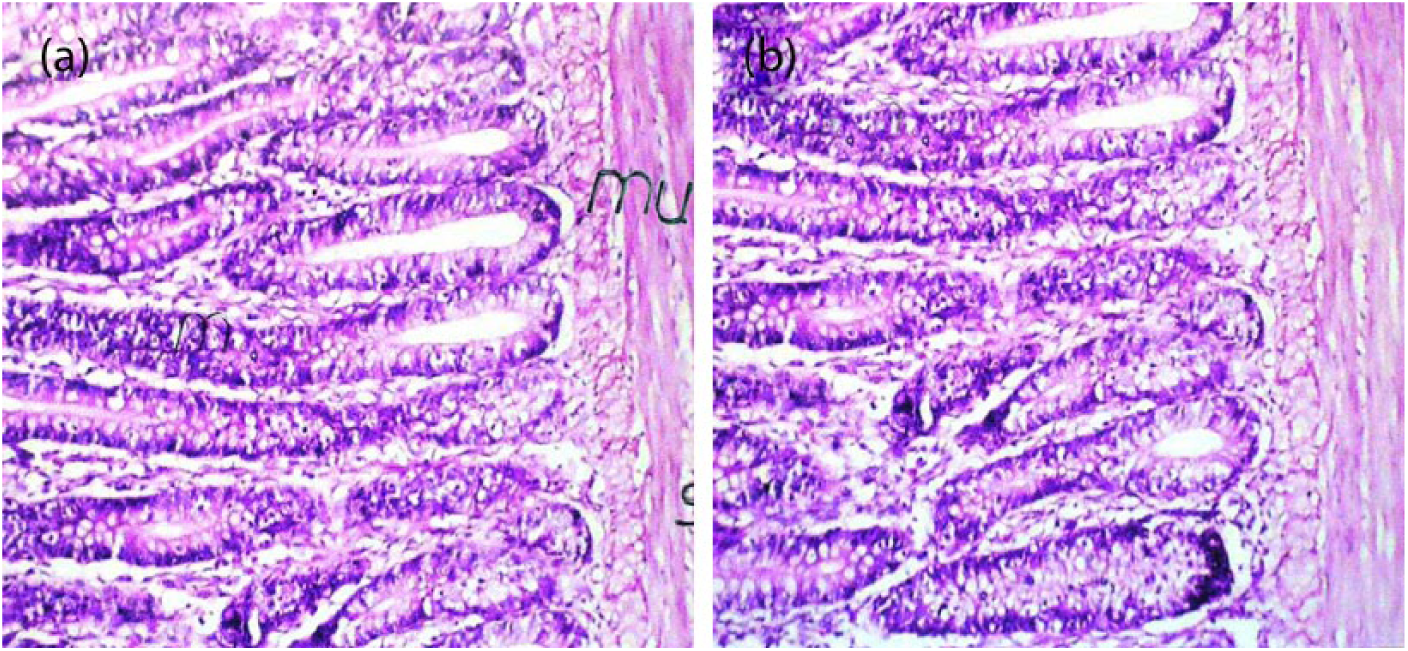
The histopathological examination of intestine (a) in rats of control group and the effect of Lactobacillus acidophilus probiotic supplementation (b) on the histological structure of the liver of albino rats. (n = 10).
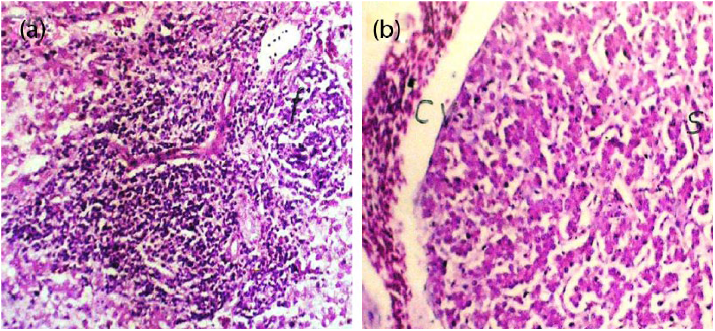
Effect of experimental infection with Salmonella typhimyrium (a) and the ameliorating effect of Lactobacillus acidophilus probiotic supplementation (b) on the histological structure of the liver of albino rats. (n = 10).
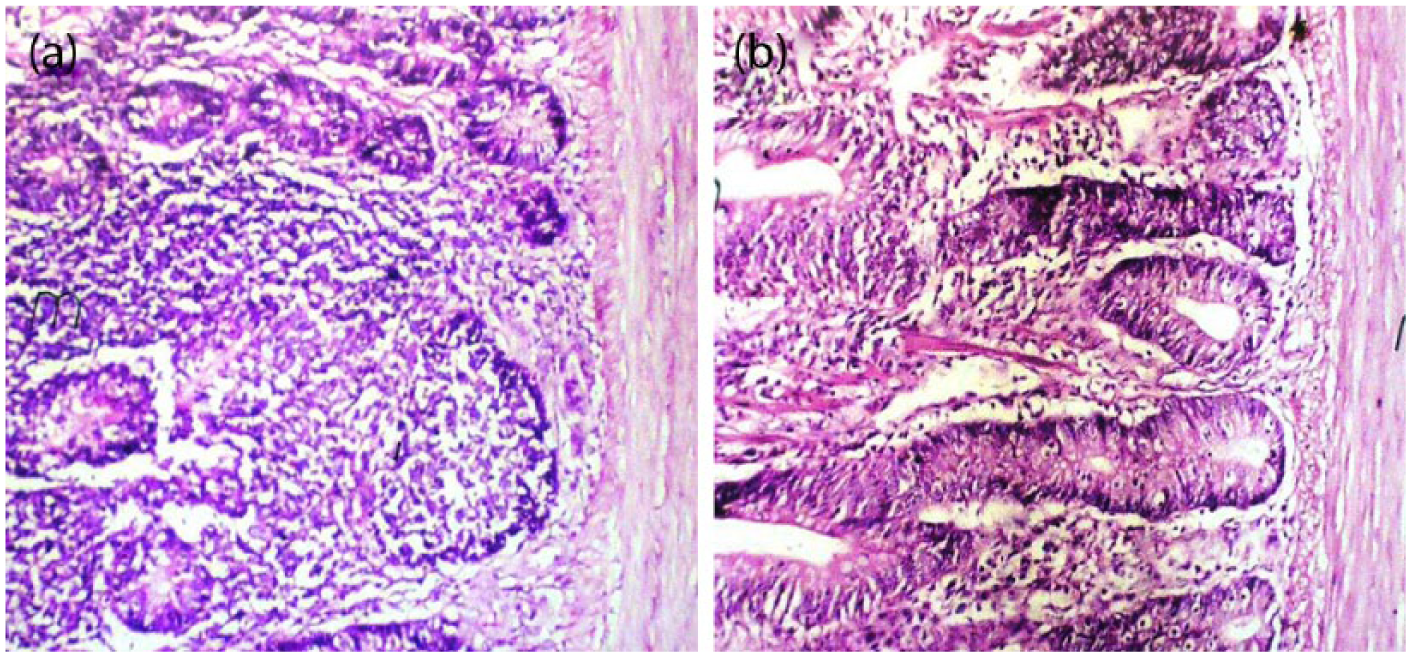
Effect of experimental infection with Salmonella typhimyrium (a) and the ameliorating effect of Lactobacillus acidophilus probiotic supplementation (b) on the histological structure of the intestine of albino rats. (n = 10).
Discussion
In Table 3 the significant increase of fibrinogen may be attributed to the inflammation and tissue damage caused by Salmonella typhimurium as fibrinogen is one of plasma proteins secreted by hepatocytes and its level is increased (positive APP) approximately 90 min after onset of a systemic inflammatory reaction. 33 Group D showed a significant decrease on the 24th and 34th days post infection. This may be related to the role of Lactobacilli in reducing the fibrinogen in blood where the consumption of probiotics can help in decreasing the level of interleukin-6 (IL-6), which in turn leads to reduction in the fibrinogen level in the blood. 34 ESR results revealed a significant increase in group C, which may be expected after increment of fibrinogen, where during the acute phase response, plasma viscosity increases as a result of the total changes in total blood protein concentration, among which is an increase of fibrinogen, which influences the ESR. 35 As for group D, the ESR result revealed a significant decrease. Such a decrease might be related to the observed decrease of fibrinogen concentration in plasma, and this may be due to the role of Lactobacillus probiotics in decreasing the degree of inflammation and infection.
In Table 4, hypoproteinemia in group C may be due to loss of protein through inflamed intestine and decreased liver production of protein. These results are in line with those recorded in intestinal and hepatic lesions, while hyperproteinemia in group D may be due to the role of probiotics in ameliorating the lesions in the liver and intestine as shown in Figures 3b and 4b, and thus may improve absorption and metabolism of protein. Concerning albumin concentration, the decrease in albumin concentration in group C is expected after the recorded decrease of plasma total proteins, and these results confirmed by Aldred and Schreiber 36 who mentioned that the demand for amino acids for synthesis of the positive APP is markedly increased, which necessitates reprioritization of the hepatic protein synthesis and albumin synthesis is downregulated and amino acids are shunted into synthesis of positive APPs. Also, hypoalbuminemia may be also related to loss of albumin through the lesioned intestine as observed in Figure 4a and also as a result of decreased production of albumin by liver due to hepatic lesions as observed in Figure 3a. The results of group D showed a significant increase in serum albumin concentration on the 34th day post infection. It is possible that this increase occurs as a result of increased plasma total proteins that were observed in this group. Also, it may be attributed to the improvement of absorption via intestine and synthesis via the liver of albumin as a response of ameliorating hepatic and intestinal lesions (Figures 3b and 4b) upon reaching a lower degree of inflammation.
As shown in Table 5, a significant decrease in serum concentration of total iron in rats in group C, which may be due to that during acute phase response a series of changes can be measured as: decreased serum levels of iron, 14 while the increase of iron concentration in group D on the 34th day post infection is possible after the improvement of the microscopic lesions of the examined liver and intestine (Figures 3b and 4b), that the intestine is the site of absorption and the liver is the site of storage of iron. As for the significant increases of TIBC and UIBC in group C, these results agree with the those of Tietz et al., 26 and these increments in TIBC and UIBC were accompanied with total iron decline. However, on the 34th day post infection we noted significant decreases in TIBC and UIBC in group D.
The results of transferrin saturation percent (Table 6) revealed a significant decrease in group C. This result agrees with Ritchie et al., 37 who reported that transferrin acts as negative APP which decreases during inflammation. Lesions in liver are an additional cause as the liver is the major source of serum transferring. Also, this decrease may be observed after a decrease of iron concentration and increase of TIBC because serum saturation of transferrin is inversely proportional to TIBC which is the direct indicator of serum transferrin concentration and directly proportional to total iron concentration in serum as the equation of measurement of transferrin saturation percent is the ratio of serum iron and total iron-binding capacity, multiplied by 100. 30 Our data showed a significant increase on the 34th day post infection in group D. This increase may be expected upon the improvement of liver lesions. The result may be also related to the action of Lactobacillus probiotic in improving the lesions in the intestine with consequent increase of the iron absorption and thus increase of transferring.
The present study revealed a significant increase in ceruloplasmin concentration in group C as noted in Table 6. The aforementioned results agree with that achieved by Al-Saadi et al., 38 who mentioned that ceruloplasmin rises in plasma after tissue injury, and acts as a protecting tool for the organism from the effects caused by the release of free radical oxidation products; while in group D a significant decrease in ceruloplasmin concentrations may be associated with the reduction of the degree of inflammation by Lactobacillus probiotics as clarified by histopathological data. The reduced tissue injuries will be associated with lowered free radical product releasing and decreased ceruloplasmin synthesis by the liver where there is no need for the antioxidant activity of ceruloplasmin.
In conclusion, we reported beneficial role of probiotics on the modification of acute phase parameters during the inflammation such as decreased fibrinogen, ESR, TIBC, UIBC, and ceruloplasmin and on one hand increased albumin, total protein, iron, and transferrin saturation percentage. Further studies will be necessary to clarify and explain the mode of action of probiotics on acute phase parameter modification.
Footnotes
Acknowledgements
We are thankful to Dr Farhat Afrin for her time and effort in correcting this manuscript and her inputs in language correction; and Mr. Mohamed Ali Abdelrahman, MD, for his assistance with language checking.
Declaration of conflicting interests
The author(s) declared no potential conflicts of interest with respect to the research, authorship, and/or publication of this article.
Funding
This research received no specific grant from any funding agency in the public, commercial, or not-for-profit sectors.