
Abstract
Adenomyosis is a uterine condition in which endometrial glands and stroma are commonly pathologically observed in the myometrium. In this study, we sought to determine the effect of resveratrol on the progression of adenomyosis. Adenomyosis was induced in mice given tamoxifen neonatally. All mice were subjected to body weight measurement and hotplate testing every four weeks beginning four weeks after birth. All mice with adenomyosis were randomly separated into 3 groups at 16 weeks: untreated, low-dose resveratrol (25 mg/kg), and high-dose resveratrol (50 mg/kg). After 3 weeks of treatment, final hotplate test and body weight measurement were performed, and the uterine horn blood samples were collected. Adenomyosis in mice caused body weight loss and uterine weight gain, reduced hotplate latency, and progression of endometrial fibrosis. The underlying biological process could be coupled with the overexpression of many cells’ proliferation and immune-regulation–related genes. Resveratrol treatment could slow the progression of adenomyosis by enhancing hotplate latency, lowering endometrial fibrosis, and restoring cell proliferation- and immune-regulation-associated gene expression levels in endometrium and plasma. However, resveratrol treatment also reduced the body weight and uterine weight. In conclusion, Resveratrol might be a potential compound for treating patients with adenomyosis.
Introduction
Adenomyosis, like endometriosis, is a prevalent gynecologic condition, and the pathogenesis of adenomyosis is poorly understood.1,2 A recent paper revealed that tissue injury and repair is shown to be the most reasonable explanation for the emergence of the adenomyosis because the etiology of archimetrosis is linked to the evolution of the stratum vasculare. 3 Adenomyosis exhibits many similarities to endometriosis in terms of estrogen reliance, symptomology, progesterone resistance, and various molecular abnormalities, but it differs in risk factors, onset age, and etiology.4,5 Our current understanding of the mechanisms causing adenomyosis-related pain is severely poor. As a result, adenomyosis treatment has proven challenging. 6 Hysterectomy has been used as a preferred therapy for severe symptomatic adenomyosis.7,8 Medical treatment such as chronic usage of oral contraceptives, high-dose progestins, selective estrogen receptor modulators, aromatase inhibitors, and gonadotrophin receptor hormone agonists can temporarily reverse adenomyosis and alleviate symptoms.4,9 A novel therapy for adenomyosis is clearly an unmet medical need that has yet to be satisfied.
We previously demonstrated that treatment of mice with induced adenomyosis using andrographolide, a nuclear factor κB (NF-κB) inhibitor, extracted from the medicinal herb Andrographis, or levo-tetrahydropalmatine, an extraction from analgesia, was able to decrease uterine contractility, reduce myometrial infiltration, and mitigate generalized hyperalgesia in mice with adenomyosis.10,11
Resveratrol is a polyphenol commonly found in various berries, grape skins, and deep-colored fruits and vegetables. It has been demonstrated to exert its beneficial effects (i.e., anti-cancer, anti-oxidation, or anti-inflammation) via modulating a variety of pathways, making it a multitargeted treatment for chronic illnesses.12-14 In addition to its antiangiogenic effect, as shown in cancer research fields, the resveratrol has various other favorable features. It has been reported that resveratrol can suppress DNA methylation and deacetylase activity through inhibition of DNA methyltransferases (DNMTs), methyl-CpG-binding domain protein 2, and pan-histone deacetylase. 15 Resveratrol was able to activate progesterone receptor isoform B and inhibit NF-κB activation and cyclooxygenase-2 expression, leading to blocked cell proliferation of ectopic endometrial tissues.16,17 Notably, numerous preclinical studies have proven that resveratrol might be a potential agent for the prevention and treatment of endometriosis.
We hypothesized that resveratrol might suppress the progression of adenomyosis through modulating genes that are typically elevated in endometriosis and adenomyosis. This research was conducted to prove this hypothesis.
Materials and Methods
Mouse Model of Adenomyosis
Pregnant ICR mice at the gestational age of around 15–16 days were acquired from Shanghai Laboratory Animal Corporation (Shanghai, China). Each mouse was kept in a single cage for the remainder of the gestation, delivery, and nursing period. Their pups were sexed one day after birth, and all the female pups were chosen for this study. The dam and her offspring were housed in the same cage until they were weaned. Experiments were approved by the ethics committee of Wenzhou Medical University.
Adenomyosis was stimulated in female neonatal mice by orally administering 1 mg/kg tamoxifen suspended in a mixture of condensed milk/peanut oil/lecithin at a 3:2:0.2 ratio by volume at a dosage volume of 5 μl/g bodyweight from post-delivery day 2 to day 5. In contrast, the control neonate mice were given a solvent without tamoxifen. These female mice were weaned and separated from their dams when they reached the age of 3 weeks. Starting at four-week age, all mice were given a hotplate test at a four-week interval. At sixteen (16)-week of age, all tamoxifen-treated female mice were randomly divided into 3 groups. The untreated group of mice received the vehicle. Another two groups were intraperitoneally (i.p.) given either low-dose resveratrol (25 mg/kg) or high-dose resveratrol (50 mg/kg) daily for 3 weeks. After the final hotplate test and body weight measurement were performed, the blood samples were collected. All mice were sacrificed by perfusing formalin into heart. Each mouse’s uterus was weighed, and the uterine horn was collected.
Immunohistochemistry Staining and Masson Staining
The Immunohistochemistry (IHC) method was described previously. 18 Briefly, the ectopic endometrium tissues were embedded in the optimal cutting temperature (OCT) compound. For each block, serial 6 μm slices were performed on a cryostat. The sections were immersed in block buffer (goat anti-mouse serum) for 15 mins before being incubated with primary antibody overnight at 4°C. After rinsing with phosphate-buffered saline (PBS), the sections were incubated with secondary antibodies for one hour before being washed with PBS and mounted. The primary antibodies were purchased from Abcam.
The fibers in endometriotic lesions were quantified using Masson trichrome staining. The OCT embedded endometrium tissues were stained with Trichrome Stain Kit (Connective Tissue Stain) (ab150686) (Abcam, Shanghai, China) according to the manufacturer’s instruction. Image Pro-Plus v6.0 was used to calculate the areas of the blue collagen fiber layer compared to the overall ectopic endometrium area.
Enzyme-Linked Immunosorbent Assay (ELISA) Assay
Following the manufacturer’s instruction, the plasma levels of hyaluronic acid (HA), high mobility group box 1 (HMGB1), and osteopontin were measured using HA (R&D Systems, Minneapolis, MN), HMGB1 (MyBioSource, San Diego, CA), osteopontin (R&D Systems), and ELISA kits, respectively.
Statistical Analysis
Data were presented as mean ± standard deviation (SD) with scatter plots. The difference between different groups was calculated by Student’s t test with Welch’s post hoc test, or one-way analysis of variance (ANOVA) with a Tukey’s multiple tests. P values of less than .05 were considered statistically significant.
Results
Resveratrol Treatment Reduced Body Weight and Uterine Weight
The body weight of mice was monitored at 4 weeks, 8 weeks, and 12 weeks, before and after resveratrol treatment. The results in Figure 1(A)-1(E) showed that the body weight in mice with adenomyosis (tamoxifen-treated group) was significantly and constantly lower than that in control normal mice at all time points. Furthermore, the uterine weight was markedly higher in mice with adenomyosis than in control mice (Figure 1(F)). These results of body weight and uterine weight shrinkage in mice with adenomyosis were consistent with our previous publications, partially confirming the successful establishment of mice with adenomyosis. The mice with adenomyosis were subjected to vehicle, resveratrol treatment (25 mg/kg) or (50 mg/kg) starting at week 16 until the end of the experiment. We observed that the body weight and uterine weight of mice that received resveratrol treatment (25 or 50 mg/kg) were considerably reduced when compared with untreated mice and control mice, suggesting resveratrol treatment at doses of 25 mg/kg and 50 mg/kg might exert unfavorable effects on mice with adenomyosis, leading to reduced body weight and uterine weight. Effects of resveratrol treatment at the doses of 25 or 50 mg/kg on the body weight at indicated time point and uterine weight of the mice. Body weight was measured at week 4 (A), week 8 (B), week 12 (C), before treatment (D), and after treatment (E). (F) Uterine weight was determined after the experiment. The data are mean ± SD with scatter plots. Control group: n = 10; untreated group: n = 10; 50 mg/kg resveratrol group: n = 9; 25 mg/kg resveratrol group: n = 10. *P < .05, **P < .01, ***P < .001 vs control group; #P < .05, ##P < .01, ###P < .001 vs untreated group.
Resveratrol Treatment Upregulated Hotplate Latency
The hotplate latency of mice was measured at 4 weeks, 8 weeks, and 12 weeks, before and after resveratrol treatment. Compared with control normal mice, the hotplate latency was significantly decreased in the mice with adenomyosis at weeks 4, 8, 12, and 16 (before treatment) (Figure 2(A)-2(D)). After 3 weeks of resveratrol treatment, the hotplate latency was notably upregulated compared to the vehicle treatment (Figure 2(E)). Effects of resveratrol treatment at the doses of 25 or 50 mg/kg on the hot plate latency at indicated time point in the mice. The hot plate experiments were conducted at week 4 (A), week 8 (B), week 12 (C), before treatment (D) and after treatment (E). The data are mean ± SD with scatter plots. Control group: n = 10; untreated group: n = 10; 50 mg/kg resveratrol group: n = 9; 25 mg/kg resveratrol group: n = 10. *P < .05, ***P < .001 vs control group; ###P < .001 vs untreated group.
Resveratrol Treatment Mitigated Extent of Lesional Fibrosis
Masson staining and hematoxylin and eosin (H&E) staining were conducted using ectopic endometrium tissues from four groups of mice. As shown in Figures 3(A) and 3(B), the extent of lesional fibrosis was dramatically upregulated in mice with adenomyosis compared with control normal mice and was considerably reduced after being treated with resveratrol at 25 mg/kg and 50 mg/kg. Effects of resveratrol treatment at the doses of 25 or 50 mg/kg on the histopathology and fibrosis in the mice. (A) H&E staining of the ectopic endometrium tissues in the mice, and the histopathological scores in the four experimental groups. (B) Masson staining of the ectopic endometrium tissues in the mice, and the fibrosis scores in the four experimental groups. The data are mean ± SD with scatter plots. Control group: n = 10; untreated group: n = 10; 50 mg/kg resveratrol group: n = 9; 25 mg/kg resveratrol group: n = 10. *P < .05, ***P < .001 vs control group; ###P < .001 vs untreated group.
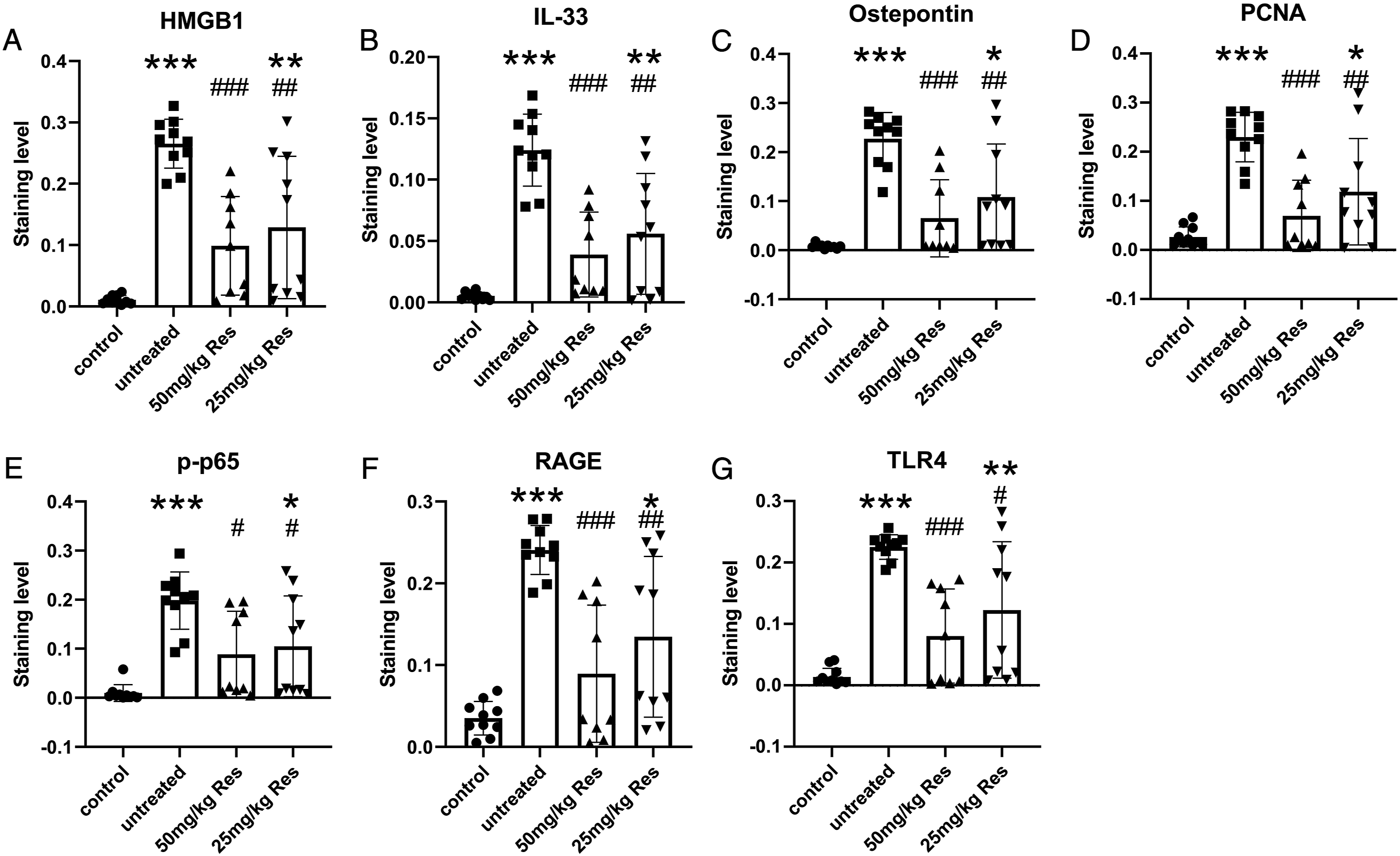
Resveratrol treatment decreased the expression levels of HMGB1, IL-33, osteopontin, PCNA, p-p65, RAGE, and TLR4
Our previous studies have shown that upregulation of several biomarkers [i.e., HMGB1, p-p65, osteopontin, receptor for advanced glycation end-products (RAGE), interleukin (IL)-33, and proliferating cell nuclear antigen (PCNA)] was revealed in the endometrium tissues of patients with endometriosis.
19
However, their expression in endometrium tissues from mice with adenomyosis was unknown. Hence, in this study, we tested the expression of HMGB1, IL-33, osteopontin, PCNA, p-p65, RAGE, and toll-like receptor 4 (TLR4) in the endometrium tissues of four groups of mice using IHC staining (Figure 4). Not surprisingly, we observed that when compared to the endometrium tissues from control normal mice, the expression of HMGB1, IL-33, osteopontin, PCNA, p-p65, RAGE, and TLR4 were substantially upregulated in the endometrium tissues from mice with adenomyosis (Figure 5(A)-5(G)). Resveratrol treatment at 25 mg/kg and 50 mg/kg were able to reduce the expression levels of HMGB1, IL-33, osteopontin, PCNA, p-p65, RAGE, and TLR4 in the endometrium tissues, suggesting that resveratrol treatment might mitigate the progression of adenomyosis through downregulation of these adenomyosis-related gene expressions. The high dose (50 mg/kg) of resveratrol exhibited a more potent gene expression inhibition effect than the low dose (25 mg/kg) of resveratrol. Representative immunohistochemistry staining of the HMGB1, IL-33, Ostepontin, PCNA, p-p65, RAGE, and TLR4 in the endometrium tissues in the mice. Immunohistochemistry staining results to indicate the staining levels of the HMGB1 (A), IL-33 (B), Ostepontin (C), PCNA (D), p-p65 (E), RAGE (F), TLR4 (G) in the mice. The data are mean ± SD with scatter plots. Control group: n = 10; untreated group: n = 10; 50 mg/kg resveratrol group: n = 9; 25 mg/kg resveratrol group: n = 10. *P < .05, **P < .01, ***P < .001 vs control group; #P < .05, ##P < .01, ###P < .001 vs untreated group.
Resveratrol Treatment Suppressed the Plasma HMGB1, HA, and Osteopontin
We recently reported that plasma HMGB1, HA, and osteopontin serve as promising biomarkers for endometriosis.
20
Whether they were able to be used as potential biomarkers for adenomyosis was not addressed. Therefore, we measured the plasma HMGB1, HA, and osteopontin levels from four groups of mice using ELISA assay. The results in Figure 6(A)-6(C) depicted that the plasma HMGB1, HA, and osteopontin levels were remarkably enhanced in mice with adenomyosis compared to control normal mice. Intriguingly, administration of resveratrol at 25 mg/kg and 50 mg/kg exerted similar effects on the downregulation of plasma HMGB1, HA, and osteopontin levels in mice with adenomyosis. Plasma concentrations of HA (A), HMGB1 (B), and Ostepontin (C) in the blood of the experimental mice, determined by ELISA assays. The data are mean ± SD with scatter plots. Control group: n = 10; untreated group: n = 10; 50 mg/kg resveratrol group: n = 9; 25 mg/kg resveratrol group: n = 10. ***P < .001 vs control group; ###P < .001 vs untreated group.
Discussion
In this study, we have shown that starting just four weeks after the induction, the mice displayed a significantly higher body weight gain rate and progressive deterioration of hotplate latency when compared to control normal mice without the induction. Furthermore, mice with adenomyosis exhibited extensive fibrosis in the ectopic endometrium, as demonstrated by Masson staining and H&E staining. In contrast, no fibrotic tissue was observed in the ectopic endometrium of control normal mice. These results were consistent with our several previous publications, confirming the successful establishment of a mouse model of adenomyosis.17-22 Remarkably, we further revealed that compared with the untreated group, resveratrol treatment was capable of enhancing hotplate latency and reducing fibrosis in the ectopic endometrium of the mice with adenomyosis, although the restoration effect of resveratrol was not complete as compared to the control normal mice. Surprisingly, the resveratrol treatment resulted in substantially reduced body weight and uterine weight as compared to either the control group or the untreated group, raising potential unfavorable safety concerns regarding resveratrol to mice and humans.23,24
Our previous study reported the significant upregulation of HMGB1, TLR4, RAGE, p-p65, PCNA, IL-33, and osteopontin in endometriotic tissues from mice with endometriosis and these results were further confirmed in ovarian endometriomas from patients with endometriosis. 20 Interestingly, our results illustrated that the expression levels of HMGB1, TLR4, RAGE, p-p65, PCNA, IL-33, and osteopontin in endometriotic tissues from mice with adenomyosis were significantly higher than those in endometriotic tissues from normal mice, confirming that adenomyosis exhibits many similarities to endometriosis. Importantly, resveratrol treatment, especially high dose, potently suppressed the expression of HMGB1, TLR4, RAGE, p-p65, PCNA, IL-33, and osteopontin in endometriotic tissues from mice with endometriosis, suggesting resveratrol might decrease the progression of adenomyosis via reducing these gene expression. Because activation of RAGE, IL-33, p-p65, PCNA, and osteopontin play critical roles in promoting cell proliferation of osteoclasts and fibroblasts, and immune cell activation and infiltration.25-30 The resveratrol-induced inhibition of these gene expression might result in reduced proliferation of osteoclasts and fibroblasts and suppressed myometrial infiltration, leading to attenuating the progression of adenomyosis.
HA is a glycosaminoglycan generated by numerous mesenchymal cells and tumor cells, and it is critically important in the recruitment and activation of inflammatory cells. 31 Osteopontin is reported to be involved in the development of angiotensin II-induced fibrosis. Osteopontin has been shown to interact with a variety of extracellular matrices (e.g., collagen and fibronectin), implying a potential function in matrix organization and stability. 32 HMGB1 has a diverse set of immunological functions, including the activation of cell proliferation, differentiation, and cytokine secretion.33,34 Accumulating evidence has linked HMGB1 to liver fibrosis. As potential indicators, the plasma concentration of HA, HMGB1, and osteopontin have been associated with fibrosis.35,36 We previously demonstrated that plasma HA, HMGB1, and osteopontin could be promising biomarkers for detecting endometriosis. 37 In line with this finding, we also observed upregulation of plasma HA, HMGB1, and osteopontin concentration in mice with adenomyosis. Resveratrol treatment significantly reduced the plasma HA, HMGB1, and osteopontin concentrations, further confirming that resveratrol can slow the progression of adenomyosis.
There are several limitations should be noted. First, the adenomyosis-related gene expression is not explored here. Second, the protective effects of resveratrol in other adenomyosis models established by different methods could be verified. Last, it would be of much clinical relevance and importance if the effects of resveratrol in clinical patients with adenomyosis are studied.
Conclusion
The current study further confirms many previous reports that the induction of adenomyosis in mice results in body weight loss and uterine weight gain, reduced hotplate latency, and progressive ectopic endometrium fibrosis. The underlying molecular mechanism might be attributed to the upregulation of several cell proliferation- and immune-regulation-associated genes, such as HMGB1, TLR4, RAGE, p-p65, PCNA, IL-33, and osteopontin. We proved that administration of resveratrol was able to attenuate the progression of adenomyosis as manifested by increasing hotplate latency, decreasing fibrosis status of endometrium, and restoring cell proliferation- and immune-regulation-associated gene expression levels in endometrium and plasma. However, it also should be noted that resveratrol treatment reduced the body and uterine weight, which may be its side effects in the treatment of adenomyosis.
Footnotes
Declaration of Conflicting Interests
The author(s) declared no potential conflicts of interest with respect to the research, authorship, and/or publication of this article.
Funding
The author(s) disclosed receipt of the following financial support for the research, authorship, and/or publication of this article: This research was supported by Zhejiang Provincial Science and Technology Bureau Foundation (LGF19H040014).
Ethical Approval
Experiments were approved by the ethics committee of Wenzhou People’s Hospital. This study was performed in strict accordance with the NIH guidelines for the care and use of laboratory animals (8th edition, NIH).