
Abstract
The current study was aimed to analyze the therapeutic effect of selected medicinal plants, that is, Curcuma longa, Zingiber officinale, Trigonella graceum-foenum, Nigella sativa, and Syzygium aromaticum against carrageenan-induced oxidative stress and inflammation in rats. Phytochemical analysis revealed the presence of diverse range of bioactives. IC50 values for antioxidant assays including DPPH (2,2-diphenyl-1-picrylhydrazyl), metal chelating, ABTS scavenging (2, 2′-Azino-Bis-3-Ethylbenzothiazoline-6-Sulfonic Acid), β-carotene bleaching, and H2O2 (hydrogen peroxide) scavenging ranged from 37-294, 71-243.4, 69.66-191.8, 98.92-228.5, and 82-234.9 μg/mL, respectively. All tested plants extract were found active against tested pathogenic microorganisms with lowest minimum inhibitory concentrations. Oral administration of tested plants extracts in different doses (250, 500, and 1000 mg/kg b. w) did not exhibit any toxicological effects on hemato-biochemical profile of treated rats in comparison to control group rats. Further, plants extract exhibited considerable anti-inflammatory activity in rats paw inflammation and decreased cellular infiltration to inflammatory site in dose dependent manner. Pretreatment of animals with tested plants extract (100, 200, and 400 mg/kg b. w.) caused significant alteration in total antioxidants, oxidants, and enzymes activities in paw tissue homogenate and the effect was more pronounced at higher concentration (400 mg/kg b. w.). Results showed that tested plants extract are rich source of diverse classes of phenolics and have therapeutic potential against oxidative stress and inflammation.
Introduction
Inflammation is the body’s protective reaction against noxious stimulus, chemicals, and invading microbes, which is normally characterized by pain, redness, swelling, and heat. 1 Several studies reported that inflammation is involved in the development of several diseases including, cardiovascular disorders, cancer, aging, and others life-threatening diseases. 2 Acute inflammation is linked with the overproduction of reactive oxygen and nitrogen species, production of pro-inflammatory and inflammatory mediators, and activation of enzymes complexes. 3 The carrageenan-induced rat paw inflammation is a well-established acute model of inflammation that is broadly used to screen novel anti-inflammatory drugs. 4 Injecting carrageenan into the subplantar region of rat paw initiates a biphasic edema. 5 The earlier phase (0-1 hour) is characterized with the release of bradykinins, serotonin, histamine, and to a smaller extent PGs (prostaglandins) produced by cyclooxygenase (COX) enzymes. While the later phase observed after 1 hour is attributed to cellular infiltration (neutrophils) and PGs production. 6
Antioxidant and anti-inflammatory processes are inter-linked, which can be involved in various pathological conditions. 7 Overproduction of reactive oxygen species (ROS) during oxidative stress can be the result of inadequate antioxidant defense system. 8 Similarly, increased production of ROS occurs during inflammation which had a critical role and stimulates the formation of inflammatory mediators such as pro-inflammatory cytokines, chemokines resulting in migration of inflammatory cells. 9 Active compounds from plants are capable of modulating the oxidative stress and may act as anti-inflammatory agents contributing to the decreased concentration of inflammatory mediators. 10 Free radicals cause oxidative stress in the cells leading to infectious and inflammatory condition. 11 Phagocytes including mononuclear cells (lymphocytes and macrophage) and polymorphonuclear leukocytes (eosinophils and neutrophils) produce excessive quantity of ROS which play a major part in the defense mechanism of the host. 12 These ROS dysregulate the cellular events leading to cellular and tissue damage, which can enhance inflammation. 13
Researchers are searching for natural antioxidants with less side effects. In order to use natural antioxidants in pharmaceutical preparations and food supplements, still the search is going on to find out effective, newer, and safe antioxidants. Medicinal plants as potential supplier of natural antioxidants and bioactive compounds, provide cost effective and safe reservoir of therapeutic and preventive alternatives. Through the continuous efforts of scientific community, several plant-derived medicinal compounds have been tested and developed into effective modern drugs. Examples of these agents include plant alkaloids vincristine and vinblastine, which are extensively used in chemotherapy and originally derived from Catharanthus roseus. Etoposide is another example of non-alkaloidal anticancer agent which is derived from Podophyllum species. It is expected that with more studies focusing on medicinal plants, effective therapeutic agents with less side effects could be identified.
Materials and Methods
Preparation of Plants Extract
Medicinal plants (C. longa, Z. officinale, S aromaticum, T foenum, and N sativa) used in current work were purchased from Faisalabad’s local market and identified by the department of Botany, University of Agriculture, Faisalabad. The plants materials were grounded into fine powder to easily pass through 100 mm sieve and then extracted with green solvent ethanol/water (70:30) mixture under constant shaking for 72 hours. The filtrates were collected and reduced to semisolid form rotary evaporator (Rotavapor, Buchi R-215, Switzerland). 14
High Performance Liquid Chromatographic Analysis of Plants Extracts
HPLC was used to detect different phenolic compounds present in tested plants extract. A Shim-pack CLC ODS C-18 (2.5 cm × 4.6 mm, 5 μm diameter) column was used. Plants extracts (10 mg/mL) were prepared in methanol and acetonitrile (30:70). A 20 μL of plants extract was eluted with mobile phase consisting of A (pH = 2.27, H2O:acetoacetate, 94:6) and B (Acetonitrile 100%) which has conditions of 15% B at 0 min, changing to 45% B in 15-30 mins, and 100% B in 35-40 mins with flow rate of 1 mL/min and UV-Visible detector spectra was recorded at 280 nm of all samples. 15
Qualitative Phytochemical Analysis
Several phytochemicals including alkaloids, steroids, terpenoids, tannins, flavonoids, saponins, cardiac glycosides, phlobatannins, phenols, coumarins, sterols, and quinines were analyzed in hydroethanolic extracts of tested medicinal plants. 16
Quantitative Phytochemical Analysis
Total phenolic contents of hydroethanolic extracts of tested plants were measured by following the protocol of Liu et al. 17 Absorbances of reaction mixtures were noted at 760 nm. Results were calculated from calibration curve of gallic acid as milligram of gallic acid equivalent (mg/GAE) per gram of plant extracts. Total flavonoids contents (TFC) of tested plants extract were determined through aluminum chloride method described by Liu et al. 17 TFC were presented as microgram of CE (catechin equivalents) per gram of plants extract.
Ferric reducing power assay
Ferric reducing antioxidant ability of hydroethanolic extracts of tested plants was assessed by following the procedure described by Rani et al. 18 Absorbances of reaction mixtures were measured and ferric reducing ability of plants extract was calculated as milligram of ascorbic acid equivalents (mg/AAE).
Phosphomolybdenum Assay
The assay is based on molybdenum reduction by the plants extract which result in the development of green color complex of phosphate molybdate. 19 The reaction mixture was heated in water bath set at 95°C for 90 mins, cooled to room temperature, and absorbances of colored mixture was measured at 695 nm.
In vitro Antioxidant Activities
DPPH Inhibition Assay
DPPH (2, 2-diphenyl-1-picrylhydrazyl) radical scavenging ability of tested plants extract was evaluated by following the method of Sidiq et al. 20 The percent inhibition of DPPH was calculated against blank containing DPPH solution only.
ABTS scavenging Assay
ABTS scavenging ability of tested plants extract was measured by following the method of Abu et al. 19 Stock solution of ABTS was freshly prepared by mixing ABTS solution (7 mM) and K₂S₂O₈ (2.45 mM) and incubated for 16 h in the dark. The results of ABTS scavenging ability were presented in terms of trolox equivalents.
β-Carotene bleaching assay
β-Carotene bleaching assay was performed by following the procedure of Ismahene et al. 21 The absorbance of reaction mixture was noted at 0 min and after 2 h subsequently against blank containing emulsion without β-carotene at 470 nm.
Metal chelating ability
Metal chelating activity of tested plants extract was investigated by following the protocol of Liu et al. 17 Equal volume of plant extract, .1 mM FeSo4, and .25 mM ferrozine was mixed. The absorbances of reaction mixtures were recorded at 562 nm against blank after 10 min incubation at 37°C. Ethylenediaminetetraacetic acid (EDTA) was used as reference standard.
H2O2 scavenging assay
The H2O2 scavenging potential of plants extract was evaluated by following the protocol of Bhatti et al. 22 Different concentrations of tested plants extract (.4 mL) were mixed with .6 mL of H2O2 solution (2 mM) and phosphate buffer (50 mmol/L, pH 7.4). Absorbances of reaction mixtures were noted at 230 nm after 10 min incubation. Ascorbic acid was used as standard.
Antimicrobial Activity
Inoculum Preparation
The antimicrobial activity of tested plants extract was tested against different bacteria and fungi, that is, Pasteurella multocida, Escherichia coli, Staphylococcus aureus, Bacillus subtilis, and 3 pathogenic fungi (Fusarium solani, Aspergillus niger, and Aspergillus terreus). All microbial strains were provided by the Medicinal Biochemistry Lab., University of Agriculture, Faisalabad, Pakistan. Bacteria were pre-cultured on Muller Hinton broth at 37°C for 24 h and turbidity of overnight bacterial culture was adjusted to .5 McFarland standard with sterile nutrient broth. Sabouraud dextrose broth was used for sub-culturing of fungus at 30°C for 48 h. 23
Agar Well Diffusion Assay
Fresh agar plates were developed by pipetting 1 mL of fresh microbial culture in autoclaved agar medium and dispensing into sterilized Petri plates. After solidification, 6 mm wells were made and different concentration of each plants extract (100 μL) was added. Fluconazole and ampicillin were used as standards. The antimicrobial activity was represented in terms of zone of inhibition in millimeter (mm). 24
Determination of Minimum Inhibitory Concentration
To determine minimum inhibitory concentration of plants extracts, resazurin-based microdilution assay was performed. For MIC, 100 μL of each plant extract (50 μg/mL) was dispensed into first column of each well while 50 μL of the nutrient broth was added in column 2-10. Column 11 and 12 contained growth control (diluted standardized inoculum) and nutrient broth only (for broth sterility control). A multichannel pipette was used to transfer plant extract from column 2-10 such that each well has twice the concentration of final test. A 50 μL of bacterial suspension was added in each well. After 24 h incubation at 37°C, resazurin solution (30 μL) was added into each well and again incubated for 2-4 h. The lowest concentration before color change was scored as minimum inhibitory concentration while column with blue color were scored as above the MIC value. MBC was determined by plating contents of each well directly into sterilized agar plates. Broth microdilutions were achieved precisely according to the protocol of Clinical and Laboratory Standards Institute (CLSI). 25
In vivo Assays
The in vivo animal study was approved by Institutional Animal Care and Use Committee, University of Agriculture Faisalabad, Pakistan. Healthy albino rats of both sexes (150-200 g) were used. The animals were kept in polypropylene cages under standard conditions of 24 ± 2°C, 12 h light/dark cycle, and 35-60% humidity. Animals were provided free access to standard rodent diet with water ad libitum. All animals were acclimatized to the laboratory settings for 1 week before experimentation. 26
Evaluation of acute and subacute toxicity
For evaluation of acute toxicity analysis, rats were randomly divided into 6 groups, with twelve rats in each group. Group I (GI) labeled as control (received saline solution only), and the remaining groups (GII-GIV) were labeled as treatment groups and received respective plant extract at 2000 mg/kg BW (single dose). Plant extracts treatment was given only once at 0 day and the rats were observed for 14 days. The general behavior of treatment and control group animals was recorded for 1 h continuously and then occasionally for 4 hours and then after 24 h daily for 14 days. For subacute toxicity analysis, rats were randomly allocated into 6 groups with twelve rats in each group. Group I served as control group and received normal saline (.9%) solution only while remaining groups were further divided into 3 subgroups and treated with different doses (250, 500, and 1000 mg/kg body weight) of each tested plant extract. Animals were given plant treatments daily for 28 days orally after 24 h. Clinical observations were performed daily, and weight of animals was recorded weekly. On 28th day, animals were sacrificed, and blood samples were collected for hematological and biochemical parameters. Weight of different organs (kidney and liver) was also recorded, and both organs were stored in 10% formalin solution for histopathological examination. Effect of plants extract on serum biochemistry and hematological parameters were estimated through automated hematology analyzer and semi-automated chemistry analyzer. 27
In vivo Anti-inflammatory Potential of Tested Plants Extract
Carrageenan induced paw inflammation
The rats were randomly placed into 6 groups with 9 rats in each group. Group I as placebo control receiving saline solution only (.9%). Animals in group II served as disease control treated with .1% carrageenan only. Group III as reference standard group and received reference drug indomethacin at a concentration of 20 mg/kg b. w. dissolved in distilled water. Group-IV served as low dose plant treatment group and administered orally with 100 mg/kg b. w. of each plant extract. Group V as medium dose treatment group administered orally with 200 mg/kg b. w. of plant extract. Group VI served as high dose plant treatment group and pre-treated orally with plant extract at a dose of 400 mg/kg b. w. After 1 hour of plant extract treatment, inflammation was induced in right paw of each rat at subplantar surface by injecting of .1% carrageenan (.1 mL) solution prepared in normal saline. The paw volume of rat was measured after each hour of carrageenan injection was measured till 6 hours, and then next day at 24 h. After the experiment the rat paw were fixed in 10% formaldehyde for histopathological analysis. 28
Oxidative stress markers and lipid peroxidation assay
Samples from rat paw tissue were homogenized in 4-(2-hydroxyethyl)-1-piperazineethanesulfonic acid (HEPES) (.05 M, pH 7.0), and phenylmethylsulfonyl fluoride (PMSF) (.2 mM) buffer and centrifuged for 8 min at 800 g. Obtained supernatant was further centrifuged at 5000 g for 15 min and stored at −80°C. Total oxidant status (TOS), total antioxidant status (TAS), 29 malondialdehyde (MDA), CAT (catalase), SOD (superoxide dismutase), and myeloperoxidase (MPO) 30 levels in rat paw tissue homogenate were evaluated.
Statistical Analysis
Data was presented as mean ± SD. Obtained data was analyzed with one-way analysis of variance (ANOVA) followed by multiple comparisons through “Tukey’s test.” IC50 values were calculated using Graphpad prism 7 (Graphpad Software Inc. San Diego, USA) software.18,30
Results
HPLC Analysis
HPLC analysis of tested plants extract exhibited the presence of large number of phenolic compounds including quercetin, p-coumaric acid, cinnamic acid, chlorogenic acid, syringic acid, vanillic acid, benzoic acid, caffeic acid, m-coumaric acid, ferulic acid, and sinapic acid. Quercetin was the chief phenolic compound detected in almost all plant extracts in significant quantity. Quercetin, ferulic acid, syringic acid, and p-coumaric acid were the main phenolic compounds detected in C longa extract, while in Z officinale, high concentrations of benzoic, sinapic, m-coumaric, caffeic acid, and quercetin were detected (Figure 1). In T foenum, after quercetin, chlorogenic acid, and cinnamic acid were present in significant quantity. Similarly, benzoic acid, cinnamic acid, quercetin, vanillic acid, and syringic acid were detected in significant quantities in N sativa. Likewise, in S aromaticum the major phenolics recorded were quercetin, ferulic acid, benzoic acid, p-coumaric acid, and sinapic acid (Table 1). HPLC chromatogram of Z. officinale showed quercetin (Rt = 3.073), caffeic acid (Rt = 12.293), benzoic acid (Rt = 14.580), m-coumaric acid (Rt = 19.680), and sinapic acid (Rt = 26.440) as major phenolic compounds. Quantification of Different Phenolics Compounds (Ppm) in Hydroethanolic Extracts of Tested Plants Extract. Data presented as mean ± S. D.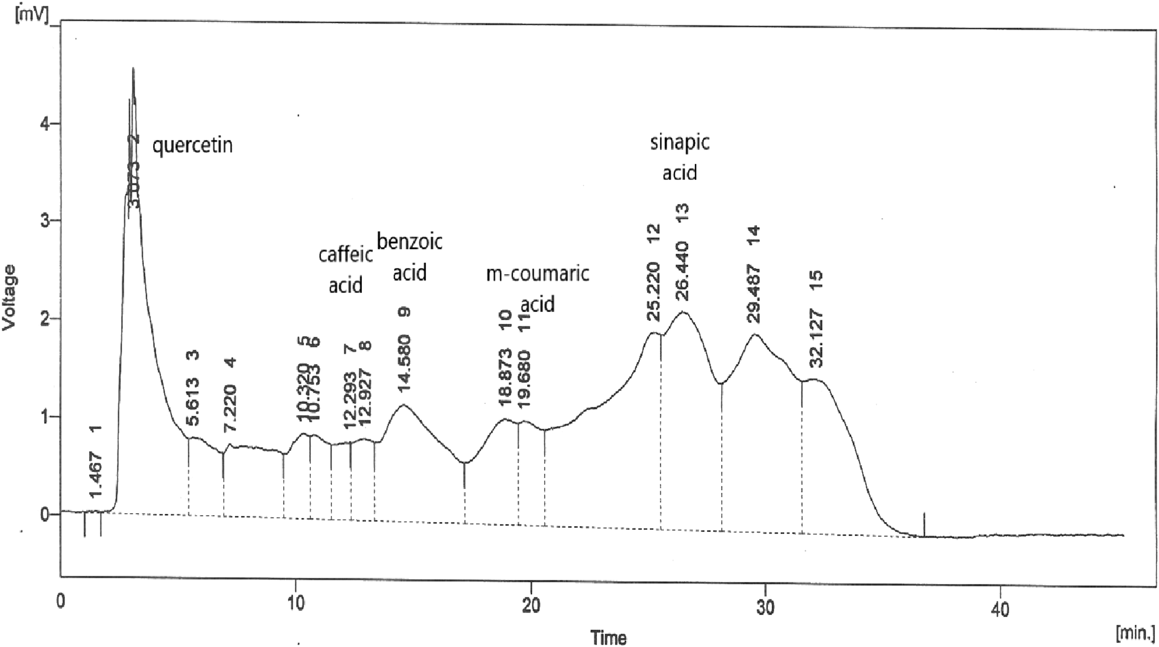

Qualitative and Quantitative Phytochemical Analysis
Qualitative Phytochemicals Analysis of Hydroethanolic Extracts of Tested Medicinal Plants.

(+) present, (−) absent, (++) significant concentration.
Total Phenolics, Flavonoids, Reducing Power, and Antioxidant Capacity of Selected Hydroethanolic Extracts of Tested Plants.

Data presented as mean ± S.D of triplicate determinations. Mean with different superscript within same column show significant (P<.05) differences among treatments, TAC; total antioxidant capacity, AAE; ascorbic acid equivalents, CE; Catechin equivalents, GAE; Gallic acid equivalents
In vitro Antioxidant Profiling
IC50 Values (μg/mL) of Different Antioxidant Activities of Tested Plants Extract.

Data presented as mean ± S. D. Mean with different superscript within same column show significant (P<.05) differences among treatments.
Antimicrobial Potential of Tested Plants Extracts
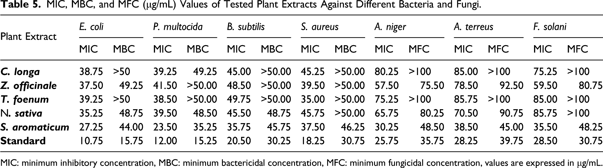
Results obtained from antimicrobial activity shown that hydroethanolic extracts of tested plants extract possess significant antimicrobial activity against all tested pathogens (Figure 2). The experiment exhibited that tested plants extract showed a variable level of antimicrobial activity in concentration dependent manner, varying from the smallest inhibition value of 7 mm to the highest value of 32 mm. Similarly, the MIC against bacteria obtained ranged from 20.50 to 49.75 μg/mL, and MBCs ranged between 30.75 to >50 μg/mL (Table 5). Hydroethanolic extracts of S aromaticum showed maximum antibacterial activity towards all tested microorganisms with MIC values of 27.25, 23.50, 35.75, and 37.50 μg/mL against E coli, P multocida, B subtilis, and S aureus, respectively. Similarly, in case of fungal strains all other extracts showed MIC of >50 μg/mL except S aromaticum which showed MIC values of 32.50, 30.75, 38.50, and 35.50 μg/mL against A niger, A terreus, and F solani. Against fungi, MIC of tested plant extracts ranged from 30.25 to >100 μg/mL while MFC ranged from 42.50 to >100 μg/mL. S aromaticum showed considerable antibacterial and antifungal activity against all fungi as compared to standard ampicillin and fluconazole (Table 5). Effect of different concentrations (50-250 μg/mL) of tested medicinal plants against antimicrobial activity of (A) E. coli, (B) S. aureus, (C) P. multocida, (D) B. subtilis, (E) F. solani, (F) A. niger, and (G) A. terreus. Ampicillin and fluconazole were used as standard. MIC, MBC, and MFC (μg/mL) Values of Tested Plant Extracts Against Different Bacteria and Fungi. MIC: minimum inhibitory concentration, MBC: minimum bactericidal concentration, MFC: minimum fungicidal concentration, values are expressed in μg/mL.
In vivo Experimentation
Acute and subacute toxicities
The acute toxicity analysis was performed in accordance with the OECD (Organization for Economic Corporation and Development) guideline. No mortality or treatment linked toxic effects or symptoms were recorded after the treatment with tested plant extracts at dose of 2000 mg/kg. Moreover, the animals did not show any treatment-related alteration in behavior, eyes, skin and fur examination, temperature, food and water intake, and breathing. Consequently, the tested plant extracts were considered safe to use at dose of 2000 mg/kg and the lethal dose (LD50) was supposed to be more than 2000 mg/kg BW. The subacute toxicity study of the tested plant extract was performed in accordance with OECD guideline. All treatment animals were administered orally at the doses of 250 mg/kg (group I) and 500 mg/kg (group II) and 1000 mg/kg (group III) for 28 days. The plants extract treatments did not produced any unfavorable effects in experimented animals and no signs of toxicity were recorded in treatment group in contrast to control group.
Effect of tested plant extracts on body and relative organ weight
Effect of Tested Plants Extract on Body Weights of Male and Female Rats.

values are presented as mean ± S.D, Results are significant at.
aP < .05.
bP < .001.
cP <.01 and.
Relative Organ Weight (g/g BW) of Rats After Treatment With Tested Plants Extract.

Values are expressed as mean ± S.D Results are significant at P < .05, P <.01 and P< .001.
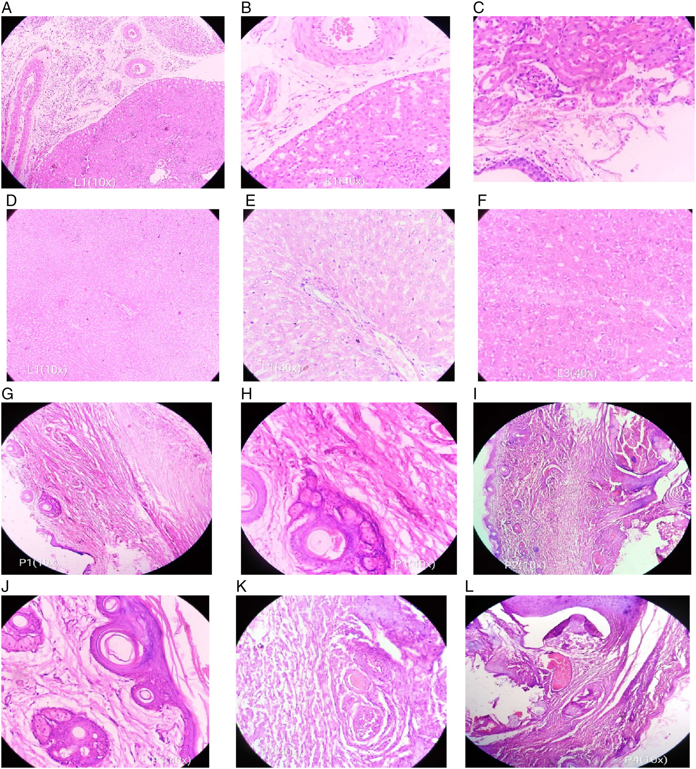
Histopathological photographs of kidney sections treated with (A) Z. officinale, (B) N. sativa, and (C) S. aromaticum at 1000 mg/kg b. w, liver sections treated with (D) Z. officinale, (E) N. sativa, and (F) S. aromaticum at 1000 mg/kg b. w. and rat paw tissues treated with (G) Indomethacin at 20 mg/Kg, (H) Z. officinale, (I) N. sativa and (J) C. longa, (K) N. sativa, and (L) S. aromaticum at 400 mg/kg b. w.
Effect of tested plants extract on hematological profile
Hematological Profile of Rats After Treatment With Tested Plants Extract.
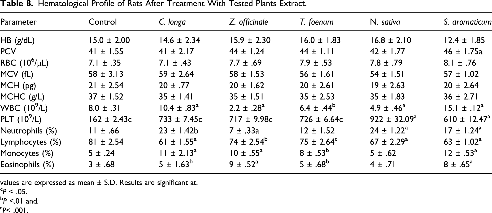
values are expressed as mean ± S.D. Results are significant at.
cP < .05.
bP <.01 and.
aP< .001.
Effect of tested plants extract on biochemical profile
Biochemical Parameters in Serum of Rats After Treatment With Tested Plants Extract.
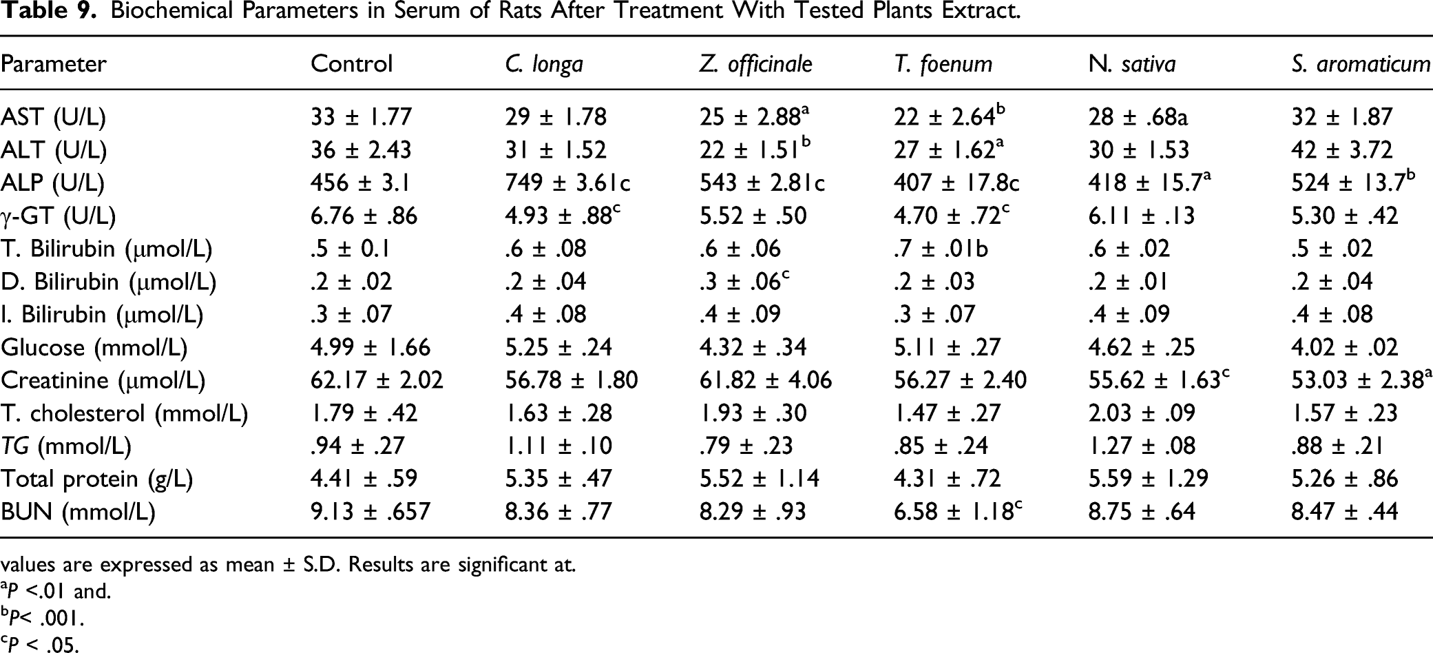
values are expressed as mean ± S.D. Results are significant at.
aP <.01 and.
bP< .001.
cP < .05.
Carrageenan-Induced Rat Paw Inflammation
Effect of Different Doses of Tested Plants Extract on Carrageenan Induced Paw Inflammation.
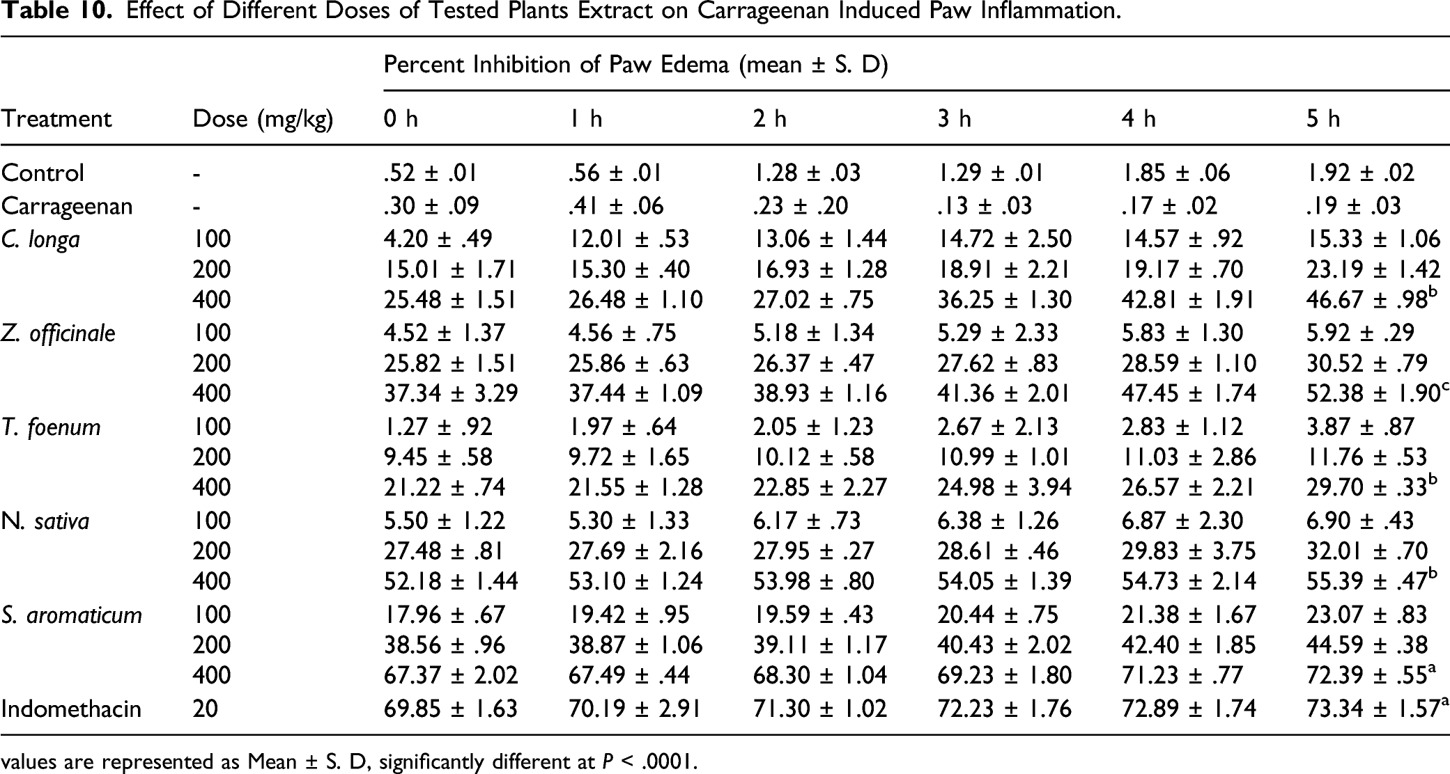
values are represented as Mean ± S. D, significantly different at P < .0001.
The results showed that plant extracts inhibited the paw inflammation in time and dose dependent manner. The percentage inhibition of inflammation (PI) at fifth hour of carrageenan injection in C longa, Z officinale, N sativa and S aromaticum treatment group (400 mg/kg b. w.) was 29.70%, 46.67%, 52.38%, 55.39%, and 72.39%, respectively (Table 10). Histopathological examination of rat paw tissues was also performed, and results are represented in Figure 3. A microscopic analysis of paw biopsies showed pronounced penetration of cellular infiltrates in connective tissue in the dermis and epidermis of paw skin in carrageenan treated rats. The epidermis displayed a sponge-like appearance and developed bullae. A decrease in inflammatory response (decrease of cellular infiltration) was seen in the paw biopsies of animals treated with indomethacin and tested plant extracts at different doses. Moreover, the inflammatory cells were decreased in number and confined near the vascular regions in treatment groups relative to the carrageenan-treated group.
Oxidative stress markers and lipid peroxidation assay
Levels of enzymatic antioxidants and stress markers were measured at 6 h of carrageenan induction in rat paw tissue homogenate and level of these biochemical markers altered significantly as compared to non-treated tissues. Level of MDA and MPO increased noticeably (P < .001) in tissue homogenate of carrageenan treated rats and reduced significantly (P < .001) in plant treatment group (Figure 4). Likewise, significant increase in CAT and SOD levels was observed in carrageenan treated group and tested plant treatment significantly (P< .01) restore the tissue enzymatic antioxidant levels therefore increase the total antioxidant status (TAS). Similarly, TOS levels was found increased significantly (P < .01) in carrageenan treated group while treatment of tested plants extract decreased the total oxidant status (TOS) of the paw tissues significantly (P < .01) in dose dependent manner. Effects of different doses (100-400 mg/kg b. w.) of S. aromaticum extracts on MDA, MPO, SOD, CAT, TOS, and TAS in rat paw tissue homogenate after 6 h of carrageenan injection. Data expressed as mean ± S. D.
Discussion
The development of novel, safe, and effective alternative anti-inflammatory drugs is a major challenge of the date. 31 Natural compounds derived from plants have been regarded as an alternative potential source of pharmacological products with decreased side effects. 32 Phytochemical studies give basic evidence about the therapeutic significance of medicinal plants. 33 The qualitative phytochemical screening of hydroethanolic extracts of tested plants extract exhibited the existence of alkaloids, terpenoids, phenolics, flavonoids, tannins, steroids, phenols, coumarins, sterols, and quinines which might be possible reason of antimicrobial and anti-inflammatory activities of tested plants. 11 Plant alkaloids have been reported as potential antioxidant and analgesic agents. Cardiac glycosides improve the cardiac output by increasing the force of contraction of heart muscles. Coumarins increase blood flow in the veins and decrease capillary permeability. 16 Saponins have ability to deal with bacteria, fungi, and pests. 34 Phenolics and flavonoids are the compounds which have vigorous scavenging role in oxidation, cancer, and inflammation.35,36 Flavonoids are the main polyphenolic substances having significant antioxidant and antimicrobial potential. 17 Both flavonoids and phenolic acids hold potential antioxidant, anticancer, cardioprotective, and anti-inflammatory properties and also protect skin from damaging effects of UV-radiation, and have immune system promoting effects. These molecules are exciting candidate for medical and pharmaceutical applications. 18 HPLC analysis of tested plants extracts showed presence of wide range of phenolic compounds with quercetin as major compound detected in all extracts. Quercetin is well known for its anti-inflammatory properties and expressed on variety of cell types in both humans and animals. Further quercetin decreased the level of inflammatory mediators, that is, leukotrienes and prostaglandins through inhibition of inflammatory enzymes cyclooxygenase and lipoxygenase.37,38 Antioxidant activities of medicinal plants majorly contribute to their therapeutic potential. Phenolic compounds are well-known antioxidants and hold substantial ability to decrease oxidative damage. They can neutralize free radicals through a series of coupled reactions with antioxidative enzymes.39,40 All tested plants extract exhibited strong antioxidant activities with best IC50 values of DPPH inhibition, ABTS cation scavenging, β-carotene bleaching, and H2O2 scavenging potential. Hydroethanolic extract of S aromaticum showed excellent antioxidant activities which is comparable to standard ascorbic acid.
Resistance to available antibiotics is a major dilemma for health care sector. 41 The development and dispersion of multidrug resistance pathogens have considerably compromised the current antibiotic therapy which led to the search for novel phytocompounds with antimicrobial activity. 42 For antimicrobial activity, a panel of clinically relevant, multidrug-resistant microorganisms was tested. Most of the tested plants extract exhibited dose dependent antimicrobial activity with good MICs values. Hydroethanolic extracts of S aromaticum showed excellent antimicrobial activities as compared to standard ampicillin and fluconazole. Flavonoid compounds specifically quercetin has broad spectrum antimicrobial effects and reported to inhibit the growth of several microbes including E coli, S aureus, and A. flavus. 41 The perception that natural products are effective and safe has led to the increased consumption of herbal remedies particularly among rural areas, where such remedies can be used for long time without consideration of dose which can cause side effects. 43 Therefore, acute and subacute toxicity studies are necessary which assist in determining the dose ranges for future investigation. In acute toxicity analysis, no change in behavioral, neuronal, and motor functions for all subjected extracts was observed. The acute oral toxicity study did not reveal any sign and symptoms in treatment groups at a dose of 2000 mg/kg. During acute toxicity study, no morbidity and mortality was noted in rats treated with tested concentration, therefore LD50 of all administered extracts was supposed to be >2000 mg/kg. Furthermore, body weight, food, and water consumption were remained unaffected after treatment with tested plants extract. However, for the confirmation of complete toxicological profile of a natural product, 14 days trial and toxicological assessment is not sufficient. This could not be adequate to assess the toxicological changes linked with the administration of plants extract in traditional medicine for a long period of time. Therefore, long-term study is required for detailed safety evaluation of natural extract. In sub-acute toxicity, different dosages (250, 500, and 1000 mg/kg b. w.) of tested plants were administered orally, and the effect of plants extract on weight, utilization of food, water intake, liver and kidney function indices, hematological, and histopathological alterations were recorded. Variation in body weight is regarded as an initial sign of toxicity upon treatment with plants extract and substantial loss of body weight is considered to be 1 of the most important indicators of declining health situation. 44 However, continuous treatment of rats with tested plant extracts over 28 days revealed no significant (P > .05) variations in body weight when compared with the weight of nontreated rats. The findings indicate that tested plants extract had no harmful effects on health and growth of animals.
Hematopoietic system is a vital indicator of pathological and physiological status for humans as well as animals. 45 The results further propose that some plants extract may include some compounds which have capability to increase defensive population of WBCs and hence to boost the immune system. 46 Further, an increase in Hb, packed cell volume and RBCs count was observed in experimental rats treated with tested plants extract. However, these changes are not much significant and within the normal ranges of specie (P < .05). Liver and kidney being vital organs, exhibit toxic properties after exposure to potent harmful compounds as these organs are principally engaged with the process of detoxification. 47 High level of liver biomarkers is a valid indicator of liver damage. 48 The administration of plants extract for 28 days did not cause any noxious effect on liver and liver biomarkers as compared to the untreated group. The little alteration has no clinical importance because values recorded are within the normal range. High levels of ALP and γ-GT are indicators of cholestatic induction and hepatobiliary obstruction. 49 Total proteins in serum are measured as a nonenzymatic biomarker of liver damage. 50 The blood glucose and lipid profile were also recorded for evaluation of metabolic states in relation to metabolism of lipids and carbohydrates. 51 Increased concentrations of BUN and creatinine in blood are a sign of renal toxicity due to improper removal of waste substances by the damaged kidneys. 52 Treatment of rats with tested plants extract has not caused variation in serum BUN and creatinine level as compared to control or untreated group, suggesting no significant (P > .05) alterations in kidney function after treatment with tested plants extract. Consequently, the findings exclude the toxic properties on kidney functions by tested plants extract.
The carrageenan-induced rat paw inflammation is a well-recognized model of acute inflammation and mostly used to assess the anti-edematous effects of natural compounds. 53 Paw edema model induced by carrageenan is biphasic response which involved variety of inflammatory mediators. 54 ROS play a crucial role in inflammatory process through activation of NF-κB (nuclear factor kappa B) and transcriptional factor such as iNOS (inducible nitric oxide synthase). 55 All tested plants extract showed anti-inflammatory effects in a concentration dependent manner against carrageenan-induced rat paw edema. Cellular infiltration (neutrophils) plays a major role during inflammation. Our findings indicated that tested plant extracts produced a marked reduction in the cellular infiltration of neutrophils and leukocytes into the carrageenan-treated rat paws (Figure 3).
Under normal physiological conditions, antioxidant defense systems prevent oxidant-induced tissue damage. 56 Antioxidants in our diet detoxify the free radicals produced in body during normal metabolism. 57 Most important antioxidant defense systems in our body comprises of non-enzymatic (vitamin C and vitamin A) and enzymatic (superoxide dismutase and catalase) antioxidants. 58 The total antioxidant and oxidant status of the body is usually measured in terms of total oxidant status (TOS) and total antioxidant status (TAS). 59 Oxidative stress is associated with pathogenesis of several diseases especially inflammation. 60 During inflammation large number of free radicals are produced at the inflammatory sites due to existence of leucocytes. MDA and MPO are used as oxidative stress and inflammatory marker for both acute and chronic inflammation. 61 Leukocytes upon activation produced MPO which have bactericidal activity.62,63, Pretreatment of rats with tested plant extracts significantly (P <.01) restored the tissue enzymatic antioxidants therefore TAS and decrease the TOS as compared to the carrageenan treated rats only. In vivo antioxidant effects of tested plants extract might be due to the existence of high phenolic and flavonoid contents which have well established anti-inflammatory potential. Based upon the phytochemical and antioxidant evaluation of tested plant extracts it could be concluded that these plants contained a broad range of phytocompounds and antioxidant compounds which contribute to their significant in vivo antioxidant and anti-inflammatory activity. Moreover, therapeutic potential of hydroethanolic extracts of tested plants extract is dose dependent. However, more scientific contributions are needed to isolate the novel active compounds from these therapeutic plants to compete NSAIDs (non-steroidal anti-inflammatory drugs).
Conclusion
On the basis of these findings, the study concluded that hydroethanolic extract of tested plants are potential source of phenolic compounds and have significant antioxidant, antimicrobial, and anti-inflammatory potential. Furthermore, plants extracts are effective against acute inflammation and efficiently reduce the lipid peroxidation induced by oxidative stress during carrageenan-induced inflammation. The anti-inflammatory and antimicrobial effects of tested plants extracts are likely to be mediated through their antioxidant compounds particularly phenolics and flavonoids. However, detailed studies are requisite to justify these findings.
Footnotes
Acknowledgments
The authors acknowledged the Medicinal Biochemistry Lab., Department of Biochemistry, University of Agriculture, Faisalabad, Pakistan for providing support to complete this research work. This research article is derived from the thesis of a PhD student.
Declaration of Conflicting Interests
The author(s) declared no potential conflicts of interest with respect to the research, authorship, and/or publication of this article.
Funding
The author(s) received no financial support for the research, authorship, and/or publication of this article.