
Abstract
The vast majority of cancers are treatable when diagnosed early. However, due to the elusive trace and the limitation of traditional biopsies, most cancers have already spread widely and are at advanced stages when they are first diagnosed, causing ever-increasing mortality in the past decades. Hence, developing reliable methods for early detection and diagnosis of cancer is indispensable. Recently, extracellular vesicles (EVs), as circulating phospholipid vesicles secreted by cells, are found to play significant roles in the intercellular communication as well as the setup of tumor microenvironments and have been identified as one of the key factors in the next-generation technique for cancer diagnosis. However, EVs present in complex biofluids that contain various contaminations such as nonvesicle proteins and nonspecific EVs, resulting in the interference of screening for desired biomarkers. Therefore, applicable isolation and enrichment methods that guarantee scale-up of sample volume, purity, speed, yield, and tumor specificity are necessary. In this review, we introduce current technologies for EV separation and summarize biomarkers toward EV-based cancer liquid biopsy. In conclusion, a novel systematic isolation method that guarantees high purity, recovery rate, and tumor specificity is still missing. Besides that, a dual-model EV-based clinical trial system includes isolation and detection is a hot trend in the future due to efficient point-of-care needs. In addition, cancer-related biomarkers discovery and biomarker database establishment are essential objectives in the research field for diagnostic settings.
Introduction
Extracellular vesicles (EVs), as phospholipid vesicles secreted by cells, 1 can be classified into 3 categories: apoptotic bodies, microvesicles (MVs), and exosomes. 1 –5 Among them, MVs originate from the buddings of the plasma membranes of tumors, neutrophils, and platelets 1 with the diameter larger than 200 nm, 6 while exosomes are derived from the inward buddings of the plasma membranes 7 with the size between 30 and 200 nm. 8 Firstly, the inward buddings of the membrane inside the endosome produce the multivesicular bodies (MVBs), 9 then exosomes are released because MVBs fuse with the plasma membranes of various cells. 10 –12 In virtue of appropriate isolation technologies, EVs can be easily extracted from blood, 13 urine, 14 breast milk, 15,16 saliva, 17 and cerebral spinal fluid body fluids, and contain proteins, 18 –20 messenger RNAs (mRNAs), microRNAs (miRNAs), 21 –23 and DNA 24 with similar genetic characteristics of their parenting cells. 25,26 Hence, EVs can be used for diagnostic settings, especially for cancers. 27 –30 In addition to EVs, circulating tumor cells (CTCs) and circulating mRNA can be also used for liquid biopsy. 31,32 However, their undesirable maneuverability and high minimum amount for detection limit further application. In this review, we will highlight the EV-based liquid biopsy.
Comparing with traditional tissue biopsies that require complex sampling procedures and are invasive, 31 EV-based liquid biopsy shows advantages of monitoring tumors with time evolution, 33 -36 long-term treatment response of the tumors, repeated sampling, 37,38 and easy sampling managements. 39,40 Moreover, traditional biopsies based on symptoms may fail to detect early-stage cancer because some symptoms only show at late-stage cancers; on the other hand, EV-based liquid biopsy, on genetic level, can waive the symptoms limitations and detect early-stage cancer. Extracellular vesicle–based liquid biopsy is also important for liver diseases, 41,42 immune diseases, 43,44 neuro diseases, 45 and Parkinson disease. 46 We only focus on cancer diagnostic in this review paper.
Extracellular vesicles show potential cancer diagnostic functions; however, EV-based liquid biopsy is limited in identifying EVs since they have size heterogeneities (30 nm to 1 mm) 25 and present in various human biofluids that contain outside vesicular small molecules (eg, RNAs and proteins). 47 Hence, efficient EV isolation methods are necessary before clinical analysis. In addition, normal EVs secreted by host cells influence the specificity and sensitivity of tumor EVs, 48 which can be possibly resolved through tumor biomarker immunoaffinity, so defining reliable cancer biomarkers is essential.
This review article presents recent EV isolation and characterization techniques and highlights the diagnosis progresses among various cancers. We compare the isolation methods by purity, recovery rate, processing time, and tumor specificity and evaluate the characterization techniques. Even though some reviews have published, we believe this review is helpful because of compiling latest diagnostic progresses for various cancers.
Extracellular Vesicle Isolation and Enrichment
Various isolation methods have been developed in the past decades and have been evaluated by recovery rate, purity, integrity, tumor specificity, and processing time. 49 In this review, we present bulk isolations that rely on size, density, coprecipitation, and affinity binding, 49 and subpopulation isolation methods that isolate and enrich subpopulation by targeting antibodies against EV surface biomarkers include common biomarkers (eg, CD9, CD63, and CD81) 50,51 and tumor-specific biomarkers (eg, HER2, EpCAM, EGFR, EGFRvIII, and GPC1). 22,52 –54
Size-Based Isolation
Ultracentrifugation
Ultracentrifugation (UC) is the gold standard protocol for isolating EVs.
55
Ultracentrifugation can scale-up sample volume and isolate EVs with high centrifugal force (100 000g),
56
while expensive equipment, skill dependent, time consuming, low purity, and yield are the drawbacks.
Density-Based Isolation
Sucrose density gradient UC separates EVs in a continuous size-based UC mode; serial ultracentrifugations are applied to remove living cells and cell debris, then the EV and proteins are separated based on different flotation densities under UC. 62 Despite the high purity, sucrose has lengthy problem due to multiple centrifugations, and the recovery rate is low due to multiple centrifugations.
Coprecipitation Isolation
Coprecipitation reduces the solubility of EVs by adding polymer or reagent (eg, ExoQuick) and causes precipitation, and EVs are isolated from precipitation with lower centrifugal forces later. 63,64 Comparing with UC, coprecipitation saves processing time but lacks scale-up and EV specificity due to additives.
Affinity Binding–Based Isolation
Membrane affinity binding
Recently, commercial kits (eg, exoEasy) use membrane-based affinity binding to isolate EVs,
65
which are selected based on generic, biochemical feature of vesicles and fixed on the affinity spin column that can be washed and eluted with buffers. The advantages of kits are easy handling, high purity, and extremely fast (25 minutes); however, low throughput and recovery rate are the shortcomings. Macías et al tested the kit and demonstrated it had low EV number and weak CD63 and CD9 Western blotting signal.
66
Immunoaffinity
One of the advantages of immunoaffinity compared to bulk isolation is the EV subpopulation isolation with high recovery rate and specificity. The immunoaffinity is the specific affinity between an antibody and antigen, which normally means the bindings between a capture antibody, a EV surface antigen, and a detection antibody. 68,69 Shao et al applied immunomagnetic beads to extract glioblastoma multiforme (GBM) EVs with 93% specificity, and they detected EPHA2, EGFR, and PDPN mRNA from patients with GBM successfully. 70 Recently, Sharma et al used the monoclonal antibody (mAb) with magnetic beads to capture tumor EVs from patients with melanoma that expresses CSPG4 epitope. 71 Masud et al applied gold-loaded ferric oxide nanocubes functionalized with antibodies that work as “dispersible nanocarriers” on separating population of EVs. 72 Bai et al have captured lung cancer EVs through queued beads functionalized with antibodies and combined with quantum dots in a microarray, which showed distinctive lung cancer marker detection level. 73 Immunoaffinity can isolate EV subpopulation, especially for tumor EVs, while limited sample processing volume is a big challenge for the immunoaffinity.
Comparison
Different isolation techniques are compared in Table 1 based on processing time, purity, recovery rate, sample scale, and tumor specificity. High recovery rate and purity are important because of clinical sensitivity and accuracy. 47 Flexible sample scale is another key point for clinical settings since various biofluids require different pretreatment, for example, urine must be in large scale to obtain enough EV number, while serum and plasma must be in small scale due to collecting limitation. It is worth to note that immunoaffinity isolation methods can be directly used for point-of-care diagnostics since tumor subpopulation capture. 74 However, immunoaffinity is highly dependent on the quality of antibodies. Until then, a platform that can guarantee high yield, purity, rapid time, and tumor specificity is still missing.
Comparison of EV Enrichment, Separation, and Purification Methods.

Extracellular Vesicle Detection and Characterization
Microscopy Quality Characterization
Scanning electron microscopy
Scanning electron microscopy (SEM) is a high-resolution technique in the EV field.
22,75,76
Extracellular vesicles are fixed by chemicals such as glutaraldehyde and dehydrated by critical point dry in ethanol; osmium tetroxide can be used to increase contrasts. Scanning electron microscopy scans EV surface with a focused beam of electrons, normally a thin sputter gold coating is required for focusing, and generates EV topography image due to the interaction between electrons and atoms in the EV sample. Majorities of EVs present spherical-shaped or cup-shaped morphologies under the SEM.
77
Quantitative Characterization
Dynamic light scattering
Dynamic light scattering (DLS) shows EV size distribution based on the intensity of the scattered light.
81
The suspended EVs are illuminated by a laser, and the intensity of the light fluctuates over time since EV goes through Brownian motion.
81
–83
The effective size of EVs is calculated by Strokes-Einstein equation based on the transformation between fluctuation rates and diffusivities of the EVs. However, the intensity of the scattered light is also associated with the size of the EV; larger EV size reflects higher intensity, which may influence accuracy.
Protein Analysis
Pierce bicinchoninic acid protein assay kit
Bicinchoninic acid is used to quantify total protein concentration including membrane proteins and intravesicular proteins.
88
–90
It is a highly sensitive and rapid method based on colorimetric solution but cannot characterize EV protein.
91
Nucleic Acids Analysis
Precipitation and spin columns
Both precipitation and spin columns complete total RNA extraction and are commercially available.
22,98
Precipitation relies on phase separation; RNA is suspended into the aqueous phase and is recovered through ethanol precipitation.
98
The spin column is based on solid-phase extraction that relied on silica and RNA binding with chaotropic agents.
Cancer Clinical Applications of EV
Conventional biopsies, such as tissue biopsy, are not only invasive 100 but have limitations to profile tumors due to tumor heterogeneous characteristics, and they cannot reflect the whole tumor information. 101,102 Hence, liquid biopsy shows advantages since it is noninvasive and on genetic level that can provide comprehensive information. Extracellular vesicles play an important role in the cancer liquid biopsy since they carry all types of genetic information from original tumors, 103,104 and they also obtain attention because of preinvasive and early state diagnosis. 105 Extracellular vesicle–based diagnostic relies on tumor exosomal biomarkers, so defining and discovering reliable biomarkers is vital. In this review, we will present the lasted EV diagnostic based on exosomal biomarkers among different cancers (Table 2).
Potential Biomarkers of EVs for Cancer Diagnostic Application.

Abbreviation: N/A, not applied.
Breast Cancer
Breast cancer (BC) plays the second position of cancer mortality in women. 106,107 Common breast X-ray screening is invasive and radioactive, thus EV, diagnostic setting is important. Both blood samples and breast milk contain reliable EV resources, while collecting milk requires specific time point even though it is simpler than serum. 108 There are several BC biomarkers; proteins such as HER2, ER, and Ki67 are highly expressed. 106,109 –111 Yang et al said the expression of TGF-β in the breast milk increased the risk of BC. 112 van’t Veer et al claimed that CD24 was abundant in patients with late-stage BC, 113 and Wang et al demonstrated CD82 expression level was negatively associated with patients with BC. 114 As for miRNA, miR-10b and miR-145 are abundant in patients with BC. 115 Hannafon et al demonstrated both miR-21 and miR-1246 expressed in patients were higher than healthy donors. 108 Recently, Zhai et al have used Au nanoflare probe to detect miR-1246 in plasma samples successfully (Figure 1A). 116 Alba et al claimed miR-105 was higher in patients with metastasis BC than in healthy donors. 117 Jong et al detected miR-21, miR-222, and miR-200c with high sensitivity with their surface-enhanced Ramen scattering sensor. 118

A, Schematic of Au nanoflare probe to detect miR-1246. Fluorescence-treated probes enter the exosomes and bind to the targets after incubation with exosomes from breast cancer cells. B, Comparison of miR-1246 expression level in patients with breast cancer (n = 46) and healthy controls (n = 28). Patients with breast cancer showed higher exosomal miR-1246. P < .0001. Reprinted with permission from Zhai et al. 116 Copyright © 2019 American Chemical Society. C, Lung cancer liquid biopsy–related exosomal biomarkers. Reprinted with permission from Cui et al. 125 Copyright © 2019 from Elsevier Ltd.
Lung Cancer
Lung cancers (LCs) are the most common and high death leading type of cancer because the majority of LCs are at late stage and go through metastasis that cannot be cured when they are first found. 119 Non-small cell lung cancer (NSCLC) is the most common type of LC and only shows symptoms at the late stage 120 ; hence, early-stage detection is essential. EFGR, MET, PIK2CA, ALK, KRAS, MAP2K1, HER2, BRAF, AKT1, CD151, CD171, and tetraspanin 8 were revealed to be highly associated with LC. 121 Ueda et al found CD91 was a powerful surface biomarker in advanced stage LCs. 94 Jakobsen et al showed CD317 was able to distinguish patients with LC with 75% accuracy. 122 Niu et al found patients with NSCLC expressed a high level of α-2-HS-glycoprotein (AHSG), the extracellular matrix protein 1 in the serum compared to healthy donors. 123 Li et al found α-2-glycoprotein (LRG1) was strongly expressed in urinary EVs from patients with NSCLC (Figure 1B). 124 Recently, Castellanos-Rizaldos et al improved the detection sensitivity and specificity of EGFR T790M from the plasma of the patients with NSCLC by combining exoRNA/DNA and circulating free tumor DNA. 125 Jin et al found let-7b-5p, let-7e-5p, miR-23a-3p, and miR-486-5p were related to early-stage NSCLC. 126 Xu et al demonstrated miR-21 and miR-155 were higher in patients with NSCLC with recurrence than without recurrence and healthy donors. 127 Moreover, Li et al found lncRNA GAS5 was downregulated in early-stage patients with NSCLC compared with healthy donors. 128
Ovarian Cancer
Ovarian cancer (OC) is difficult to be detected until it has spread within the pelvis and abdomen at the late stage, so early-stage detection is necessary. 129 Symptoms of early-stage OC are rare and nonspecific even in the advanced stage, so EV diagnostic setting on genetic levels has advantages. Protein biomarkers such as claudin-4, HSP70, HER2, and TrkB derived from exosomes from patients showed different expression compared with healthy controls (Figure 2A). 130 –133 CD24 and EpCAM are also possible biomarkers for OC. 137 Exosomal miRNAs are much more powerful for OC diagnosis. Overexpressed level of miR-21, miR-200b, miR-100, miR-320 miR-141, miR-125b, miR-1246, miR-375, and miR-93 differed between OC patients and healthy donors. 138 The miR-1290 also showed the possibility of diagnosis on high-grade OC. 139 The miR-30a-5p was highly expressed in the urine samples of patients with OC, 140 while miR-145, miR25, and miR148a were under expressed. 141 Xu et al found miR-101 was expressed lesser in patients with OC than in healthy donors. 132 Qiu et al found metastasis-associated lung adenocarcinoma transcript 1 (MA-LAT1) was positively associated with OC. 142

Molecular components (long noncoding RNAs, microRNAs, and membrane proteins) in exosomes from patients with ovarian cancer. Reprinted with permission from Yang et al. 130 Copyright © 1999-2019 John Wiley & Sons, Inc. B, Histogram and boxplot of fluorescence intensity of exosomes (Target: Prostate-specific membrane antigen (PSMA) positive) from patients with prostate cancer and healthy donors detected by superparamagnetic conjunctions and molecular beacons (SMC-MB) platform. Reprinted with permission from Li et al. 134 Copyright © 2019 American Chemical Society. C, Comparison of glypican-1 expression level in patients with pancreatic cancer (n = 20), benign pancreatic disease (n = 7), and healthy controls (n = 11). Glypican-1 in patients with pancreatic cancer were elevated. Reprinted with permission from Lewis et al. 135 Copyright © 2019 American Chemical Society. D, Quantitative reverse transcription polymerase chain reaction analysis of exosomal H19 from patients with bladder cancer, benign disease, and healthy controls. P < .001. Reprinted with permission from Wang et al. 136 Copyright © International Scientific Information.
Prostate Cancer
Prostate cancer (PCa) is one of the most common types of cancer in men. 143 Some types of PCa grow slowly with minimum harmful effects, while some types are aggressive. 144 The early stage of the PCa that may be defined as the prostate gland is easy to cure, but they show no signs or symptoms. So the liquid biopsy based on exosomal molecular contents is important. Bhagirath et al used the nCounter technology and found miR-1246 was a promising aggressive PCa biomarker. 145 Donovan found PCA3 and ERG mRNAs predicted high-grade PCa. 146 Li et al used an ultrasensitive and reversible nanoplatform to detect PSA, PCA3, and mRNA successfully in urinary exosomes from patients with PCa (Figure 2B). 134 However, PSA has limitation because it may not differ cancer and benign prostatic hyperplasia (BPH). 146 Some miRNAs, such as miR-141 and miR-375, from serum EVs were associated with metastatic PCa, and the miR-19b distinguished PCa with 100% specificity and 93% sensitivity. 147 As for long noncoding RNAs, Wang et al found SAP30L-AS1 was related to tumor invasion, and SChLAP1 was expressed higher in PCa compared with BPH and healthy controls. 148
Pancreatic Cancer
Early-stage PC can only be detected in people with pancreatic cysts or family history of PCs, 149 but it can seldom be detected in other conditions. 150 Serum cancer antigen 19-9 (CA19-9) is a possible biomarker for PC 151 ; however, it fails to show sensitivity and specificity of early-stage PC. Zhou et al compared 216 patients with PC with 220 healthy controls and found miR-122-5p, miR-125b-5p, miR-1192-5p, miR-193b-3p, miR-221-3p, and miR-27b-3p were significantly higher in patients with PC. 152 Goto also found higher expression levels of miR-191, miR-21, miR-451a in PC. 153 Lewis et al developed an AC electrokinetic microarray chip that was capable of differing 20 patients with PC from healthy donors based on glypican-1 and CD63 expression levels with 99% sensitivity (Figure 2C). 135 Li et al designed an ultrasensitive polydopamine bifunctionalized Surface-Enhanced Raman Scattering (SERS) immunoassay with a detection limit of one exosome in 2 mL, and they discriminated patients with PC based on GPC1, MIF, and EGFR surface biomarkers successfully. 154 Jin et al claimed ZIP4 promoted PC growth and could be a novel diagnostic biomarker for PC. 155 Recently, Lux claimed the combination of CA19-9 and c-Met improved sensitivity test of patients with PC. 156 They found PD-L1-positive patients showed shorter postoperative survival time that can be used as a negative prognostic factor. Moreover, the combination of CD104, Epcam, Tspan8, and some miRNAs such as miRNA-1246 improved diagnostic sensitivity and specificity of patients with PC. 157
Bladder Cancer
Bladder cancer is usually in the bladder but can show in other parts that belong to the urinary tract drainage system. 158 Around 50% of the patients with muscle-invasive bladder cancer will go through metastasis and die in 2 to 3 years, even patients with non-muscle-invasive BC usually have recurrence rate. 159 The symptoms of bladder cancer include hematuria, pelvic pain, and urination pain, 160 while these symptoms normally show at the middle or late stage; hence, the early diagnosis of bladder cancer is very important. Cystoscopy is a gold standard diagnostic tool for non-muscle invasive bladder cancer, 161 but it is expensive and time-consuming since subsequent cystoscopy is necessary once the result is negative, 161 and it fails to provide sensitive surveillance information. Hence, EV diagnosis with novel biomarkers is essential for patients with bladder cancer and suspected individuals. Both blood and urine provide reliable EV source for bladder cancer diagnosis; miR-148b-3p, miR-141 were increased, but miR-27a-3p, miR-100, miR-92a, and miR-99a were decreased in serum and plasma. 162 Compared with blood, urine shows advantages because urine contacts with bladder directly and can be collected with various time point easily to reflect different stages of the diseases, and EVs are able to cross the basement membrane into the urine with miRNAs. The miR-375 was decreased in high-grade bladder tumors, while miR-146a-5p was increased, especially in low-grade tumors. 162 Wang et al also found serum exosomal H19 expression level was higher compared to healthy controls and benign disease patients (Figure 2D). 136 Zhan et al found PCAT-1 and SPRY4-IT1 were capable of the bladder cancer diagnosis. 163 Zhang et al claimed UBC1 and SNHG16 identified by multivariate logistic regression model also provided high diagnostic accuracy. Moreover, the high UBC1 expression level was associated with low recurrence-free survival. 164
Melanoma Cancer
Melanoma is the most serious type of skin cancers, but it can be treated successfully in the early stages. 165 Melanomas can occur in any areas of the skin that are exposed to excess UV light. 166 Sharma et al isolated melanoma tumor-derived exosomes successfully with an mAb 763.74 that captured the chondroitin sulfate peptidoglycan 4 (CSPG4+) that are expressed on the surface of exosomes. 71 Chen et al claimed that exosomal PD-L1 was associated with anti-PD-1, which has shown promise in treating melanoma tumors. Hence, detection of PD-L1 on the exosomes is essential. 166
Glioblastoma Multiforme
Glioblastoma multiforme is an aggressive type of cancer that occurs in the brain or spinal cord, and it tends to occur in older adults. 167,168 Curing GBM is seldom possible but effective treatment can slow the progression of cancer and relief the symptoms. 18 However, treating early-stage GBM guarantees minimum dissatisfactory effects. Normal diagnosis methods include the neurological examination, imaging tests (magnetic resonance imaging), and tissue tests. Nevertheless, compared with EV liquid biopsy, they are expensive and invasive. EGFRvIII was a well-known GBM-related biomarker, and it was highly expressed on the surface of EVs from GBM patients. 22 Lan et al found miR-301a was a potential biomarker for GBM. 169 Tan et al also found serum lncRNAs HOTAIR was significantly higher than in the healthy controls. 170 Ebrahimkhani et al found miR-182-5p, miR-328-3p, miR-339-5p, miR-340-5p, miR-485-3p, miR-486-5p, and miR-543 were most likely stable for GBM classification after modeling and data comparisons among 26 relative microRNAs (Figure 3). 171 Santangelo also claimed miR-22 and miR-222 were expressed higher in high-grade GBM patients. 172
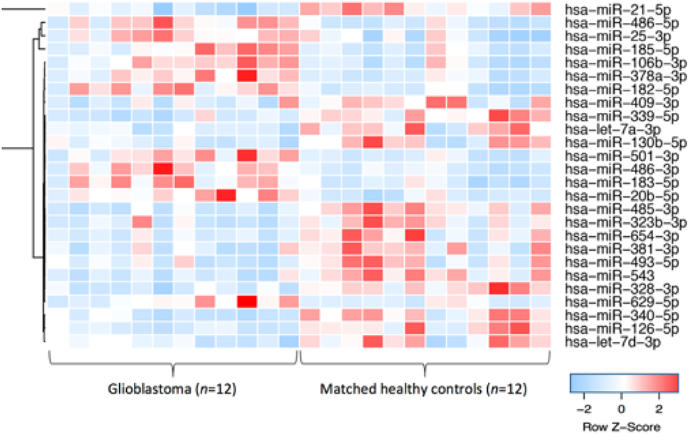
Hierarchical clustering of 26 microRNAs shows differences in GBM and healthy control exosomal profiles (fold change ≥2 or ≤0.5). Reprinted with permission from Ebrahimkhani et al. 170 Copyright Springer Nature Publishing AG.
Conclusion and Perspective
Extracellular vesicles play an important role in tumor microenvironment; besides carrying parenting genetic information, the specific tumor-related genetic information can be used for diagnosis and immunotherapy, while size and biomolecular heterogeneities of EVs bring problems on isolating EVs that challenge the EV diagnostic setting, which requires EV isolation in a pure and rapid manner. Hence, standard sample collection, storage procedures, and efficient isolation mechanisms are important. In this review, we summarized updated isolation methods, and majorities of them enriched the bulk EVs, and immunoaffinity can isolate tumor subpopulation EV based on tumor surface biomarkers. Successful isolation is the first step of the cancer diagnosis; characterizing EVs in qualitative and quantitative way, such as microscopy, DLS, NTA, and TRPS, is also necessary. The most important thing is the molecular content characterization such as protein classification and RNA sequencing, which is the fundamental of EV-based cancer liquid biopsy because clinical trials make decision based on expression level of reliable cancer-associated RNA and protein biomarkers. However, protein biomarkers lack tumor precision and show no superiority since the same type of biomarkers can be present in multiple cancers. For example, HER2 has high expression level in breast and LCs, while CD24 is abundant in both ovarian and BCs. In that, combination of different protein biomarkers to define a specific type of cancers is necessary. Comparing with proteins, RNAs may show high specifically toward single-type cancer, but it is relatively expensive. Overall, defining and exploring specific and reliable biomarkers and compiling a cancer biomarker database are big breakthroughs for cancer diagnosis. In this review paper, we introduce EV isolation and detection separately, and we believe in the future the combination of isolation and detection methods with high efficiency is desirable and plays key role since they fulfill the point-of-care cancer diagnostic need. Moreover, even though EV-based liquid biopsy is advantageous in many aspects such as noninvasive collection, early-stage detection compared with traditional biopsy, until now, gold standard operating procedures for collecting, isolating, and detecting EVs are still missing. Also, the universality and pricing are other concerns about liquid biopsy. Up to now, only HER2 and EGFR are qualified as biomarkers tested in clinical trials. Nevertheless, EV-based liquid biopsy is still a long road ahead, and we believe it will be more reliable and standard in the future since outstanding researchers make efforts on it.
Footnotes
Author Contribution
Chunyan Ma and Fan Jiang has contributed equally to this work.
Declaration of Conflicting Interests
The author(s) declared no potential conflicts of interest with respect to the research, authorship, and/or publication of this article.
Funding
The author(s) disclosed receipt of the following financial support for the research, authorship, and/or publication of this article: This study was supported by Zhejiang Medical Technology Program (2018KY918).