
Abstract
The biological consequences of mechanical whole body vibration (WBV) on the brain are not well documented. The aim of the current study was to further investigate the effects of a 5-week WBV intervention on brain functions. Mice (C57Bl/6J males, age 15 weeks) were exposed to 30 Hz WBV sessions (10 minutes per day, 5 days per week, for a period of 5 weeks; n = 10). Controls received the same intervention without the actual vibration (n = 10). Humans (both genders, age ranging from 44-99 years) were also exposed to daily sessions of 30 Hz WBV (4 minutes per day, 4 days per week, for a period of 5 weeks; n = 18). Controls received the same protocol using a 1 Hz protocol (n = 16). Positron emission tomography imaging was performed in the mice, and revealed that glucose uptake was not changed as a consequence of the 5-week WBV intervention. Whole body vibration did, however, improve motor performance and reduced arousal-induced home cage activity. Cognitive tests in humans revealed a selective improvement in the Stroop Color-Word test. Taken together, it is concluded that WBV is a safe intervention to improve brain functioning, although the subtle effects suggest that the protocol is as yet suboptimal.
Introduction
Many vibration studies that were published in the previous century focused on the detrimental effects of mechanical vibrations in the work environment, for example, when operating tools (eg, sledgehammer and form machines) or while riding in a vehicle (eg, truck, helicopter, and tank). The latter vibrations affect the whole body and for such vibrations, the term whole body vibration (WBV) was introduced. Reviews of the literature on work-related vibrations show that exposure to such levels of vibrations mainly leads to increased health risks of the musculoskeletal system as well as the peripheral nervous system. 1,2 However, thereafter positive effects of experimentally/therapeutically induced WBV were found, suggesting that, depending on the settings, WBV is a safe and effective way to train the musculoskeletal system and to improve physical performance. For example, increased muscle strength 3 and reduced knee osteoarthritis symptoms 4 have been reported. In addition, WBV improves physiological and health-related components of physical fitness, such as higher bone density 5 and lower blood pressure. 6 In elderly patients, WBV improves mobility, balance, general health status, 7,8 as well as body composition, insulin resistance, and glucose regulation. 9
Few studies examined the impact of WBV on cognition and the brain. In an animal study, rats were exposed to 4 hours of WBV (30 Hz) per day for 4, 6, or 8 weeks. These rats showed memory impairment and signs of brain damage. 10 Positive effects of WBV on the brain were found with much shorter durations of the WBV sessions. We previously showed in CD1 mice that a 5-week WBV intervention (30 Hz, 5 or 30 minutes per day for 5 weeks) improved balance beam performance (sensory motor test) and novel object recognition (memory test depending on the cortex) in a dose-dependent manner. 11 In C57Bl/6J mice, we showed that a 5-week WBV intervention resulted in increased activity of the cholinergic system in the somatosensory cortex and amygdala, 12 brain regions innervated by the cholinergic cells of the nucleus basalis in the forebrain. Preliminary studies showed that the expression of the immediate early gene c-fos, indicative for enhanced neuronal activity, was enhanced by WBV. 13 Increased neuronal activity is directly associated with acute increased glucose metabolism, 14 whereas a decrease in basal levels of glucose metabolism is typically interpreted as indicative for brain pathology, and for instance considered an early biomarker for Alzheimer disease. 15,16 No data are available about possible changes in glucose metabolism in the brain after WBV stimulation.
18F-fluorodeoxyglucose (18F-FDG) positron emission tomography (PET) is a sensitive method for longitudinal, in vivo measurement of glucose metabolism in tissues of humans and also small rodents. Contrary to traditional methods such as indirect calorimetry and doubly labelled water, it has the advantage that it can measure tissue specific glucose metabolism in living animals and humans. 18F-fluorodeoxyglucose PET small animal imaging is routinely used in our group to evaluate the effects of interventions on brain metabolism 17 -19 and is sensitive enough to detect small physiological differences, such as time-of day differences in brain and heart glucose uptake in mice. 20 To further examine whether WBV is safe for the brain, we herein studied baseline brain glucose uptake with 18F-FDG PET before and after a 5-week WBV intervention in C57Bl/6J mice.
In human studies, mixed effects were found for cognition with some evidence toward a detrimental effect, but without indications of a dose–response relationship. Two studies found some positive acute effects of WBV. In a study of Ishimatsu et al (2016) lower reaction times during WBV versus control were found on a sustained attention go no-go task. 21 However, these lower reaction times went together with more errors suggesting a speed-accuracy trade-off. Zamanian and coworkers found improved performance on a divided attention (choice reaction time) task but not on a selective attention task, which holds true for 3 different vibration magnitudes without a specific magnitude effect. 22 One other study examined a dose–response relation in which the magnitudes of the vibrations were varied (1.0, 1.6, and 2.5 m/s2 rms), but found no differences on a short-term memory task. 23 This is to a large extent in agreement with the findings of Sherwood and Griffin (1990). 24 In a few studies, short-term effects of WBV were examined. The WBV vibration with limited durations (2-10 minutes) appeared to improve attention/inhibition measured immediately after the WBV, but effect-sizes were generally small, 25 except for young adults with attention deficit hyperactivity disorder who achieved larger improvements. 26 -28 In this study, we performed a pilot study with a 5-week WBV intervention in older adults aged >40 years.
Taken together, this study set out to examine the impact of a 5-week WBV intervention on brain functioning in mice and humans. Specific aims of the study were (1) to examine glucose uptake in the brain, (2) to test whether WBV reduces arousal-induced activity (both mouse studies), and (3) to test executive functioning and memory in humans using a WBV intervention that is adapted from the 5-week WBV intervention used in mice.
Materials and Methods
Animals and Housing
Twenty young male C57Bl/6J (age 15 weeks at the start of the experiments; Charles River, France) mice were used. The mice were divided in 2 groups, balanced for body mass: a WBV group (n = 10) that underwent a 5-week WBV protocol and a control group (sham stimulated mice, termed pseudoWBV [pWBV]; n = 10) that received pseudo stimulation. Mice were kept on a 12:12 light/dark cycle at a temperature of 21°C ± 2°C. Mice were sedentary housed. Food, RMH-B 2181 (AB diets BV, Woerden, the Netherlands), and water were available ad libitum throughout the experiment.
Whole Body Vibration Protocol for Mice
The WBV protocol for mice was adapted from our previous studies in CD1 and C57Bl/6J mice 11,29 and was described in more detail by Keijser and colleagues. 11 The WBV setup (see Figure 1A) consisted of an oscillator (LEVELL R.C. Oscillator Type TG200DMP) and power amplifier (V406 Shaker Power Amplifier). A box (44.5 (L) × 28 (W) × 16 (H) cm) was attached to the oscillator which contained 12 removable compartments for individual mice (6.5 (L) × = 7.5 (W) × 20 (H) cm). During the stimulation procedure, mice in the WBV group were placed in the compartments and subjected to low intensity sinusoidal vibrations with a frequency of 30 Hz, as described previously by Keijser and co-workers 11 for 10 minutes (amplitude 0.0537 mm; g-force (peak) 0.098 g). Mice in the pseudo stimulated pWBV group served as controls and were placed in the compartments at the same schedule and for the same amount of time, but the oscillator was not turned on. Treatment sessions were performed during the light-phase on weekdays. To prevent putative time of day and anticipatory effects, the timing of treatment sessions varied every day and was randomly distributed over the light-phase. A rotation schedule was used to place mice at varying locations in the removable compartments, correcting for location dependent small variations in the stimulation intensity of the device. 11 Mice were subjected to the WBV or pWBV protocol for 37 days, containing in total 27 stimulation days.

A, Set-up of the mouse platform: a box (2), (length: 44.5 cm, width: 28 cm; height: 16 cm) is connected to a vibrator (1). Mice are placed in separate compartments (3) to avoid social interactions (eg, fights between males); 4 = amplifier; 5 = oscillator. B, Set-up of the human platform, with a chair mounted on a vibration platform suitable for wheel chairs.
Motor Performance (Balance Beam)
The balance beam is a sensorimotor integration test, which focuses on hind limb functioning. 30 In our lab, this test has been proven to be sensitive to the effects of disease phenotype, 31 exercise 32 and also WBV stimulation, 11 in different mouse strains. This test was, therefore, included as a control for the effectiveness of the WBV protocol in this experiment. The balance beam apparatus consisted of a 5 mm wide and 100 cm long aluminum beam. The beam was elevated 50 cm above the floor. One end of the beam contained a platform on which the home cage of the test mouse was placed, as a safe exit for the mice. The top of the cage was in level with the end of the beam. Prior to the first test session mice were trained to cross the beam in the right direction by placing them consecutively 5, 10, and 40 cm from the home-cage on the beam and guiding them toward the home-cage if necessary. A test session consisted of 3 consecutive trials crossing the full 100 cm length of the beam. All trials were recorded. The video files were scored by a previously trained observer. The observer was blinded for the trial, experimental condition, and time-point of the movies by randomization of the video files. The average crossing time of 3 correct trials for each mouse was used in the analysis. The balance beam test was performed twice; in the week prior to the start of the WBV protocol, and at the end of the WBV protocol, and on the day following the day of the last treatment session.
Arousal-Induced Home Cage Activity Measurements
Besides improvements in cognition and motor performance, observations in previous experiments indicated that during WBV mice quietly explored the box, sometimes displayed rearing (against the wall of the box) or grooming, or lied down. Pseudo whole body vibration mice typically revealed more aroused behavior, continuing exploring the box during the entire pWBV session. These observations suggested that WBV reduces arousal-induced activity. We, therefore, also monitored the acute effect of WBV and pWBV in the C57Bl/6J mice. At 3 different time-points, the acute effect of the WBV/pWBV treatment on arousal was measured on day 1, after the first treatment session, on day 17, and after the last treatment on day 37. In the minute directly following the treatment session, the mice were transferred to their own home-cages, without the wire-mesh lid on. The home cages were subsequently video recorded from above, for 5 minutes, with a camera using a recording speed of 25 frames/second. The mice were individually tracked, using the center point of the body as the marker. Mice were tracked using the grey scaling method and a sampling frequency of 12.5 frames/second with the Ethovision XT 11.5 (Noldus, Wageningen, the Netherlands) software package. The total distance moved in centimeter was extracted as a measure for arousal-induced home cage activity.
Brain Glucose Uptake in Mice
Brain glucose uptake was measured by means of 18F-FDG PET scans in the week before and after the treatment protocol. The scan protocol was designed to optimize the detection of 18F-FDG brain uptake and adapted from Fueger and colleagues. 32 Mice were food deprived 6 hours prior to the scan. Mice were than briefly anaesthetized for 2 to 3 minutes with isoflurane in medical air (induction 5%, maintenance 1.5%). During this period, 18F-FDG (5.64 MBq; standard deviation [SD] 0.70) was administered intravenous via penile vein injection. During the 60-minute tracer uptake period, the mice were housed in their own home cage in a quiet environment, at 30°C ± 1°C, within the thermoneutral 33 ambient temperature zone for mice. Just before the PET scan, mice were anaesthetized again with isoflurane in medical air (induction 5%, maintenance 1.5%) and 4 mice per scan were placed, in prone position, on heating-pads, in the dedicated small animal PET camera (Focus 220, Siemens Medical Solutions, Malvern, Pennsylvania). A 10-minute static scan was acquired, starting 60 minutes after tracer injection. A transmission scan was obtained for attenuation and scatter correction, using a 57Co point source.
Positron emission tomography scans were iteratively reconstructed (OSEM2D, 4 iterations, and 16 subsets) into a single frame after being normalized and corrected for attenuation and decay of radioactivity. Images with a 512 × 512 × 95 matrix, a pixel width of 0.475 mm, and a slice thickness of 0.796 mm were obtained. Individual animal head regions, containing the brain, were cropped from the image using AMIDE 1.05 Software. 34 The images were resliced into cubic voxels (0.2 mm) and converted into %injected dose per gram (%ID/g) = (tissue activity concentration [MBq/g]/injected dose [MBq]) × 100, assuming a tissue density of 1 g/mL. The 18F-fluorodeoxyglucose uptake was not corrected for blood glucose levels . 17,35
Using VINCI 4.72 software (Max Planck Institute for Metabolism Research, Germany) the images were first automatically co-registered to each other and an average image based on all individual images was constructed. The average image was manually aligned to a C57Bl/6J magnetic resonance imaging (MRI) brain atlas. 36 Subsequently, the original individual images were then co-registered to the average image aligned with the MRI atlas. An atlas-based region of interest (ROI) was created for the whole brain, excluding the Bulbus olfactorius and the caudal part of the brainstem, because these regions are most affected by partial volume effects. The %ID/g values were extracted for this ROI in all mice and analyzed.
Whole Body Vibration Protocol for Humans
Design
In this pilot study, a double-blind randomized clinical trial was performed with an experimental group receiving an experimental WBV intervention and a control group receiving a sham intervention. Randomization was done by an independent person using random numbers and with a 1:1 allocation ratio.
Participants
Healthy participants were recruited among personnel, volunteers, and partners of inhabitants of 2 nursing homes in the North of the Netherlands. Inclusion criterion was age >40 years. Exclusion criteria were wheelchair bound, serious cardiovascular problems, cerebral trauma, epilepsy, rapidly progressive or terminal disease, degenerative neurological illness, a history of alcoholism or drugs abuse, depression, severe visual or auditory problems, and problems with the Dutch language. A total of 34 participants completed the study. The experimental group included 18 participants (mean age 65.8 years with range 42-99 years; 61.1% females; mean Mini-Mental State Examination (MMSE) score 29.1 with range 27-30), the control group included 16 participants (mean age 66.0 years with range 45-90 years; 50.0% females; mean MMSE score 28.1 with range 27-30).
Interventions
A vibration platform with chair, developed by Pactive Motion (type Rolstoelpod), was used (see Figure 1B). The platform generated vertical vibrations with a frequency of 30 Hz and amplitude of 0.5 to 1 mm for the experimental group and 1 Hz and 0.5 to 1 mm for the control group. Resulting in g-force (peak) of 0.9 to 1.8 g for the experimental group, and 0.002 g (peak) for the control group. The participants underwent the vibrations while sitting on the chair with their back against the back of the chair, their arms on the rests, and their feet (without shoes) on the surface of the platform. In both groups, vibration sessions were performed within 4 minutes per session, 4 sessions per week, and during 5 weeks, containing in total 20 stimulation days. The sessions were guided individually by well-trained students in human movement sciences.
Procedures
Baseline assessments were performed 3 days before the first WBV session. The WBV sessions were given in an exercise room. One day after the last WBV session, the postintervention assessments were performed. The assessments were performed in a quiet room. Baseline and postintervention were guided by the same assessor. Once a week, the participants rated the comfort of the WBV session on a scale from 1 (very unpleasant) to 10 (very pleasant).
Measures
Cognitive function was measured with 3 tests: the Stroop test, Digit Memory Span forward/backward, and the Trailmaking Test (TMT).
The Stroop test was used to measure selective attention and inhibition. 37 The Stroop test consisted of 3 parts. In the Word test, the participants had to name 100 names of colors (red, blue, green, or yellow) printed in black as fast as possible. In the Color-Block test, they had to name the color of 100 squares (red, blue, green, or yellow). In the Color-Word test, the participant had to name the ink color of 100 color names (red, blue, green, or yellow) printed in a color other than the name (eg, the word red was printed in yellow ink). For each task, the time to complete the task was recorded. The interference score was calculated by subtracting the score on the Color-Block test from the score on the Color-Word test.
The Digit Span Forward test was used to measure verbal short-term memory. 38 During this test, the participant was asked to repeat series of verbally presented digits. The number of digits increased by 1 digit every 3 trials. The test was stopped when 2 or more errors were made in a series with the same length. The score was the number of series correctly repeated. The Digit Span Backward test was used to measure verbal working memory. 38 The test was similar to the Digit Span Forward test with 1 exception and the participant had to repeat the series in the reversed order.
The TMT was used to measure cognitive flexibility. Besides cognitive flexibility, the TMT test was suggested to measure visuomotor speed and attention. 39 The TMT consisted of 2 parts. In part A, the participant had to draw a line between encircled numbers in the ascending order (1-25). In part B, the participant had to draw a line alternating between circles with numbers (1-13) and letters (A-L) in the ascending order (1-A-2-B-3-C). For both parts, the time needed to complete the task was recorded. In addition, the interference score was calculated by subtracting the score on part A from the score on part B.
Statistical Analyses of Mouse and Human Data
Animal data were processed and analyzed for statistical differences using Microsoft Excel (version 2016) and Systat Sigmaplot (version 12.5). Data were checked for normality (Shapiro-Wilk test) and equal variance (Levene median test) and subsequently analyzed with 2-Way-Repeated Measures analysis of variance. If a significant main effect or interaction was present, pairwise multiple comparisons using the Holm-Sidak method were performed. Data were reported as average values with the standard error of the mean (SEM), unless stated otherwise. Human data were analyzed using SPSS version 23. Differences between the experimental and control group in perceived comfort were examined with a nonparametric Mann-Whitney U test. Differences in intervention effects were investigated with analyses of covariance with the pretest to post-test gain scores on the cognitive tests as dependent variables, group (experimental and control) as between-participant factor and age as covariate. Partial η2 effect sizes were calculated. Benchmarks of 0.01, 0.06, and 0.14 were used to indicate, respectively, small, moderate, and strong effect sizes. The cut-off value used for statistical significance was P < .05 for both animal and human data.
Ethics of Mouse and Human Experiments
The animal experiments were approved by the Institutional Animal Care and Use Committee of the University of Groningen under license number: DEC6321C. The human study was approved by the Medical Ethical Committee of the University Medical Center Groningen and registered in the Dutch trial register (NTR4512). The study was performed in accordance with the Declaration of Helsinki. All participants signed informed consent. The GPS location of the studies was latitude 53º 13′ 9,01″ N; longitude 6º 34′ 0,01″ E.
Results
Mouse Study
Body mass
Mean body masses at the start of the experiment on the day of the PET scan were 25.3 g (SEM 0.50) for the WBV mice and 25.3 g (SEM 0.46) for the pWBV mice. At the end of the experiment during the final PET scan, WBV mice weighed 27.0 g (SEM 0.53) and PWBV weighed 26.8 g (SEM 0.49). There was a significant increase in body mass over time in both groups (F(1,18) = 34.8; P < .001), but there were no differences between the experimental groups before (t = 5.082× 10−015; P = 1), or after treatment (t = 0.286; P = .78).
Balance beam test
The comparison between the pretest and post-test values of the time needed to cross the balance beam revealed that mice in both groups increased their performance over time (Figure 2). The WBV mice increased performance by 21.3% and the pWBV mice by 11.9%. This resulted in a significant main effect of time in the statistical analysis (F(1,18) = 10.974; P = .004), but not of treatment (F(1,18) = 0.870; P = .363). However, post-hoc analysis revealed that only the WBV stimulated mice showed a statistically significant increase in performance. On average, the WBV mice crossed in 21.00 seconds (SEM 1.36) before treatment and in 16.5 seconds (SEM 0.98) post-treatment (t = 2.989; P = .008). The pWBV mice took on average 21.26 seconds (SEM 1.32) to cross the beam pretreatment, and 18.73 seconds (SEM 1.66) posttreatment (t = 1.695; P = .107).

Balance beam crossing time of the WBV and pWBV mice (n = 10 each), before (black bars) and after (white bars) the 5 week WBV protocol. Only the WBV animals increased their performance statistically significant (*2-way-RM-ANOVA: post-hoc Holm-Sidak for WBV: t = 2.989; P = .008). pWBV denotes pseudo whole body vibration; RM-ANOVA, repeated measures analysis of variance; WBV, whole body vibration.
Arousal-induced home cage activity
The activity measures revealed an acute suppressing effect on home cage activity immediately following the WBV treatment. Figure 3A shows the difference in the distance that the mice moved within their home cages during the 5 minutes immediately following WBV or pWBV stimulation. The average of all 3 measurements (day 1, 17, and 37) were included in the group average. The WBV mice moved on average 211 cm (SEM 10) and the pWBV moved 250 cm (SEM 13) during the first 60 seconds following treatment. This decreased over the course of 5 minutes to 172 cm (SEM 9) for the WBV group and 184 cm (SEM 9) for the pWBV group. There was a significant main effect of time on the distance moved (F(1,18) = 55.931; P < .001). Post-hoc analysis on the group level revealed that there was only a statistically significant difference between WBV and pWBV mice during the first 60 second interval (t = 2.553, P = .018). This was also reflected in the effect sizes over the analysis timeframe (Figure 3B). The effect size (Cohens d) decreased from 1.07 in the first 1-minute bin to 0.43 in the final bin. We, therefore, only used the first 1-minute bin in the analysis of home cage activity over the treatment time (Figure 3C). In the first minute after treatment, the WBV mice always showed less arousal-induced home cage activity than the pWBV group on all 3 time points during the intervention protocol, as reflected by a significant main effect of treatment (F(1,18) = 5.466; P < .031). This effect increased during the intervention. Post-hoc analysis revealed that there was a statistically significant effect (t = 2.247; P = .029) after the final treatment on day 37. The WBV mice moved on average 211 cm (SEM 15) and the pWBV mice moved 267 cm (SEM 29) on this day.

(A) Distance moved per 60 seconds bin, averaged per animal for days 1, 18, and 37. The corresponding effect sizes (Cohen d) are shown in (B). (Error bars are SEM; *two-way-RM-ANOVA: post-hoc Holm-Sidak for WBV vs pWBV: t = 2.553; P = .018). (C) Distance moved when returned to the home cage in the first minute after the first WBV session (day 1), after 2.5 weeks (day 17), and at the end of the WBV intervention (day 37) (Error bars are SEM; *2-way-RM-ANOVA: post-hoc Holm-Sidak for WBV vs pWBV: t = 2.553; P = .018). pWBV denotes pseudo whole body vibration; RM-ANOVA, repeated measures analysis of variance; SEM, standard error of the mean; WBV, whole body vibration.
Brain glucose metabolism
The 18F-fluorodeoxyglucose PET imaging revealed no substantial differences between the WBV and pWBV groups before and after the intervention protocol (Figure 4). The 18F-FDG uptake for the WBV mice was 3.77%ID/g (SEM 0.14) pretreament and 3.87%ID/g (SEM 0.10) posttreatment. The uptake for the pWBV group was 3.65%ID/g (SEM 0.13) pretreatment and 4.04%ID/g (SEM 0.16) posttreatment. There were no significant main effects (effect of treatment: F(1,18) 0.0255; P = .875, effect of time: F(1,18) 4.317; P = .052), therefore not allowing post-hoc analysis.

Overview of the average brain 18F-FDG uptake in %ID/g (bar-chart) pre- and post-treatment for the WBV and pWBV mice. The top right panel shows an overlay of the 18F-FDG PET data with the C57Bl/6 J mouse MRI brain atlas 19 in the horizontal, coronal, and sagittal plane. The light colored region indicates the brain regions included in the ROI. The darker colored regions (bulbus, caudal part of the brain-stem) are excluded from the ROI. The bottom 4 panels show the average uptake for the WBV and pWBV mice (n = 10 each) before and after the WBV intervention. 18F-FDG denotes 18F-fluorodeoxyglucose; %ID/g, %Injected Dose per gram; MRI, magnetic resonance imaging; PET, positron emission tomography; pWBV, pseudo whole body vibration; ROI, region of interest; WBV, whole body vibration.
Human Study
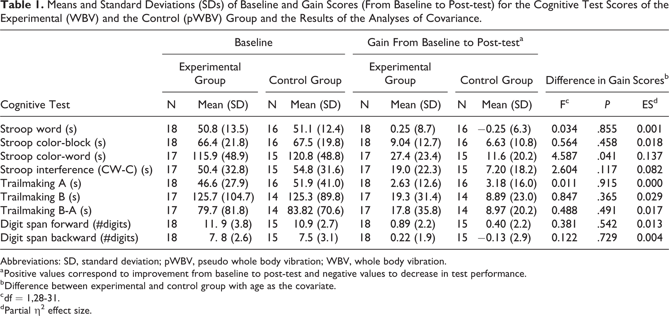
Perceived comfort was 7.0 (SEM 1.5) for the experimental group and 6.9 (SEM 1.6) for the control group (P > .05). Table 1 shows the cognitive test results for both groups at baseline and for the gain scores. Positive values on the gain scores indicate an improvement in test score from baseline to post-test. For all tests except the Trailmaking A test, the experimental group improved more than the control group. However, the differences were generally small with only a statistically significant effect for the Stroop Color-Word test with a nearly strong effect size (Figure 5). For the Stroop interference score, a statistically moderate nonsignificant effect was found.
Means and Standard Deviations (SDs) of Baseline and Gain Scores (From Baseline to Post-test) for the Cognitive Test Scores of the Experimental (WBV) and the Control (pWBV) Group and the Results of the Analyses of Covariance.
Abbreviations: SD, standard deviation; pWBV, pseudo whole body vibration; WBV, whole body vibration.
a Positive values correspond to improvement from baseline to post-test and negative values to decrease in test performance.
b Difference between experimental and control group with age as the covariate.
c df = 1,28-31.
d Partial η2 effect size.

Stroop Color-Word test scores for the experimental (WBV) and the control (pWBV) group pre (black bars) and post (white bars) intervention. Error bars are SEM; *P < .05. pWBV denotes pseudo whole body vibration; SEM, standard error of the mean; WBV, whole body vibration.
Discussion
In summary, PET imaging revealed that glucose uptake was not changed as a consequence of a 5-week WBV intervention. The WBV did, however, improve motor performance and reduced arousal-induced activity in mice. Cognitive tests in humans revealed a selective improvement in the Stroop Color-Word test. Taken together, it is concluded that our WBV intervention is a safe intervention that can improve at least some aspects of brain functioning. A limitation of the cognitive test in the human study, however, might be that we did not control for variables such as dietary supplements, caffeine intake, or sleep quality. Other factors influencing the direct comparison between mice and humans are based on the inherent differences between the species. Mice received WBV while standing on 4 legs, sitting or lying down, or a combination of these, whereas our 2-legged human participants were seated. Also the number of WBV sessions and the duration of the WBV session (respectively, 37 and 10 minutes in mice, and 27 and 4 minutes in humans) were not similar. The reason for the lower number of WBV sessions in humans was to ensure high adherence rates and to prevent to ask too much from our participants and supervisors (in case of the older participants). The shorter duration of the WBV session was based on pilot studies in which it was found that WBV sessions longer than 4 minutes were perceived as too long for the older participants as used in this study. Nonetheless, we found positive effects of WBV in both mice and humans.
The improvement in motor performance in the C57Bl/6J is in line with what we previously showed for the CD1 mice. 11 This corroborates the notion that WBV is known to improve neuromuscular performance. 40 The pWBV animals did improve as well, which could be attributed to a learning effect. Higher physical activity immediately after pWBV compared to WBV suggests that WBV reduces experimentally induced behavioral arousal. The arousal-reducing effect gradually increased in magnitude over the course of the experiment. The effect decreased within minutes after a session, as can be observed in Figure 3A, indicating a rather acute effect of WBV. If such an arousal-reducing effect also occurs in humans, it could contribute to the observed acute effects of WBV on attention. 25 Of note, the results also make clear that the 5-week WBV protocol by itself does not induce behavioral arousal in mice.
The cholinergic projection from the nucleus basalis in the forebrain to the amygdala might be involved in this arousal-reducing effect, as it is known that this projection responds to sensory input. 41 These cholinergic cells appear to be ideally located within the basal forebrain for evaluating sensory stimuli for their level of significance, via inputs from the midbrain and limbic system, and to modulate intrinsic cortical responsiveness appropriately in order to attend to sensory stimuli (see, for review, Wenk, 1997). Electrophysiological evidence has implicated cholinergic cells of the nucleus basalis in the control of attentional processes, as well as a role in the control and maintenance of arousal. We previously demonstrated that a 5-week WBV intervention results in an increased activity of the cholinergic projection of the nucleus basalis to the amygdala and neocortex. 12 Activation of the cholinergic system may, therefore, play a key role in the effects of WBV we found in mice and humans. Whole body vibration can be viewed as a form of passive exercise, which would be in line with observations that voluntary exercise can reduce arousal-induced activity and dampens anxiety-related behavior. 42,43 Even restricted exposure to voluntary exercise (a running wheel for 2 hours per day for 12 days) resulted in reduced anxiety behavior in mice. 44 Also in humans, exercise can alleviate anxiety. 45
To the best of our knowledge, this is the first 18F-FDG PET imaging study in the brain, related to WBV. The 18F-FDG PET data in our study did not reveal any significant difference in brain uptake ratio due to WBV. There was a small but not significant increase in the pWBV group posttreatment. Other (unpublished) data from our group typically indicate a much stronger brain glucose uptake over time if animals are more aroused during the tracer uptake period. The effects of brain glucose uptake are very sensitive to peripheral glucose metabolism. 32 As such it may well be that the absence of the increase in brain glucose uptake is associated with the arousal decreasing effect of the WBV protocol observed in this study. In humans, brain glucose metabolism was studied in relation to high intensity and aerobic exercise. An acute global decrease in brain metabolism has been found immediately after exercise. 46 Long-term chronic exercise interventions show more mixed patterns in glucose uptake increases, or decreases which seem very brain region specific. One study found an increase in glucose uptake in parietal-temporal and caudate regions after 12-week high intensity training. 47 A 3-month walking intervention in older women on the other hand showed differences in glucose uptake in varying brain regions between the control and the treated group, but no global differences in glucose uptake were observed. 48 Our results in mice after WBV stimulation are in line with these mixed findings in humans and indicate no major beneficial, but certainly also no detrimental effects of WBV in mice on baseline global brain glucose metabolism. It may well be that there are brain region-specific effects of the WBV stimulation in mice, but the detection of such effects in mice is hampered by the spatial resolution of PET.
The aim of the human part of this study was to explore the effects of a 5-week WBV intervention protocol in a sample of older adults without cognitive impairments. Our results showed that the experimental WBV with 30 Hz versus pWBV with 1 Hz improved the performance on the Stroop Color-Word test but not on other conditions of the Stroop test. In addition, no beneficial or detrimental effects were found for the TMT test and the Digit Span tests. These results are partly in line with prior research on the short-term effects on WBV in younger populations. The Stroop interference scores appeared to be better after WBV versus resting condition 25,26,28 and without an improvement in the Stroop Color-Block test score. 25,28 Similar to the current study, Regterschot et al. (2014) did not find an effect on the Digit Span Backward test. 25 If the cholinergic activity was also enhanced in humans by WBV as we found in mice, it could explain the improvement in the Stroop Color-Word test. This test positively correlates with cholinergic activity, 49 although it should affect other tests as well. Possibly, the used WBV protocol is not yet optimal to induce improvements in the other tests. Anyway, we should interpret this finding with caution given the small sample size of this pilot study, the large age range and the high variability in response as reflected in the standard deviations of the gain scores. Future research with larger sample size should support our findings. Also, research on potential neurobiological mechanisms underlying the hypothesis that WBV affects inhibition specifically is warranted. In addition, given the large variability in response, future studies should investigate personalized settings, in which also the repeated exposure of short bouts of WBV can be considered. Finally, more research is necessary to examine the population specificity of WBV effects. Especially people with cognitive impairments might benefit from WBV. A recent study 50 demonstrated that WBV is feasible and safe in older people with dementia, although they did not find evidence that WBV affected physical function or quality of life in this specific population. However, whether WBV affects cognitive function of people with dementia is still unknown. Taken together, it is concluded that our 5-week WBV intervention is a safe intervention to improve brain functioning, although the subtle effects suggest that the protocol is as yet suboptimal.
Footnotes
Acknowledgments
We thank Gosse Beeksma for his contribution to the literature search, Ar Jansen and Jürgen Sijbesma for their valuable biotechnical support in the animal study, and Siebrant Hendriks for his assistance in processing the PET data.
Declaration of Conflicting Interests
The author(s) declared no potential conflicts of interest with respect to the research, authorship, and/or publication of this article.
Funding
The author(s) received no financial support for the research, authorship, and/or publication of this article.