
Abstract
We reported the acceleration of skin wound healing in diabetic rats by repeated exposure to low-dose radiation (LDR). Here, we explored whether the wound healing could be further improved when LDR was combined with a topical application of basic fibroblast growth factor (bFGF) or zinc. Wounds were established on the backs of type 1 diabetic rats induced by a single injection of streptozotocin. Rats were treated daily with normal saline (Diabetes), LDR, bFGF, zinc, or combined 3 treatments for 5 consecutive days with a 2-day break between each consecutive 5-day treatment. Changes in wound size, histopathology, and microvessel density were assessed on days 5, 10, and 15, respectively, once treatment is started. All treatment regimens significantly accelerated skin wound healing, tissue remodeling, and new vessel formation compared to diabetes group. However, the combined LDR plus bFGF and zinc provided a better beneficial effect on wound healing than either one of these treatments alone. Further, we found that the effects of LDR and bFGF were similar, whereas zinc alone induced a weaker response. Our results suggest that whole-body LDR plus the topical application of bFGF and zinc can further accelerate wound healing in diabetic rats.
Introduction
Foot ulcer is a common complication in patients with diabetes, partially caused by impairments in normal wound healing. It has been reported that 3% of patients with diabetes mellitus will suffer an incurable foot ulcer, even if blood glucose levels and diet have been well controlled. 1 The etiology of impaired wound healing in patients with diabetes is very complex and attributed to numerous factors. For instance, hyperglycemia in diabetes was found to delay wound healing by decreasing the collagen content in the wound and inhibiting skin proliferation and differentiation. 2,3 The reduced levels of several growth factors in the wounds of patients with diabetes, 4 such as basic fibroblast growth factor (bFGF), 5 may also contribute to poor repair, as does an imbalance in matrix metalloproteinases (MMPs) and the tissue inhibitors of metalloproteinases, which play essential roles in wound healing by regulating wound debridement, angiogenesis, epithelialization, and remodeling of scar tissue. 6 Furthermore, impaired neovascularization due to dysfunctional endothelial progenitor cells (EPCs) also contributes to the delayed or reduced wound healing. 7,8 The levels of EPCs are decreased in the peripheral blood of diabetic patients and show a reduced ability to proliferate and differentiate at the site of healing.
Although a gold standard for the treatment of diabetic foot ulcers has been established, 9 which includes assessing the vascular status, optimizing glycemic control, debriding the wound, eliminating infection, and off-loading the ulcer, 20% to 30% of patients with diabetes will require amputation as a result of an infected foot ulcer. 10 Thus, the treatment of foot ulcers in patients with diabetes is still a challenge. As a result, several adjunct therapeutic methods have been investigated to facilitate wound healing in these patients, using methods that can theoretically address one or more of the underlying factors that lead to impaired wound healing.
The bFGF belongs to FGF superfamily. In acute wounds, an upregulation of bFGF stimulates the migration of human keratinocytes and fibroblasts as well as the synthesis and deposition of extracellular matrix. This, in turn, contributes to the formation of granulation tissue, reepithelialization, and tissue remodeling. 11 -13 However, as mentioned earlier, bFGF expression is decreased in the wounds of patients with diabetes, leading to a delay in wound healing process. 5 The topical application of recombinant human-bFGF (rh-bFGF) in diabetic wounds is therefore one of several plausible complementary or adjunct therapeutic options for treating foot ulcers in diabetes. Indeed, previous studies have confirmed that topical treatment with rh-bFGF accelerates wound healing in diabetic rats 14 and patients. 15,16 Further study disclosed that, to obtain a better therapeutic effect on wound healing, bFGF should be applied daily or using release delivery system due to its short half-life which has been proven to be less than 5 minutes in serum when used in intravenous injection in free form. 17,18
Zinc is an essential trace element in humans and interacts with many proteins, including various enzymes, to exert its biological role. 19 Zinc is reported to be involved in the wound healing process, with evidence to show that the level of zinc at the wound margin increases significantly after wounding, peaking at the time of granulation tissue formation, and epidermal proliferation. 20 Others have reported that zinc aids wound repair through the activation of MMPs. 21,22 Moreover, clinical and epidemiological studies have shown that patients with diabetes commonly have zinc deficiency. 23 -25 Therefore, topical application of zinc in diabetic wounds has shown the promise in a diabetic rat model. 26,27
Our previous study demonstrated that repeated whole-body, low-dose radiation (LDR) exposure to 75 mGy X-ray increased the proportion of CD31+/CD34+ stem cells in the bone marrow and in the circulation; promoted vessel regeneration, cell proliferation, and MMP-2 and MMP-9 expression in the wound tissue; and significantly accelerated skin wound healing in diabetic rats. 28 In the previous study, 75 mGy X-rays was used as LDR because we have shown its stimulation of bone marrow stem cells and mobilization into circulation. 29,30 This suggests the potential clinical utilization of LDR as an innovative, adjunct therapeutic method for diabetic wound healing. However, because of the etiological complexity of diabetic foot ulcers, the treatment of diabetes is complicated. We hypothesize that a combination of several complementary or synergistic therapeutic methods may be necessary for treating patients with diabetes, taking into consideration the multiple facets that contribute to the development of diabetic foot ulcers.
Thus, in the current study, we explored the therapeutic benefit of a combination of repeated whole-body LDR exposure and the topical application of bFGF and zinc on wound healing in rats with type 1 diabetes when compared to the 3 therapies alone (LDR, bFGF, or zinc). Therapeutic efficacy was measured in terms of wound healing rate, histopathological changes, and vascular regeneration.
Materials and Methods
Animals and Type 1 Diabetes Model
Male Wistar rats, weighing 180 to 220 g, from Jilin University Animal Center were maintained in light- (12:12-hour light–dark cycle) and temperature-controlled quarters (22°C) with free access to rodent chow and water. All animal procedures were approved by the Jilin University Animal Care and Use Committee, which is certified by the Chinese Association of Accreditation of Laboratory Animal Care. To induce type 1 diabetes, rats were intraperitoneally administrated with 50 mg/kg streptozotocin. From the third day after streptozotocin injection, fasting blood glucose levels of rats were measured using a Sure Step Complete Blood Glucose monitor (Abbott Diabetes Care Inc., Alameda, CA). When the measured fasting blood glucose level was higher than 16.7 mmol/L, the rats were considered diabetic and were used for subsequent experiments.
Establishment of the Skin Wound and Treatment
The cutis trauma model was established 60 days after the onset of diabetes as described previously. 28 Briefly, 120 diabetic rats were anesthetized, and the hair was removed with an electric razor and the skin disinfected with 75% ethanol. A 9-cm2 circular, full-thickness skin flap (3.4 cm in diameter) was removed from the backs of the rats. The diabetic rats were divided into 5 groups (n = 24), and treated with normal saline (Ctrl group), repeated whole-body LDR (LDR group), a topical application of bFGF (bFGF group) or zinc (zinc group), or a combination of LDR, bFGF, and zinc (multiple factors [MFs] group). As described in our previous study, 28 rats in the LDR group were exposed to whole-body X-rays at a dose of 75 mGy for 5 consecutive days with a 2-day break between each consecutive 5-day treatment. A total of 2 5-day consecutive irradiations of 75 mGy with a cumulative radiation dose of 750 mGy was given, and the rats killed on day 15 (2 × 5 irradiation days + 2 × 2 break days = 14 days, and day 15 rats were killed). For rats in the bFGF group, 50 U/cm2 bFGF diluted in normal saline was sprayed onto the surface of the wound for 5 consecutive days, with a 2-day break between 2 cycles of consecutive 5-day treatments. Usage of bFGF at 50 U/cm2 was according to our previous report, 31 where 1 to 4 µg/2.5 cm2 (equal to 40-160 U/cm2 with 100 U/µg bFGF). Rats in the zinc group were similarly treated but received 1 mL of 4% ZnSO4 solution. Topical application of zinc can be safely used with a wide range of doses. 32 -34 Particularly, considering that diabetic individuals may have certain extents of zinc deficiency, we provided 4% about 2 cycles of 5 daily applications with 2-day break between 2 cycles. Rats in the MF group were treated in series with whole-body LDR, bFGF, and ZnSO4 solution (as described above), with a 5-minute break between each treatment.
Histopathology and Masson Trichrome Staining
Rats were killed on days 5, 10, or 15 after wounding. The wounds were excised and fixed in 10% formalin. The fixed samples were embedded in paraffin and cut into 4-mm sections for hematoxylin and eosin (H&E) staining and Masson trichrome staining.
Immunohistochemical Staining for Evaluation of Vascular Regeneration
Paraffin sections were stained with antirat CD34 antibody (BD Sciences, San Diego, California) using the avidin–biotin–peroxidase complex (SABC) immunohistochemical method to detect CD34-positive cells. Any vessel with one or more CD34-positive cells was counted as a new vessel. Microvessel density (MVD) was determined by averaging 3 high-powered fields in each of the 3 slides from each animal.
Statistical Analysis
Experimental data are presented as the mean (SD). Comparisons were performed with a 2-sampled t test with equal variances using SPSS 19.0 statistical software. Differences were considered significant at P < .05.
Results
Rats in the MF Group Showed More Efficient Wound Healing
A full-thickness circular skin wound (3.4 cm in diameter) was made on the backs of the rats at 60 days after the onset of diabetes. Rats were then treated according to 1 of 5 conditions: control (saline), LDR alone, bFGF alone, zinc alone, or a combination of LDR, bFGF, and zinc. Wounded areas were analyzed at specified time points. As shown in Figure 1A and B, on day 5, the healing size increased significantly in the LDR, bFGF, and MF groups when compared to the control group; the zinc treatment alone showed no significant beneficial effect on wound healing. Moreover, the MF treatment accelerated wound healing more efficiently than either LDR alone or bFGF alone; however, there was no significant difference in healing size between these 2 single-treatment groups on day 5. By day 10; however, all of the therapeutic treatments significantly accelerated wound healing, with MF still providing the highest beneficial effect on wound healing. On day 15, MF resulted in an obvious reepithelialization on the wound and again showed the most significant effect on healing in the diabetic rats, with weaker responses, including the healing size and reepithelialization, seen for rats treated with LDR alone or bFGF alone. Rats in the zinc alone group again failed to show any significant increases in the healing size of the wound when compared to the control group; albeit, an increased tendency was observed. Moreover, the reepithelialization process was still not observed on the wound of diabetic rat in the control group, further confirming that the healing process of diabetic wound was delayed. These results indicate that wound healing in diabetic rats can be accelerated by LDR, bFGF, or a combination of factors (MF).
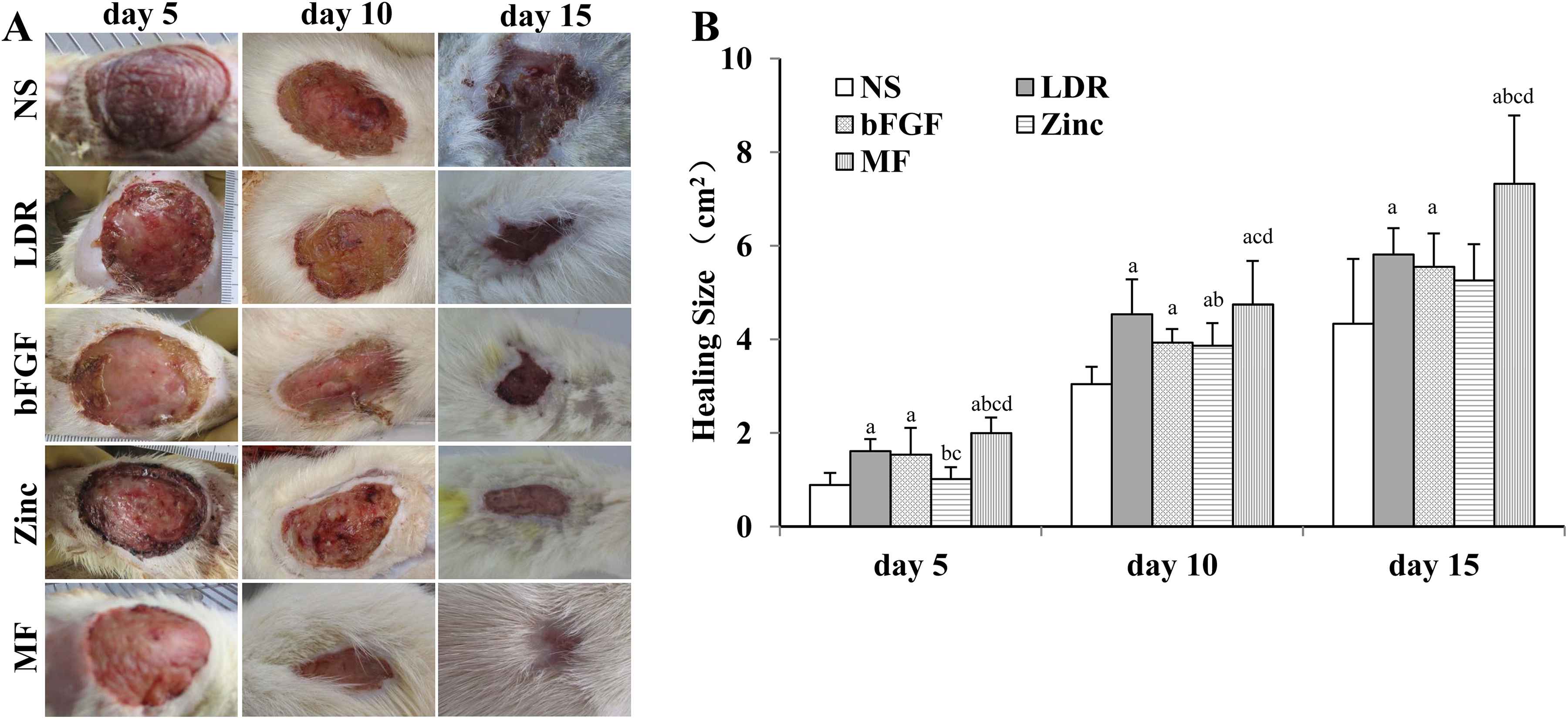
Skin wound and wound healing size. A, Representative images of skin wounds on days 5, 10, and 15 after wound formation. B, Healing size was calculated by determining the differences between the initial wound size (9 cm2) and the remaining unhealed size at the specific time point. Data are presented as the mean (SD; n = 8). LDR indicates low-dose radiation; MF, multiple factors; NS, normal saline. a P < .05 compared with NS; b P < .05 compared with LDR; c P < .05 compared with bFGF; d P < .05 compared with zinc.
Histopathological Examination of the Wounds of Diabetic Rats After Treatment
The H&E staining (Figure 2, left panel) at day 5 showed an apparent inflammatory response accompanied by large inflammatory exudate on the wound surface, and minimal granulation tissue formation in all groups. Moreover, the newly synthesized collagen in the wounds of all rats was thin, with an irregular and sparse arrangement. Among the groups, rats in the control group showed the most severe inflammatory response, with minimal granulation tissue and the presence of collagen fibers. On day 10, all of the treatment groups showed an increase in granulation tissue formation and an increase in the synthesis of collagen fibers in the wounds, with rats in the MF group showing the most beneficial effect. On day 15, rats in all of the treatment groups showed increased epidermal thickness and more regularly arranged collagen fibers, leading to improved scar healing, with the MF and LDR groups showing the best wound healing response.
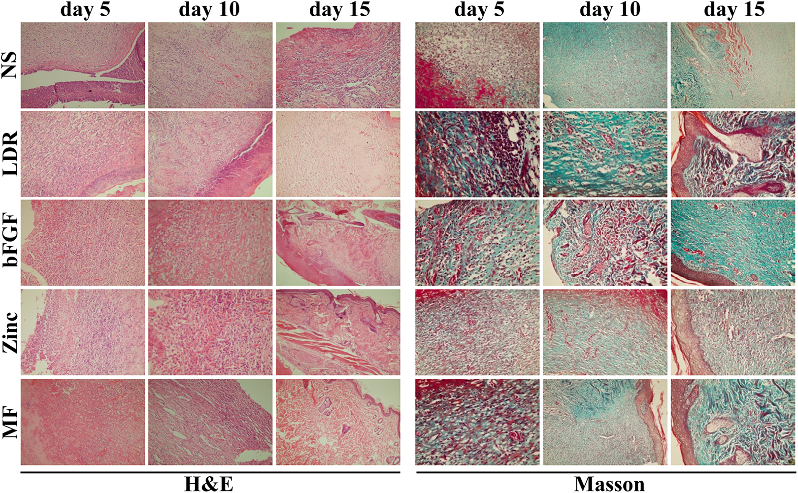
Histological examination. On days 5, 10, and 15 after wound formation, wound tissues from of mice each group were subjected to H&E (left panel) and Masson trichrome stain (right panel) staining and examined under a light microscope. The images (×200) are the representatives of each group (n = 8). H&E indicates hematoxylin and eosin; LDR, low-dose radiation; MF, multiple factors; NS, normal saline.
We also performed Masson trichrome staining of the skin to show collagen remodeling and maturation in the wounds. As shown in Figure 2 (right panel), the newly formed collagen on day 5 was arranged in the form of network within the wounds for all of the groups. By day 10, the content and arrangement of collagen fibers within the wounds had improved in all treatment groups, among which rats in the MF group showed the best improvement. Treatment with bFGF or zinc showed the least improvement, but the response was still better than the untreated control wounds, which showed scant, thin, and irregular collagen within the wounds. On day 15, we observed well-arranged collagen fibers in the wounds of rats treated with MF or LDR, with less regularity noted for collagen fibers in the wounds of rats treated with bFGF or zinc alone, as compared with the control.
A Combination of Factors Accelerates Vascular Regeneration in the Wounds of Diabetic Rats
Vascular regeneration is an important event during wound healing. We therefore sought to analyze the MVD, an index of new vessel formation in the wound. This was achieved by first measuring endothelial cell numbers using anti-CD34 immunohistochemical staining. As shown in Figure 3A, CD34-positive cells were detected in the wounds of all the rats at all of the examined time points. We found a higher level of new vessel formation on day 5 in the wounds of rats in the LDR, bFGF, and MF groups when compared to the control and zinc groups. On days 10 and 15, we found a significant increase in the MVD in all the treatments groups as compared with the control group; rats in the MF group showed the highest rates of neovascularization followed by rats in the LDR group, then rats in the bFGF and zinc groups (Figure 3B).

New vessel regeneration in wound tissue. A, On days 5, 10, and 15 after wound formation, wound tissues of mice from each group were subjected to immunohistochemical staining for CD34 and examined under a light microscope. The images (×200) are the representatives of each group (n = 8). B, Microvessel density (MVD) was calculated by counting the vessels with single or multiple CD34-positive cells in the high-magnification field (×200). LDR indicates low-dose radiation; MF, multiple factors; NS, normal saline. Data are presented as the mean (SD; n = 8). a P < .05 compared with NS; b P < .05 compared with LDR; c P < .05 compared with bFGF; d P < .05 compared with zinc.
Discussion
Foot ulcer is a serious and devastating complication of diabetes, aggravating the patient’s condition and posing a significantly increased economic burden to these individuals and to all national health-care systems across the globe. Although multiple adjunct therapies (eg, LDR, bFGF, or zinc) have been experimentally or clinically validated to accelerate wound healing in patients with diabetes, the treatment for diabetic foot ulcer is far from satisfactory, particularly given that many patients with diabetic foot conditions will finally require amputation. 10 In the current study, we demonstrated that a combination of multiple adjunct therapies, including LDR and the topical applications of bFGF and zinc accelerated wound healing more effectively than any of these 3 therapies alone. Histopathological examination and MVD analysis, to measure tissue reconstruction and vascular regeneration in the wound, respectively, further confirmed the superiority of the combinatorial approach.
We previously investigated whether LDR could accelerate diabetic wound healing, based on previous findings that LDR can mobilize hematopoietic progenitor cells into the peripheral blood circulation. 29 We found that multiple whole-body LDR accelerated wound healing, likely through mobilization of the bone marrow-derived stem cells. 28 More interestingly, the findings that LDR significantly reduces bacterial burden in the blood, spleen, and kidney of mice with sepsis 35 and that bacterial burden in diabetic wound contributes to impaired healing 36 indicate that multiple whole-body LDR might also promote wound healing of diabetic mice through enhancing bacterial clearance of wound. These findings encouraged us to further explore whether the combination of LDR with other therapeutic agents could induce a stronger positive effect on diabetic wound healing.
Zinc plays a key role during wound healing, and zinc deficiency is commonly found in patients with diabetes. Indeed, zinc deficiency has been attributed to poor or slowed wound healing in patients with diabetes. 23 -25 Similarly, serum zinc in diabetic rat was also found to be significantly lower than normal control, 37 and zinc-deficient rat induced by a low-zinc diet showed reduced wound breaking strength, 38 which indicated that the zinc deficiency of diabetic rat was also involved in impaired wound healing. A previous study showed that topical zinc through the use of transdermal iontophoresis can increase the breaking strength in skin scars and accelerate wound healing in diabetic rats. 27 Furthermore, it has been reported that topical application of zinc oxide in wound of diabetic rat significantly inhibited bacterial growth in granulation tissue, which disclosed one of the potential underlying mechanisms through which zinc supplement promote wound healing of diabetes. In the current study, we found that spraying a topical 4% ZnSO4 solution could promote vascular regeneration and tissue reconstruction in wound healing, thereby accelerating wound healing in diabetic rats. However, these positive effects were seen at a later time point than treatment with the other agents and were of a lower intensity than treatment with LDR alone.
Like zinc, studies have reported increased bFGF levels during acute wound healing under normal conditions, with bFGF reported to stimulate granulation tissue formation. 11 -13 Because low levels of endogenous bFGF are expressed in patients with diabetes, 5 we surmised that the topical application of rh-bFGF in diabetic wounds would be a rational adjunct approach to improve treatment. In a previous study by Uchi et al, 15 topical bFGF treatment of nonischemic diabetic ulcers for 8 weeks dose dependently accelerated wound healing, confirming the clinical efficacy of bFGF for diabetic wound treatment. Our results showed that the application of 50 U/cm2 bFGF for multiple days also accelerated wound healing in diabetic rats. Yet, in terms of the end points in our study, we found that the therapeutic effect of bFGF on wound healing was similar to that of zinc supplementation and was deemed overall to be weaker than that seen with LDR alone.
Finally, as expected, the combination of LDR, bFGF, and zinc gave the most significant therapeutic effect on wound healing, with the greatest improvement in the size of the original wound, and evidence of tissue reconstruction that resembled normal tissue. We also found significant vascular regeneration when compared to LDR, bFGF alone, or zinc alone. However, the beneficial effect of the combined treatments was lower than the sum of individual effect of each treatment alone, indicating that the effect of LDR was additive with those of bFGF and zinc. Overall, our findings suggest that wound healing in diabetic rats can be enhanced by a combination of repeated whole-body LDR exposure and the topical application bFGF and zinc, thus offering a potentially novel approach for the treatment of wounds in patients with diabetes.
Footnotes
Declaration of Conflicting Interests
The author(s) declared no potential conflicts of interest with respect to the research, authorship, and/or publication of this article.
Funding
The author(s) disclosed receipt of the following financial support for the research, authorship, and/or publication of this article: This study was supported by a research grant from Science and Technology. Department of Jilin Province (20160101035JC to W-y.G.) and National Natural Science Foundation of China (81300660 to W-y.G.).