
Abstract
The radioprotective effect of Achillea millefolium L (ACM) extract was investigated against genotoxicity induced by ionizing radiation (IR) in human lymphocytes. Peripheral blood samples were collected from human volunteers and incubated with the methanolic extract of ACM at different concentrations (10, 50, 100, and 200 μg/mL) for 2 hours. At each dose point, the whole blood was exposed in vitro to 2.5 Gy of X-ray and then the lymphocytes were cultured with mitogenic stimulation to determine the micronuclei in cytokinesis-blocked binucleated cell. Antioxidant capacity of the extract was determined using free radical-scavenging method. The treatment of lymphocytes with the extract showed a significant decrease in the incidence of micronuclei binucleated cells, as compared with similarly irradiated lymphocytes without any extract treatment. The maximum protection and decrease in frequency of micronuclei were observed at 200 μg/mL of ACM extract which completely protected genotoxicity induced by IR in human lymphocytes. Achillea millefolium extract exhibited concentration-dependent radical-scavenging activity on 1,1-diphenyl-2-picryl hydrazyl free radicals. These data suggest that the methanolic extract of ACM may play an important role in the protection of normal tissues against genetic damage induced by IR.
Introduction
Ionizing radiation (IR) produces reactive oxygen species (ROS) such as OHO, H2O2, OHO, O2−. These toxic substances are highly chemically reactive and can react with cellular biomolecules including proteins, lipids, and DNA, resulting in varieties of oxidative lesions. 1 Elevated ROS level has been suggested to be involved in cellular dysfunction and death. Reactive oxygen species are able to cause DNA breaks. Radiation-induced DNA lesion is main reason for cell killing. If DNA lesions can’t be effectively repaired by endogenous defense systems, it leads to genome instability and chromosome abnormalities. 2 Radiation-induced genome aberration has a crucial role in the mechanisms underlying radiation-induced carcinogenesis. 3 With respect to radiation damage to humans, it is important to protect biological systems from radiation-induced genotoxicity. Amifostine is a powerful radioprotective agent with a thiol group in its structure, but this drug has limited usage in clinical practice due to its side effects and toxicity. 4,5 The search for less toxic radioprotectors has spurred interest in the development of natural products.
Achillea millefolium L (Compositae; ACM) is a well-known medicinal plant. 6,7 In Iranian folk medicine, several species of Achillea called Bumadaran in Persian have been used to treat bleeding, wounds, inflammation, pain, and spasmodic diseases. 8,9 Phytochemical studies on ACM have shown that this plant is rich in flavonoids and caffeic acid derivatives. 10 –12 The presence of other secondary metabolites including essential oil, sesquiterpenes, alkaloid, steroids, and triterpenes has also been reported in this plant. 13 Previous studies on the extracts of ACM have reported antioxidant, 14 antiviral, 15 antispasmodic, 16 hepatoprotective, 17 gastroprotective, 18 estrogenic, 19 immunological, 20 and anti-inflammatory 21 activities. In addition, the aqueous extract of this plant showed protective effects against cyclophosphamide-induced testicular toxicity. 22 Based on the above findings and the anti-inflammatory and antioxidant potential of ACM extract, the present study aimed to evaluate the radioprotective activity of the plant by using in vitro radiation-induced genetic damage in volunteers’ human blood lymphocytes.
Materials and Methods
Plant Material and Extraction
The aerial parts of ACM were collected from Kalat Naderi, Khorasan-e-Razavi province, Iran, at 1586 m above sea level, during the flowering stage. A voucher specimen (44246) was deposited at the herbarium of the Research Center for Plant Sciences, Ferdowsi University of Mashhad, Mashhad, Iran. Dried aerial parts of the plant were cut into small pieces and then extracted with methanol by maceration at room temperature. The extract was concentrated by using a rotary evaporator (Heidolph, Germany) and dried by a freeze dryer (Zirbus, Germany).
Genotoxicity Assay
This study was performed after obtaining permission from medical ethics committee of the university. Healthy, nonsmoking human male volunteers aged between 20 and 25 years were enrolled in this study. Twelve milliliter whole blood were collected in heparinized tubes and divided in centrifuge tube at 0.9 mL. Blood samples were treated with 100 μL solution of herbal extract at the concentrations 10, 50, 100, or 200 μg/mL (final concentrations). These samples were incubated for 2 hours at 37°C. The extract was dissolved in ethanol and diluted in cultural medium. Ethanol concentration was same in control and extract solutions (0.5%).
At each concentration and for each volunteer, tubes were irradiated at 37°C with 6 MV X-ray beam produced by a radiotherapy machine (Linear accelerator, Siemens, Primus, Germany) at a dose of 2.5 Gy with a dose rate of 190 cGy/min. Three tubes were used as the control nonirradiated sample from 3 volunteers. Subsequently, 0.5 mL of each sample (control and irradiated samples in duplicate) was added to 4.4 mL of Roswell Park Memorial Institute (RPMI) 1640 culture medium (Gibco, Paisley, UK), which contained 10% fetal calf serum. Then phytohemagglutinin (100 µL/5 mL, Gibco) was added to the cultures as lymphocyte stimulator. All cultures were incubated at 37°C in a humidified atmosphere of 5% CO2 and 95% air. Cytochalasin B (Sigma, USA; final concentration: 6 µL/mL) was added after 44 hours of culture. Following 72 hours of incubation, the cells were collected by centrifugation for 7 minutes at 3500 rpm and resuspended in cold potassium chloride. Cells were immediately fixed in a fixative solution as methanol–acetic acid (6:1) 2 times. The fixed cells were dropped onto clean microscopic slides, air-dried, and stained with Giemsa solution (10%). All slides were evaluated at 100× magnification in order to determine the frequency of micronuclei in the cytokinesis-blocked binucleated cells with a well-preserved cytoplasm. 23 At each concentration and for each volunteer, 1000 binucleated lymphocyte cells were examined from the irradiated and control cultures in duplicate to record the frequency of micronuclei.
Measurement of Free Radical-Scavenging Activity
The free radical-scavenging capacity of the methanolic extract of ACM was determined as bleaching of the stable 1,1-diphenyl-2-picryl hydrazyl radical (DPPH). 24 Different concentrations of the extract (0.05-1 mg/mL) were added, at 2 mL, to 2 mL methanol solution of DPPH (10 mg/250 mL). After 15 minutes at room temperature, the absorbance was recorded at 517 nm. The experiment was performed in triplicate, and butylated hydroxytoluene (BHT) was used as a standard antioxidant agent. Percentage of scavenging was calculated using the formula: [(Control − Test)/Control] × 100.
Statistical Analysis
For each volunteer, at each concentration, the incidence of radiation-induced micronuclei was recorded. The data were analyzed with student t test. A P value of .05 was accepted to denote significance.
Results
The percentage of micronuclei in binucleated lymphocytes in 3 donors treated with 2.5 Gy X-ray was 5.41 ± 1.25, while the percentage in nontreated control lymphocytes was 0.69 ± 0.17. It showed a significant increase of 8-fold in frequency of micronuclei in lymphocytes exposed to 2.5 Gy of X-ray (P < .01; Table 1). The frequency of micronuclei after pretreatment with ACM at doses of 10, 50, 100, or 200 μg/mL was 2.76 ± 0.23, 1.85 ± 0.05, 1.13 ± 0.06, and 0.73 ± 0.06, respectively (Figure 1). The data demonstrate that human blood incubated with ACM, and then exposed in vitro to X-ray radiation, exhibited a significant reduction in micronuclei frequency compared to blood samples incubated with X-ray alone. The extract at all concentrations exhibited significantly lower micronuclei frequency than irradiation control sample (P < .01). The values of total micronucleated binucleated cells were 49%, 66%, 79% ,and 86% fold less in the 10, 50,100 and 200 μg/mL concentrations of ACM extract (Table 1). A concentration-dependent effect for ACM was observed in the reduction of chromosome damage in lymphocytes exposed to ionizing radiation (P < .01). The ACM did not exhibit any genotoxicity in cultured lymphocytes at concentration of 200 µg/mL without exposure to radiation. It is interesting to observe that the frequency of micronuclei in ACM at a concentration of 200 µg/mL was lower than control group (P < .01).
The Frequency of Micronuclei Induced In Vitro by 250 cGy X-Ray Radiation (IR) in Cultured Blood Lymphocytes From Human Volunteers Examined at Different Doses of Achillea millefolium (ACM).

Abbreviations: BN, binucleated; SD, standard deviation.
aOne thousand BN cells were examined in each sample.

In vitro protection by Achillea millefolium (ACM) at different concentrations (10, 50, 100, and 200 µg/mL) against genetic damage induced by X-ray (IR; 2.5 Gy) in cultured whole blood lymphocyte. The data represent average ± standard deviation of 3 human volunteers. P < .001: Sample at control compared with similarly irradiated lymphocytes. P < .01: IR sample compared to 10 ACM + IR, 50 ACM + IR, 100 ACM + IR, and 200 ACM + IR samples. P < .01: 10 ACM + IR and 50 ACM + IR; 50 ACM + IR and 100 ACM + IR; and 100 ACM + IR and 200 ACM + IR samples. P < .01: between groups of 200 ACM and control samples.
In antioxidant assay, an excellent scavenging effect was observed with ACM. Scavenging effects of the methanolic extract of ACM on DPPH radicals increased with the increase in concentrations, it was 90% at 1 mg/mL which was similar to BHT (Figure 2).
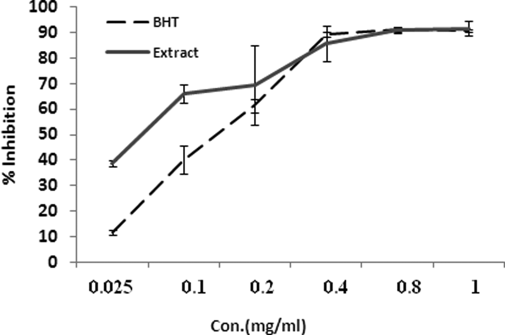
Scavenging effect of different concentrations of Achillea millefolium (extract) and butylated hydroxytoluene (BHT) on the 1,1-diphenyl-2-picrylhydrazyl (DPPH) free radical at 517 nm.
Discussion
In this study, we demonstrated that the methanolic extract of ACM has potent radioprotective effect against genotoxicity induced by X-ray in human lymphocytes. Achillea millefolium reduced the frequency of micronuclei in binucleated lymphocytes that increased by IR and exhibited protective effect at concentrations of 10, 50, 100, and 200 μg/mL by factors 1.9, 2.9, 4.8, and 7.4, respectively. Furthermore, considerable antioxidant activity with free radical-scavenging property was observed from the extract. With respect to side effects induced by IR in patients undergoing radiotherapy, the radioprotective agents have an important role for reduction of side effects in patients. Increase in the intracellular level of ROS produced by IR is a potentially toxic insult. The ROS interact with macromolecules to induce DNA damage. Natural compounds may play a role in scavenging free radicals such as hydroxyl radicals generated by ionizing radiation. In this study, ACM exhibited radioprotective effect on reducing micronuclei induced by X-ray. Treatment of whole blood with ACM for 2 hours prior irradiation reduced the frequency of micronuclei. Antioxidant activity of ACM have been reported in several studies. 11,16 Oral administration of the hydroalcoholic extract of ACM reduced gastric ulcers induced by acetic acid. The antioxidant activity of the extract may be responsible for its gastroprotective effect. 18 In other study, the aqueous extract of this plant with antioxidant and anti-inflammatory potential showed protective effect against cyclophosphamide-induced testicular toxicity. 25
In the present study, ACM extract showed a potent radical-scavenging effect against free radical DPPH. The main characteristic of antioxidants is an ability to trap free radicals. The ROS may attack DNA, proteins, and lipid that can initiate degenerative diseases. Antioxidant compounds such as phenolic acids, poly phenols, and flavanoids can scavenge free radicals and protect normal tissues against disease related to oxidative stress. Chemical analysis of the methanol extract of the aerial parts of ACM showed the presence of several phenolic compounds such as flavonoids (eg, rutin, luteolin glucosides, apigenin glucosides) and quinic acid derivatives such as chlorogenic acid. 10 Chlorogenic acid and other caffeoylquinic acid derivatives exhibit antioxidant activities and DNA damage protective effects. 22 In a research, chlorogenic acid and quinic acid showed radioprotective effects and decreased the DNA damage induced by X-ray irradiation in human blood lymphocytes in vitro. 26 Flavonoids with high antioxidant activity are considered as a radioprotector. 27 Some flavonoids such as luteolin 7-O-glucoside isolated from the whole plant of Pilea microphylla exhibited protective effect against γ radiation-induced cytotoxicity. These compounds showed significant antioxidant activity and reduced lipid peroxidation, formation of intracellular ROS, and DNA strand breaks. 28 Apigenin, as a dietary antioxidant, protected human peripheral blood lymphocytes from γ radiation-induced oxidative damages. 29
Previously, we showed that extracts of Zataria multiflora and hawthorn as medicinal plants protected human lymphocytes against genotoxicity induced by gamma irradiation. Hawthorn and Zataria extracts that have strong antioxidant activities may affect scavenging free radicals such as hydroxyl radicals generated by γ-rays in cells. 30,31 It is interesting, ACM at high concentration, 200 μg/mL, completely normalized DNA damage induced by IR on human lymphocyte. This result is promising that a natural product showed an excellent radioprotective effect.
In this study, it was found that ACM with antioxidant properties can contribute to reduce genotoxicity induced by IR in human lymphocytes as well as can be used as herbal medicine in several diseases, and it can help to protect body against side effects induced by irradiation during radiotherapy.
Footnotes
Authors’ Note
This research was the subject of a PharmD thesis of Mostafa Rostamnezhad as a student of Mazandaran University of Medical Sciences.
Declaration of Conflicting Interests
The author(s) declared no potential conflicts of interest with respect to the research, authorship, and/or publication of this article.
Funding
The author(s) disclosed receipt of the following financial support for the research, authorship, and/or publication of this article: This study was supported by a grant from Mazandaran University of Medical Sciences, Sari, Iran.