
Abstract
One-dimensional materials, such as nanowires, nanotubes, or nanofibers, have attracted more and more attention recently due to their unique physical properties. Their large length-to-diameter ratio creates anisotropic material properties which could not be reached in bulk material. Especially one-dimensional magnetic structures are of high interest since the strong shape anisotropy reveals new magnetization reversal modes and possible applications. One possibility to create magnetic nanofibers in a relatively simple way is offered by electrospinning them from polymer solutions or melts with incorporated magnetic nanoparticles. This review gives an overview of most recent methods of electrospinning magnetic nanofibers, measuring their properties as well as possible applications from basic research to single-cell manipulation to microwave absorption.
Production of magnetic nanofibers
Electrospinning technology
The electrospinning technology can be used to prepare nanofibers or nanofiber mats from polymer solutions or melts, possibly including ceramics, metallic nanoparticles, carbon nanotubes, and so on, in addition.1–3 Generally, electrospinning can be subdivided into needle-based and needleless techniques (Figure 1). In the needle-based method, a polymer solution or melt is pressed through a needle into a strong electrical field, drawing the polymer to a substrate and on this way stretching and thinning the fiber so that diameters of some ten to some hundred nanometers are reached. Needleless methods include, for example, a wire-based technique in which one wire is coated with a polymer solution or melt, which is drawn to the second wire by the strong electrical field between them. Alternatively, rotating cylinders and other geometries can be used as electrodes, modifying not only the amount of created fibers per time, but also their alignment. In both cases, the so-called Taylor cones are formed between both electrodes. This influences the fiber formation due to possible instabilities, resulting in modifications of the fiber lengths and diameters. 4 Modified electrospinning equipment in addition allows for creating core-shell or similar double-material structures, also called Janus structures,5–7 for electrospinning hollow fibers 8 or even more sophisticated hierarchical structures. 9

Different electrospinning techniques: (a) needle-based, (b) wire-based, and (c) using a rotating cylinder.
Polymers for electrospinning
Typical electrospinning polymers are, for example, water-soluble ones such as polyethylene glycol (PEG or PEO) which is often used as spinning agent (Figure 2(a)),10–12 polyacrylonitrile (PAN) which belongs to the few materials which can be electrospun from the low-toxic solvent dimethyl sulfoxide (DMSO) (Figure 2(b))13,14 and is often used as a precursor for carbon nanofibers,15,16 or poly(ɛ-caprolactone) (PCL) which is typically used in cell growth applications.17,18 Blending different polymers is as well possible as adding non-polymeric substances, such as ceramics or metals.19,20

Nanofiber mats prepared from (a) PEG and (b) PAN.
Adding magnetic properties to nanofibers is usually performed by adding magnetic nanoparticles to the polymer solution or blend. Different mechanical and magnetic properties can be reached by working with the blended fibers or calcinating them to obtain the pure metallic fibers.
Field-assisted electrospinning technology
Guarino et al. used a needle-based electrospinning setup to create magnetic nanofibers with polycarbonate-urethane (PCU) as polymer filled with nickel nanoparticles. They modified the counter-electrode with permanent magnets, creating a constant magnetic field perpendicular to the electric field, in this way preparing aligned magnetic nanofibers between the magnetic poles (Figure 3). 21

Magnetic field–assisted needle-based electrospinning.
Another magnetic field–assisted electrospinning process was described by Mei et al. In their electrospun PAN/FeCo nanofibers, the alignment degree increased with decreasing voltage and increasing FeCo content. 22
Shehata and Abdelkader 23 used a concave collector, modified by magnetic disks creating a magnetic field during electrospinning, and found the degree of fiber orientation increased, as compared with the unmodified concave or the conventional flat collector. The self-organization of a polyvinyl alcohol (PVA)/ferrofluid cone array, forming tips from which electrospinning occurred in the presence of an electric field in addition to the magnetic field, was used to create magnetic nanofibers with relatively high saturation magnetization. 24
Nickel ferrite–barium titanate core-shell nanofibers were created by magnetic field–assisted electrospinning, allowing for assembling them into disks or films in a uniform or gradient magnetic field. 25
Magnetic composite nanofibers
Generally, magnetic nanofibers can be used when the polymer material is kept after electrospinning, so that the magnetic nanoparticles are usually embedded in a polymer matrix.
PAN nanofibers with Fe3O4 (magnetite) nanoparticles were needle-electrospun from DMF (dimethylformamide) after ultrasonic dispersion for 12 h. 26 Ultrasonic treatment, however, was investigated in detail by Peer et al., using Fe-based magnetic nanoparticles in a PEO matrix. They found that a compromise must be made between the increased nanoparticle distribution in the polymer solution and the loss of possible damage of the polymer, leading to a loss of quality of the electrospun nanofibers, which they suggested to be reached after 30 min of ultrasonic treatment. 27
Using polyvinylpyrrolidone (PVP) as spinning agent, Fe3O4/PVP nanofibers were electrospun as one layer of an electrospun composite, comprising magnetic, conductive, and photoluminescent properties. 28 In combination with PCL, Fe3O4/PCL nanofibers were needle-electrospun. 29 Different amounts of Fe3O4 and Fe2O3/NiO (diiron nickel tetroxide or nickel ferrite) were electrospun with PAN by a wire-based technique. 30 Using a blend of carboxy methyl cellulose (CMC) and PVA, Fe3O4/CMC/PVA-blended fibers were needle-electrospun, indicating that fiber diameters as well as the numbers and shapes of beads along the fibers vary with the metal content. 31 Embedding Fe3O4/graphene nanoparticles in PVA nanofibers (Figure 4), Noori et al. 32 found significantly reduced nanofiber diameters and increased thermal stability, as compared to pure PVA nanofibers.

Embedding nanoparticles, for example, from graphene and Fe3O4, into a polymer solution, for example from PVA, results in composite nanofibers. 32
To avoid nanoparticle agglomerations, iron oxide nanoparticles were surface functionalized using polyethyleneimine (PEI), aminopropyltriethoxysilane (APTS), PEG, and tetraethoxysilane (TEOS) before electrospinning with polyamide (PA) 6.33,34
Hernández et al. used electrospinning to prepare Nd0.05Bi0.95Fe0.95Co0.05O3 fibers from a polymer blend solution, resulting in nanofibers with partly interesting curls. In addition, these fibers revealed conductivity above the usual ferroelectrical materials’ range. 35
Rare earth magnets are only scarcely used for electrospinning. Wang et al., 36 for example, prepared gadolinium methacrylate from methacrylic acid (MAA) and gadolinium oxide, mixed the resulting crystal powder with dicumyl peroxide and PAN, and prepared nanofibers by needle-electrospinning.
Pure magnetic nanofibers
Another possibility is calcinating the polymer after electrospinning to obtain pure magnetic or pure ceramic nanofibers.
Li et al. prepared CoFe2O4 nanofibers by first preparing a sol of Co(NO3)2·6H2O, Fe(NO3)3·9H2O, citric acid, ethanol, and distilled water. By calcinating this precursor under inert gas atmosphere, they received CoFe2O4 nanoparticles which were needle-electrospun with PEO. Afterward the fibers were again calcinated in inert gas to obtain pure CoFe2O4 nanofibers. 37
CoxZn(1-x)O nanofibers were created by needle-electrospinning Zn(CH3COO)2·2H2O, Co(CH3COO)2·4H2O, and PVP from a DMF/ethanol solution, followed by calcination of the nanofibers. 38
A hybrid material of La0.7Sr0.3MnO3 (LSMO), a ferromagnetic material with colossal magneto-resistance, and La1.85Sr0.15CuO4 (LSCO), a high-temperature superconductor, was needle-electrospun using PVA. After calcination, an additional oxygenation step was necessary to tailor the desired phase composition.39–42
SmCoFe nanofibers with different degrees of Fe substitution, as compared to pure SmCo nanofibers, were needle-electrospun using PVP as polymer, which was afterward calcinated out of the fibers. 10at% Fe substitution resulted in an increased net magnetic moment, coercive field, and saturation magnetization and thus in an increased maximum energy product.43–45
Ghazi et al. prepared magnesium-zinc ferrite nanofibers by electrospinning, using PVP as the spinning polymer, doped with zinc nitrate, ferric nitrate and magnesium nitrate hexahydrate. They found the cubic spinel structure of magnesium ferrite to be obtained also for Zn doping. After calcination at 550°C, the maximum of the particle size distribution was smallest for 10% Zn concentration. 46
NiCo2O4 (nickel cobaltite) was embedded in a polymer solution from poly(styrene-co-acrylonitrile) (SAN); the electrospun nanofibers were afterward calcinated, resulting in fractal NiCo2O4 aggregates along the nanofibers after temperature treatment. 47
To obtain the α-Fe2O3 phase from PVA/Fe2O3 nanofibers, a heat treatment was used, reducing the fiber diameter which was also influenced by the PVA content of the mixture. 48
A more complicated technique was used by Guarino et al. to prepare polydimethylsiloxane (PDMS) nanofibers with Ni nanoparticles. By coaxial electrospinning, core-shell fibers with PDMS/Ni in the core and a PVP shell were created. After removing the shell, the core exhibited interesting elasto-magnetic deflection due to transversal magnetization. Using such nanofibers, magnetic forces could be used to deform devices which may open new applications in biomedicine. 49
For the preparation of LaFe1-xMnxO3 nanofibers, Jeong et al. dissolved La(NO3)3, Fe(NO3)3, and Mn(NO3)2 with PVP in DMF and performed needle-electrospinning. The fibers were afterward calcinated to remove the polymer. With increasing Mn doping, the average crystallite size decreased, while the fiber diameters stayed similar. 50
For strontium hexaferrite nanoparticles embedded in PVA nanofibers, Murillo-Ortiz et al. 51 interestingly found a positive effect of relatively low nanoparticle densities in the polymer matrix on the fibers’ hard magnetic properties which was attributed to the vanishing inter-particle exchange interaction which made the magneto-static interaction dominant and created a new magnetic shape anisotropy along the fibers.
Magnetic carbon nanofibers
As explained above, PAN belongs to the possible precursors of carbon nanofibers. The aforementioned nanofibers either included the polymer used for electrospinning, or this polymer was calcinated out of the fibers to produce pure metal fibers. There are, however, also literature reports on a third approach, using PAN as the polymer enabling electrospinning and afterward stabilizing and carbonizing the PAN/nanoparticle-blended fibers to obtain magnetic carbon fibers (Figure 5).

PAN/nanoparticle-blended electrospun nanofibers can be stabilized and carbonized to create magnetic carbon fibers.
Huang et al., for example, used PAN and polyvinylidene fluoride (PVDF) in different ratios, combined with Fe(NO3)3·9H2O, for needle-electrospinning. These nanofiber mats were stabilized at 250°C in air and afterward carbonized at 800°C in nitrogen atmosphere to obtain carbon fibers with Fe and FeOx nanoparticles.52,53 The slow stabilization process in air is a necessary first step before carbonization can be carried out, resulting in cyclization, dehydrogenation, oxidation, aromatization, and crosslinking reactions. 16 Without this thermal pretreatment, the PAN nanofibers would be decomposed at temperatures higher than approx. 300°C instead of being carbonized.
Blending PAN and polymethylmethacrylate (PMMA) with C15H30FeO6 was used to create porous carbon/Fe nanofibers by electrospinning, followed by stabilization and carbonization. 54
By using nickel(II) acetylacetonate and cobalt(III) acetylacetonate as precursors, partly combined with multiwall carbon nanotubes, magnetic carbon fibers were created by electrospinning both magnetic precursors with PAN from DMF. Here, the authors found that the fiber diameters decreased with increasing nanoparticle concentration, which they attributed to the increased conductivity of the spinning solution. 55
A more complicated method was necessary to create magnetic nanofibers containing gadolinium (Gd) and europium (Eu). To create Gd2O2S nanofibers doped with dysprosium (Dy) and Eu, Gd2O3, Eu2O3 and Dy2O3 were dissolved in HNO3 (nitric acid). After heating and thus drying the solution, lanthanide nitrides were gained which were put into DMF in which afterward PVP was dissolved. Needle-electrospinning resulted in nanofibers which were afterward sintered at 700°C. These pre-products were then sulfurized using the dual-boat sulfurization method, placing the samples beside a porcelain boat with sulfur powder in the furnace, resulting in the desired Dy- and Eu-doped Gd2O2S nanofibers. 56
Abdalla et al. also added NiFe2O4 nanoparticles and partly additional multiwall carbon nanotubes to carbon nanofibers, produced by electrospinning them from a solution of PAN in DMF, and afterward stabilized and carbonized at 850°C. Adding the multiwall carbon nanofibers to the spinning solution resulted in an increase of the amount of prepared nanofibers by nearly a factor of 2. 57
Multifunctional magnetic nanofibers
Sometimes, magnetism is not the main function of magnetic nanofibers, but the magnetic material serves as a base for other functionalities. Most recently, Wang et al. 58 produced CuFe2O4 fibers in which they could tailor the amount of CuO nanoparticles on the fiber surfaces by modifying the ratio of Co to Fe precursor, making such fibers applicable for catalytic oxidation.
In other cases, other physical properties are desired besides ferromagnetism. In this case, Janus fibers are often useful to avoid undesired interdependencies between both functionalities (Figure 6).

Janus fibers can be produced by electrospinning from two syringes to combine desired material properties.
Conjugate electrospinning was used to prepare Janus microfibers which combined magnetic with fluorescent properties, but strictly separating both materials responsible for the respective properties which avoided undesired influences of each material on the other one. 59
Janus nanostructures with tunable magnetic and photoluminescent properties were prepared from Eu(benzoic acid)3 1,10-phenanthroline, Tb(benzoic acid)3 1,10-phenanthroline, Fe3O4 nanoparticles, and PVP as the polymer used as a spinning agent. 60
Another method was described by Wang et al. They also used PVP in ethanol with Tb(benzoic acid)3 1,10-phenanthroline to create fluorescence properties, but added Fe3O4 and PVP in ethanol for the magnetic part. Conjugate electrospinning was performed using two syringes with plastic spinnerets for the two solutions and a rotating drum as collector, resulting in aligned nanofibers with similar diameters. 61
Xi et al. 62 created luminescent, magnetic, and electrically conductive Janus fibers via a homemade coaxis/monoaxis spinneret, preparing a coaxial Fe3O4/PVP core and a Eu(benzoic acid)3 1,10-phenanthroline/PVP shell as one part of the fiber, and adding the conductivity by the second half of the fiber, consisting of polyaniline/PVP.
Janus fibers from CoFe2O4/PAN and 1,8-naphthalene anhydride/PVP were electrospun, revealing good magnetic and fluorescent properties. 63
Fan et al. 64 combined magnetic and fluorescent properties by combining Fe3O4/PAN and Eu(benzoic acid)3 1,10-phenanthroline/PAN electrospun nanofibers and found higher fluorescence than in mixed Fe3O4/Eu(benzoic acid)3 1,10-phenanthroline/PAN nanofibers.
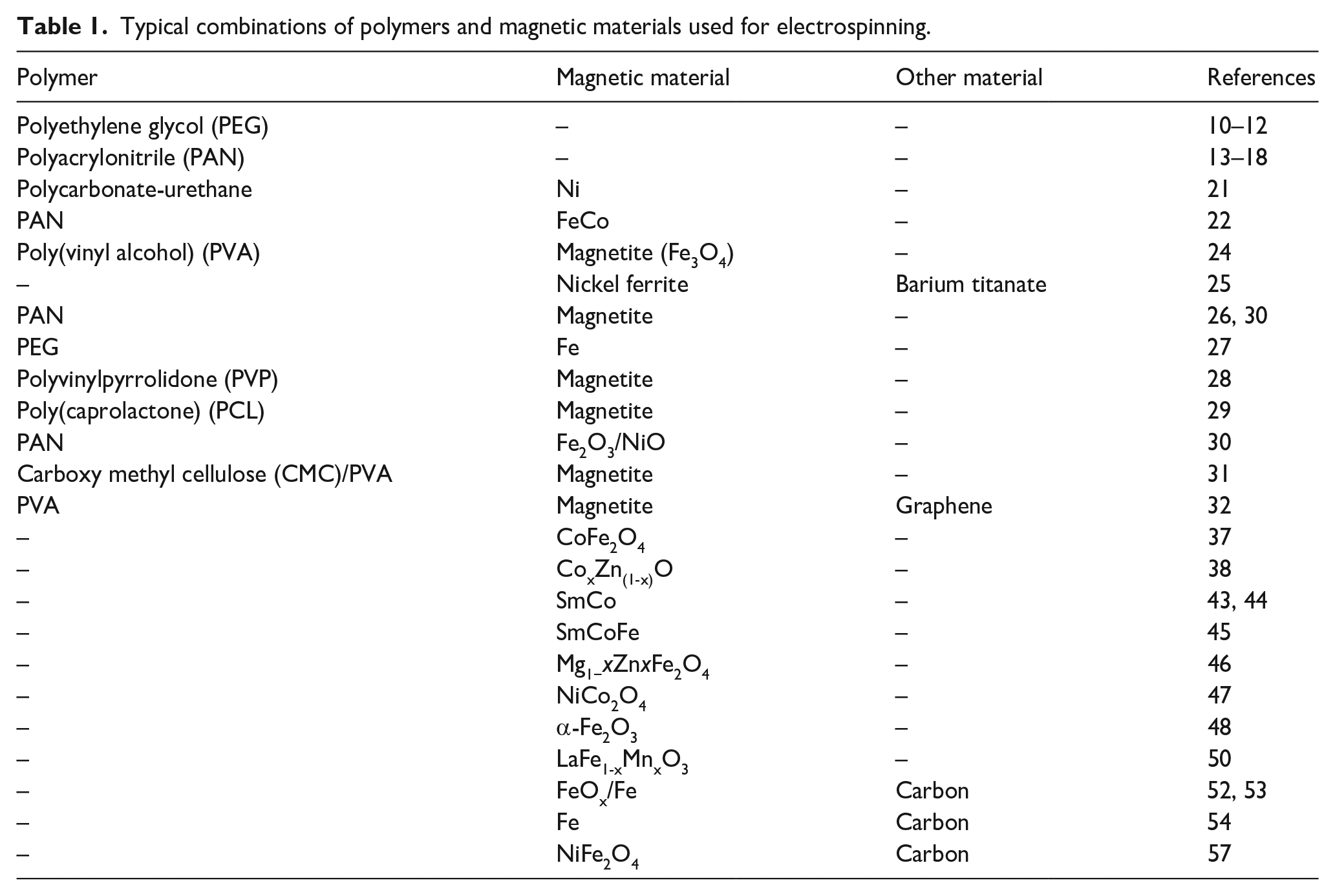
An overview of typical recent materials of magnetic electrospun nanofibers is given in Table 1. As shown here, nanofibers from a broad range of magnetic materials can be electrospun, either in the form of a composite with a polymer, or purely or as a blend with carbon.
Typical combinations of polymers and magnetic materials used for electrospinning.
Magnetic properties
Measuring magnetic properties can be done by most common instruments which are used to examine magnetic thin layers or nanostructures, such as superconducting quantum interference device (SQUID), 65 vibrating sample magnetometer (VSM), 60 or alternating gradient magnetometer (AGM). 66 Optical methods, such as magneto-optical Kerr effect (MOKE), have not yet been reported in the literature for investigations of electrospun nanofiber mats; the roughest materials examined in this way are to the best of our knowledge paper coated with magnetic flakes. 67
SQUID measurements were, for example, used to examine the magnetic properties of pure and Mn-doped electrospun barium titanate nanofibers which were found to be diamagnetic and paramagnetic, respectively. 68
LSMO nanofibers as well as LSMO/LSCO nanowire networks were investigated by SQUID, revealing their soft magnetic properties.41,42
Gd2O2S nanofibers, partly doped with Dy and Eu in different ratios, were found to be strongly paramagnetic, using SQUID measurements. 56
SQUID measurements can also be used to measure zero field-cooling/field cooling curves in a broad temperature interval, allowing for detecting magnetic phase transitions. Such blocking temperatures were, for example, measured for Fe3O4/CMC/PVA nanofibers, where the blocking temperature was found to be only slightly dependent on the Fe3O4 concentration in the fibers, indicating that nanoparticles aggregate already at low magnetite concentrations. 31
VSM measurements were used to investigate magnetic nanofibers containing ferromagnetic Ni nanoparticles, indicating a difference between measurements parallel and perpendicular to the axes of the aligned nanofibers. 17 Similarly, VSM measurements revealed about a factor of 2 between saturation magnetization and remanence along and perpendicular to the aligned PAN/FeCo nanofibers, electrospun by a magnetic field–assisted technique. 22
For Co-doped ZnO nanofibers, VSM measurements found a strong dependence of the magnetic properties on the amount of Co, with higher Co content resulting in larger coercivities and a maximum saturation magnetization for an intermediate Co fraction, 38 while a previous study of the same system underlined the presence of a Co3O4 phase in samples with Co contents higher than 2% and a large paramagnetic signal in addition to the weak ferromagnetism at room temperature. 69 Huang and Or 70 reported that the dielectric loss and magnetic loss properties were significantly better for Co-doped ZnO nanofibers, as compared to nanoparticles of the same material.
VSM measurements were also applied to examine the influence of Fe substitution in SmCo nanofibers, showing changes in the maximum magnetization, squareness of the hysteresis loop and coercivity depending on the amount of Fe substitution. 45 Similarly, Zn doping of MgFe2O4 was investigated by VSM. Here, the doping decreased the net magnetization which was attributed to the decrease of the number of Fe3+ ions on the B-site of the spinel structure and the Mg2+ ions on the A-site. 46
For nanofibers produced from Nd0.05Bi0.95Fe0.95Co0.05O3 in a polymer matrix, ferromagnetic behavior was revealed at room temperature, using a VSM. 35
Low-temperature measurements by VSM showed a spin-glass state in electrospun La0.33Pr0.34Ca0.33MnO3 nanofibers below approx. 50 K, in addition to a colossal magnetoresistance of up to 95%. Furthermore, a transition from a paramagnetic insulator to a ferromagnetic metal phase was found at approx. 70 K. These properties made such nanofibers promising for new microelectronic devices. 71 Samarium–cobalt nanofibers were investigated by VSM, finding decreasing saturation magnetization and increasing coercivity with increasing Sm content. 43
Applications
Typical applications of nanofiber mats are based on their large surface-to-volume ratio and range from filter materials72,73 to catalysts,74,75 and from cell growth and tissue engineering to medical wound dressing.76–78
Absorption of microwave and other irradiation
Magnetic nanofibers can, for example, be used for microwave absorption (Figure 7). Li et al. 37 found the microwave absorption performance of CoFe2O4 nanofibers strongly enhanced and the absorption bandwidth broadened, as compared to bulk material, which was attributed to the electromagnetic coupling between the nanofibers and the microwaves. Huang et al., 38 Huang and Or, 70 and Qiao et al. 79 used Co-doped ZnO nanofibers as microwave absorbers. Ni-Zn spinel ferrite nanofibers were suggested by Hou et al. 80 as microwave absorbers for the range of 2–18 GHz. Porous carbon/Fe nanofibers were also shown to have good electromagnetic wave absorption properties.54,81 In the range of 5.4–18 GHz, carbon/NiFe2O4 nanofibers with additional carbon nanotubes included in the spinning solution showed good microwave absorption. 57 Electrospun Co3O4/carbon nanofiber mats also showed good electromagnetic wave absorption between 4.5 and 14.4 GHz, 82 similar to Co/C nanofibers. 83 Using Fe3O4/graphene oxide/PVDF nanofibers, Samadi et al. 84 could increase the microwave absorption and in addition show piezoelectric properties of these fibers. Electrospun Fe3O4/SiO2 nanofibers showed good resistance against acids and high heat and flame resistance in addition to good microwave absorption between 7 and 18 GHz. 85 Fe3O4/PVP nanofibers showed good electromagnetic shielding for the frequency range between 8.2 and 12.4 GHz. 86 Using polycarbosilane and ferric acetylacetonate as precursors, Hou et al. 87 prepared electrospun Fe3Si/SiC nanofibers which were found suitable for microwave absorption between 8.5 and 16.5 GHz. Mixing Ni0.5Zn0.5Fe2O4 and SrFe12O19, a soft and a hard magnetic material, in porous electrospun composite fibers, Zhou et al. 88 found that by tailoring the mass ratio of soft and hard phases, the reflection loss and the bandwidth could be optimized for wide-band microwave absorption. A similar material was applied by Xiang et al. 89 who used Ni0.4Co0.2Zn0.4Fe2O4/BaTiO3 nanofibers for microwave absorption and found a broad effective absorption bandwidth between 7.8 and 17.3 GHz. Moradipour et al. 90 used electrospun nanofibers consisting of Cu2Fe2O4, Fe2O3, Fe3O4, and Al2O3 to examine microwave absorbance in the frequency range of 5–12 GHz. FeNi/C nanofibers showed different microwave absorption properties depending on the carbonization time. 91 Similarly, Bayat et al. 92 found the electromagnetic shielding properties of electrospun Fe3O4/C nanofibers related to the carbonization temperature and the magnetite nanoparticle diameter.
Magnetic nanofiber mats can be used for microwave absorption.
Absorption of thermal neutrons was investigated for PAN containing gadolinium, showing that the addition of this rare earth magnetic material significantly reduced the neutron transmissivity even through a thin shield. 36 Europium, on the other hand, was used as dopant for porous TiO2 nanofibers, increasing the photoluminescence emission intensity with the europium content. 93
Removal and decomposition of contaminants
Fe3O4/PAN nanofibers mats were coated with horseradish peroxidase, a bio-enzyme with photocatalytic properties. This composite was used for phenol removal from wastewater. 26 Another approach to create photocatalytically active nanofibers was investigated by Chang et al. They produced CoFe2O4 nanofibers and decorated them afterward with BiOI nanoplates. These magnetic fibers showed high visible-light photocatalytic properties, degrading rhodamine B fast, could be re-used several times and collected in an external magnetic field after use. 94 Similarly, Chang et al. 95 found Fe2O3/BiOI electrospun hollow nanofibers to show high catalytic performance even under visible light for rhodamine B decomposition. Good catalytic properties and cycle stability was also recognized for PVA/poly(acrylic acid)/Fe3O4/MXene@Ag nanoparticle composites, produced by electrospinning, 96 as well as CuO-NiO/carbon nanofibers which were carbonized after electrospinning 97 and Bi2MoO6/ZnFe2O4 heterojunction nanofibers which were tested by rhodamine B degradation. 98 Electrospun magnetic nanofibers from a Fe3O4/PVA core and a poly(3-hydroxybutyrate) and PCL shell were decorated with TiO2 nanoparticles by electrospraying, resulting in good photocatalytic activity under UV light. 99
Removal of Cr(VI) from drinking water, investigated with magnetic carbon fibers containing Fe and FeOx, was found to strongly depend on Fe and N co-doping. 52 Methylene blue was degraded in the presence of H2O2 and UV irradiation by PCL/Fe3O4 nanofiber mats 29 or by ZrO2 hollow nanofibers with iron nanoparticles added along the inner surface. 100
Zhang et al. 101 developed PLA/γ-Fe2O3 nanofibers with dual-scaled pores for motor oil absorption and found high absorption capacity combined with high oil permeation flux under gravitational force, making this system useful for continuous oil/water separation in wastewater treatment. Electrospun PVDF and polysulfone nanofibers with embedded Halloysite nanoclay and CoFe2O4 nanoparticles were found suitable for oily liquid sorption. 102 Blending γ-Fe2O3 (maghemite) with a carbon precursor to create γ-Fe2O3/C electrospun core-shell nanofibers was suggested for the creation of an electrochemical sensor for trace cadmium ions. 103
For the decolorization of amaranth as a model dye, Lin et al. used cobalt ferrite nanoparticles electrospun from a PAN solution and afterward stabilized and carbonized to create CoFe2O4/carbon nanofibers. This material worked as a catalyst for the activation of peroxymonosulfate which then generated sulfate radicals and thus decolorize amaranth in an aqueous solution. 104 For the activation of peroxymonosulfate, Zhang et al. 105 found cobalt/carbon nanofiber composites suitable, with cobalt revealing higher catalytic performance than Co3O4 or carbon nanotubes. Fe3O4/graphene/PAN nanofibers were successfully used for removal of Basic Red 46 from aqueous solutions. 106 Electrospun Sr2+ doped BiFeO3 nanofibers showed varying photocatalytic properties, as measured by methylene blue degradation, and photoluminescence; the photocatalytic properties were found significantly higher than in nanoparticles of the same material. 107 Doping BiFeO3 with Sm, on the other hand, resulted in nanofibers with high magnetization, large dielectric constant, and polarization than pure BiFeO3 over a wide temperature range. 108 Doping this material with Mn instead resulted in soft magnetic behavior with increased remanent magnetization. 109
Biotechnology and biomedicine
Even for cell growth applications, magnetic nanofibers are of interest. Polylactic acid–glycolic acid (PLGA) nanofibers with Fe3O4 nanoparticles were coated with pyrrole, resulting in polypyrrole (PPy)/Fe3O4/PLGA fibrous scaffolds with electrical and magnetic properties, thus enabling delivering electrical and magnetic signals. Pre-osteoblasts grown on these scaffolds were stimulated electrically and magnetically, resulting in higher cell viabilities than the reference cells, stimulated only with one method or not at all. Magnetic stimulation was even able to align cells on the magnetic scaffolds, making this material highly applicable in bone tissue engineering. 110 Similarly, 3D growth and viability of bone marrow mesenchymal stem cells were found to be significantly supported by core-shell magnetic structures based on electrospinning. 111 Brito-Pereira et al. 112 found silk fibroin nanofibers filled with CoFe2O4 or Fe3O4 to be not cytotoxic and thus applicable for tissue engineering. Tissue engineering is one of the possible applications of electrospun PMMA nanofibers in which carbon nanotube/Fe3O4 fibers were embedded. 113
For drug delivery or bioseparation, nanofibers with strong paramagnetic properties prepared by the coaxial technology (Figure 8), such as Gd2O2S, possibly doped with Eu or Dy, were suggested. 56 Combining doxorubicin (DOX), an anticancer drug, NaX nanozeolites and Fe3O4 in a PLA/chitosan spinning solution enabled controlled release of the DOX to kill H1355 carcinoma cells. The magnetic particles allowed for investigating the influence of a magnetic field, showing that in this way the antitumor activity could be increased to 82% dead tumor cells after 1-week treatment in an external magnetic field. 114 Demir et al. examined Fe3O4 nanoparticles in PCL nanofibers, produced by electrospinning, for drug delivery. Comparing the delivery of hydrophobic and hydrophilic model systems from such blended fibers with pure PCL fibers, they found different influence of the magnetic nanoparticles on release of hydrophobic and hydrophilic dyes, respectively. 115 PCL/Fe3O4 electrospun nanofibers were used to deliver heat and anticancer drugs simultaneously, enabling killing HeLa cancer cells efficiently. 116

Utilization of the coaxial electrospinning technology for drug delivery from nanofiber mats.
Hyperthermia therapy applications were also based on magnetic nanofibers. Matos et al. developed electrospun nanofiber mats of cellulose acetate with included magnetic nanoparticles and measured their heating ability in comparison to the effect of a pure polymer nanofiber mat with magnetic nanoparticles coated on the fibers. While the latter showed a better heating capability, it had also the disadvantage of possible cytotoxic effects, making the nanofiber mat with embedded magnetic nanoparticles a responsible choice. 117 Sarier et al. 118 suggested magnetite/fatty acid embedded in electrospun poly(methacrylic acid-ethyl acrylate) nanofibers.
Superparamagnetic γ-Fe2O3 nanoparticles were electrospun with polyurethane and used for hyperthermia treatment, exhibiting a high temperature increase in an alternating magnetic field. 119
Magnetic sensors
Electrospun and calcinated Janus nanowires, containing barium titanate and cobalt ferrite, were used as magnetic field sensors. For this, they were placed on patterned metal electrodes on a silicon substrate, in this way connecting several nanowires in parallel. In this way, a magnetic field sensitivity of approx. 0.51 mV/Oe could be reached at 1 kHz. 120 Large magneto-electric coefficients were also found in barium titanate–cobalt ferrite and lead zirconate titanate–nickel zinc ferrite Janus nanowires. 121
As an UV detection sensor as well as for photocatalysis, electrospun Fe2O3/TiO2 nanofibers were used by Liu et al. The metal nanoparticles shifted the absorption range of TiO2 from the UV to the visible range, enabling photocatalysis under visible light. 122 Chemical adsorption of Hg(II) ions could be performed and measured by nanofibers containing fluorescent carbon quantum dots and Fe3O4 nanoparticles, indicating the Hg(II) sorption by a declining fluorescence signal. 123
Applications in physics
In the area of neuromorphic computing, PAN nanofibers with Fe3O4 and Fe2O3/NiO nanoparticles were suggested for data storage and transport.30,124
For creation of a triboelectric nano-generator (Figure 9), Im and Park used nanofibers prepared from Fe3O4 nanoparticles in PVDF. They showed that for nanofiber contents above 17%, the particles became visible outside the fibers, correlated with a decrease in the triboelectric performance. 125 Garcia-Marquez et al. 126 suggested Fe3N and Fe3C electrospun nanocomposites as magneto-active membranes, in addition to the more common application in magnetic field–assisted separation. Due to their bio-compatibility, Guarino et al. 127 suggested nickel-polyurethane nanofibers as magneto-active components for biomedical purposes as well as sensors and actuators. Sensing acetone was performed with Pt-CuFe2O4 electrospun nanotubes. 128

Electrospinning can be used to create a triboelectric nano-generator. 125
For hydrogen production by splitting water, usually precious metals are used as electrocatalysts. Due their high costs, more inexpensive alternatives are necessary. For this application, Zhang et al. 129 prepared CoFe2O4 by electrospinning these nanoparticles in PVP which was calcinated at different temperatures afterward, and found reasonable oxygen evolution reaction efficiencies.
For the use in supercapacitors, Bhagwan et al. 130 created spinel-NiMn2O4 electrospun nanofibers, showing good energy densities and power densities which could be supported by red light irradiation.
Conclusion
As the large number of scientific publications dealing with electrospun magnetic nanofibers even during the last 2 years underlines, this topic is of high recent interest in basic research as well as for diverse applications.
While the magnetic properties attract especially attention due to their possible strong differences in comparison with bulk or thin film materials, based on the usual shape anisotropy of magnetic materials as well as the special production process which results in different grain structures, they offer also new functionalities. Electrospun magnetic nanofibers can be used, for example, for microwave absorption, photocatalysis, biotechnological and medical applications, and even as sensors and actuators.
Our review gives an overview of the most recent basic examinations and applications and will support other research groups working on electrospinning to find suitable spinning techniques and materials as well as applications for their magnetic nanofiber mats.
Footnotes
Declaration of conflicting interests
The author(s) declared no potential conflicts of interest with respect to the research, authorship, and/or publication of this article.
Funding
The author(s) disclosed receipt of the following financial support for the research, authorship, and/or publication of this article: This research was funded by Volkswagen Foundation grant “Adaptive Computing with Electrospun Nanofiber Networks,” No. 93679.