
Abstract
This article presents the strength, diameter, elongation and tenacity variations of banana pseudo-stem fibres extracted from five common Sri Lankan cultivars. The banana plants that were selected represent the dry and wet climatic zones of Sri Lanka. Mixtures of all layers of stem from the selected cultivars were subjected to three different extraction methods. Decortication technique was used for mechanical extraction, sodium hydroxide was used for chemical extraction and pectinase enzyme was used for biological extraction. The mechanical extraction method resulted in coarser fibres, chemical extraction resulted in a 30% reduction in the fibre diameter, while the biologically extracted fibre presented a 40% increase in fibre strength compared with the mechanically extracted fibres. Scanning electron microscopy analysis revealed the presence of damaged fibrils in mechanical extraction and removal of binder material in the chemical and biological extraction methods. Furthermore, Fourier transform infrared spectroscopy analysis proved the removal of lignin in chemical and biological extraction. Different cultivars presented different behaviours in extensibility, which is common in natural fibres.
Keywords
Introduction
Banana is a member of the genus Musa (belongs to the family Musaceae). The Swedish botanist, Carolus Linnaeus, named the species ‘Musa’ after Antonius Musa, a Roman physical. 1 Four sections of species were identified in Musa and were reduced to two main sections based on molecular analyses, which were considered as parents of the cultivated types, namely Musa acuminate (AA) and Musa balbisiana (BB). The corresponding contribution of M. acuminata and M. balbisiana to the cultivar is denoted as As and Bs, respectively.
Further classification is obtained based on the presence of one or more sets of chromosomes (known as ploidy level). For instance, an AB is a diploid, an AAB is a triploid and ABBB is a tetraploid. Triploid (AAA, AAB and ABB) hybrids are the most familiar, seedless, cultivated varieties. 2
The Sri Lankan banana varieties are classified as follows:
AAB genome/Mysore: Ambul and Seeni;
AB genome/Kolikuttu: Kolikuttu, Suwendel and Puwalu;
AAA genome group/Cavendish: Anamalu, Embon, Bin Kehel, Nethrapalam and Rath Kehel;
ABB genome/plantain varieties: Alu kehel, Etamuru, Hambanpuwalu, Marathamana, Mondon and Sambel.
Banana (Musa acuminata Colla) is an extensively cultivated fruit throughout Sri Lanka. 3 Nearly 50,000 ha of land is under banana cultivation and the annual production is about 45,000 tonnes. 4 The increasing demand for banana in the international market has resulted in an increase in acreage and production, thus generating a considerable amount of pseudo-stem biomass waste. 3 The waste banana stems cannot be left dumped in the field as they cause emission of toxic gases, such as CO2, and also facilitate the growth of harmful fungi which may attack the remaining banana trees. Furthermore, they cannot be used as animal feed. 5
All varieties of banana stems and almost every part of the banana plant contain fibres of different strength, colour and staple length, which can be used for various applications. Therefore, the pseudo-stem can be successfully utilized for numerous applications and products. 3
In the recent past, banana fibres had limited application and were predominantly used to manufacture hand-crafted products such as ropes, mats and other composite materials. The emerging trend of green products, concern for the environment and eco-friendly fabrics have resulted in recognizing the natural banana fibre for all of its favourable properties, with its usage increasing in all the sectors of textile and apparel applications. 6
Banana fibre has favourable physical and chemical characteristics which facilitate its use in textile and apparel products. The chemical structure of banana fibre is composed of cellulose, hemicellulose and lignin. The cross section of the banana fibre consists of a number of smaller fibres bundled together. These smaller fibres may vary from as few as 10 to about 100 individual fibres. Each of these individual fibre cells is similar in shape and size to cotton fibres, as far as cross sections are concerned. These fibres are bound with lignin and give a hemicellulose structure. 7
Traditionally, banana fibres were extracted manually. This process is time-consuming and the extracted quality is based on the skilled practice of the labourer. 8 Banana fibres are now extracted by chemical, mechanical or biological methods. Mechanical extraction fails to remove the natural binding material from the interspaces of the fibre in the fibre bundle while chemical extraction causes environmental pollution. However, increased fibre yield can be obtained using biological extraction compared to the other two techniques, with minimum harm to the environment. However, natural retting is a time-consuming and tedious process. Degumming is essential after extraction to obtain the optimum utilization of fibres. This process removes the heavily coated non-cellulosic natural binding material from the cellulosic part of the fibre. 9
The main mechanical extraction methods are the Bacnis and Leonit processes. In the Bacnis process, a simple stripping action is used to pull apart the trunks and to undress the sheath. The fibre is obtained by pulling and crushing of the ribbons (tuxy) and removing the pulpy material. In the Loenit process, a sharp pointed tool is used to obtain the ribbons from the sheath. A comb-like device is used to separate the fibres. The latest mechanical extraction method is the decortication process. In this process, cut banana stems of 100–200 cm length are crushed between two drum rollers to obtain the fibres after removing the pulpy part. The machine increases fibre production by 20–25 times compared with the manual process. 10
Biological natural retting can be performed by soaking the stem in an open container containing fresh water. It should be kept for a few days or weeks to activate a combination of several microorganisms. 2 Furthermore, enzymes are used for fibre extraction. The selection of enzyme is based on the type of substrate, composition, size and lignin content. The common enzymes used are cellulases and pectinases. Cellulase enzymes increase the smoothness by removing fibrils from the surface. Hence, there is a reduction in the mechanical properties due to the damage occurred in the fibres. Pectinases remove the inter-lamellar pectin, which acts as a natural adhesive substance between fibres. 9 Furthermore, a consortium of bacteria species, namely Bacillus cereus, Bacillus licheniformis, Bacillus subtilis and Bacillus polymyxa, is isolated and used for extraction and processing of fibres from banana and plantain stems using the controlled system tank retting (CSTR) method. 3
Chemical extraction methods have been experimented in researches. Alkali treatment is used in chemical extraction. Alkali treatments facilitate fibrillation, where the composite fibre bundle is broken down into smaller fibres.
The alkali (sodium hydroxide (NaOH)) reduces the fibre roughness and results in good quality fibre. In addition, sulphuric acid, hydrogen peroxide, protease and sodium citrate can also be used for chemical extraction. 11
Analysis of molecular orientation is essential for explaining the physical behaviour of a macromolecular system. Fourier transform infrared spectroscopy (FTIR) is one of the important tools to examine the molecular structure at the macro level. The molecular-level investigations with FTIR mainly focus on the crystal structure analysis. According to the orientation of polymer chains, natural fibres consist of two regions: amorphous and crystalline. 12 Furthermore, FTIR indicates the presence and removal of molecular bonds. Scanning electron microscopy (SEM) scans the sample with a focused electron beam and provides captured images with detailed information about the sample topography and composition.
The objective of this article is to analyse the effect of the fibre extraction method on the physical behaviour of fibres extracted from five common Sri Lankan banana cultivars. Finally, the findings will be used to develop a commercially viable processing technique that will yield textile grade fibre. Tensile strength, tenacity, elongation and fibre diameter were measured after extracting fibre using different extraction methods and the relationship between the fibre properties and the extraction method was determined. Further analysis to predict the structural behaviour is done using SEM and FTIR.
Materials and methods
Test specimens
Banana stems were collected from cultivation fields located in the dry and wet climatic zones of Sri Lanka, which possesses a considerable amount of land used for cultivation. Two stems were randomly selected to represent a cultivar, whereas 10 samples were randomly selected representing 5 common cultivars with different genomes. Stems were conditioned to the standard atmospheric conditions before subjecting to treatment.
The stems were cut into two with a stem cutting machine. The stems consist of layers that can be separated manually using a hand-held knife. The layers were selected representing all sections such as the xylem, vascular cambium, phloem and periderm. The tested samples were 30-cm-long strips of stem.
Fibre extraction
Mechanical extraction was done using a fibre decortication machine. Cut strips of banana stem were fed in between a squeezing roller and a scraper roller and the fibres were extracted. Manual combing was done to remove the excess pith from the fibres.
Chemical extraction was done using NaOH. The cut strips of banana stem were immersed in a bath with 5% NaOH under standard atmospheric conditions.
Biological extraction was done using 5% pectinase enzyme, where cut strips of banana stems were immersed in the bath and kept under standard atmospheric condition.
Finally, all the treated strips of stem were manually combed using a soft comb to separate the individual fibres and were dried in the sun.
Conditioning and testing
The extracted fibres were conditioned and tested under standard atmospheric conditions. Zoning technique was used to select the fibres for the tests. A handful of fibres were selected from all the layers and divided into two parts, of which one was discarded and the other was retained. The retained fibres were again divided into two parts, of which one was discarded. This process was repeated until 5 g of fibre was obtained as a representative sample for each test. The selected fibres were used for testing.
Tensile testing
The tensile properties of the fibres were measured with Instron Tensile Testing machine having a load cell of 10 N, which operates under the principle of constant rate of elongation. The test was conducted according to the standard method of ASTM D3822, with a specimen length of 5 cm. The cross-head moving speed was adjusted to give a fibre breaking time of 20 ± 3 s. The load values were measured in Newton (N) with an accuracy of four decimals and the extension values were measured in millimetres (mm) with an accuracy of two decimals. A total of 50 tests were carried out and the average values were obtained.
Diameter
In all, 10 fibre samples were tested and 5 readings were obtained from each sample. The average values of 40 readings were obtained. The diameter values were measured using SEM images with a magnification factor of 300×–1500×.
Fibre tenacity
Fibre count was calculated using the cut and weigh method. The tenacity was calculated using the measured count and strength values.
Morphological analysis
All the 10 samples were tested and SEM images were obtained to analyse the morphological structure. The specimen containing the fibres were arranged on a pin stub and then positioned on a holder that can be inserted into the SEM. High acceleration voltage (10 kV) and 100 μA beam current were used to obtain high-resolution imaging. The longitudinal views of fibres were analysed.
FTIR spectroscopy
The macromolecular behaviour was examined using Bruker/ALPHA FTIR spectrometer equipped with an attenuated total reflection (ATR) attachment. The fibre samples were analysed following ASTM E168-06 and they were placed directly under the crystal and ATR attachment.
Results and discussion
SEM images were obtained to analyse the topography of the fibre. Mechanically extracted fibre diameters are comparatively higher than biological and chemical extraction methods. Hence, they were observed under the magnification factors of 300× and 600×. Magnification factor of 1500× was not possible due to blurred and smudged appearance of the captured images. However, in the case of biological and chemical extraction, images were obtained under the magnification factors of 600× and 1500× for improved clarity.
Physical tests
Figure 1 displays the tensile strength (breaking load) of a single fibre (technical fibre, which consists of several elementary fibres) extracted from five different cultivars using three different extraction methods. It illustrates that the biological extraction method results in the highest fibre strength among all the cultivars, while the chemically extracted fibre has the lowest strength among three cultivars. However, AAB (Ambul) and AB (Kolikuttu) cultivars exhibit comparatively higher fibre strength with chemical extraction than mechanical extraction. Chauhan and Sharma 13 state that biological extraction with enzymes increases the strength of the fibre as it breaks only the lignin component which acts as a binder, but mechanical extraction weakens the fibre as the cellulose component could also be damaged.

Tensile strength (breaking load) of single fibres extracted from five different cultivars under different extraction methods.
This was confirmed and is evident in the SEM analysis shown in Figure 2. In the mechanically extracted fibre, the damaged fibrils are protruding out of the fibre surface, as shown in Figures 6 and 7. However, in the biological extracted fibre the binding material is removed but the fibrils are intact. Furthermore, Robles et al. 14 explain that the process of mercerization involves a partial destruction of the intermolecular bonds, due to the penetration of NaOH into the swollen amorphous regions of the fibre where the cellulose molecules are held by hydrogen bonding, which leads to the formation of sodium cellulose and sodium alcoholates. A large part of the residual hemicellulose is removed and rupture of some C–C bonds reduces the lengths of the chain. Hence, chemically extracted fibres exhibit lower strength. It is evident that the average fibre strength has increased by 40% in biological extraction compared to mechanical extraction.

SEM analysis of fibres extracted from different extraction methods and cultivars.
Figure 3 illustrates the diameter of single fibre extracted from selected cultivars under different extraction methods. Mechanically extracted fibres exhibit comparatively the highest diameter. Biologically extracted fibres exhibit a 10% reduction in diameter compared with mechanically extracted fibres, while in the chemical extraction method, the reduction in diameter is 30% compared with mechanically extracted fibres.

Diameter of single fibres extracted from five different cultivars under different extraction methods.
Mukhopadhyay et al. 7 state that the cross section of banana fibre comprises a number of smaller fibres bundled as a collection. The number of smaller fibres may vary from 10 to 100 and each of these individual fibrils is similar to cotton fibrils. These fibrils are bound together with lignin and give a hemicellulose structure. This phenomenon can be further confirmed through the observations of SEM analysis.
Analysis of the SEM images showed that the partially separated binder materials were available on the surface of the chemical and biological extracted fibres. This indicates the removal of binder material, hence facilitating more compact packing of fibrils, which result in the reduction of the overall diameter of the bundle of fibrils. Furthermore, with SEM analysis, empty spaces between the fibrils are visible, which confirms the removal of binder materials. This may be due to the deep penetration of enzyme or NaOH into the fibre bundle, leading to breakdown of the binder material, so that the fibrils are separated away from the bundle leading to the reduction in diameter. Therefore, it can be concluded that the mechanically extracted fibre diameter is higher due to the remaining binder material, which occupies some space in the fibre bundle. Biological and chemical extractions remove the binding material and reduce the space utilized for binders and hence result in the reduction of diameter.
Figure 4 indicates that there is no interrelationship of extensibility with different fibre extraction methods. However, the variation in extension may be due to the rearrangement of molecular chains. Kulkarni et al. 15 state that there is a minor variation of fibre extensibility with the changing diameter. However, the extensibility exhibits large variations among the selected cultivars. Patel and Patil 16 investigated the extensibility of cotton fibre with different cotton varieties and showed that the variations in extensibility varies from 5% to 13% and the extensibility variation is also influenced with factors such as the linear density and moisture content of the fibre. Furthermore, Warwicker and Hallam 17 state that the alkali treatment on cotton fibre also alters the fibre extensibility. Hence, the similar variation in extensibility in banana fibre may also be due to the variations of other related factors with varying cultivars. However, further studies need to be done to confirm the factors leading to the variation in extensibility.

Extension of single fibres extracted from five different cultivars under different extraction methods.
Figure 5 illustrates the fibre tenacity variations along with the different extraction methods for selected Sri Lankan banana cultivars. The biological extraction method exhibits the maximum tenacity values, whereas the tenacity variation in the other two extraction methods varies with the cultivars. Further reduction in diameter in the biological and chemical extraction has influence on reducing the tenacity. The reduction in diameter and increase in strength values indicate the enhanced distribution of strength throughout the fibre and results in higher tenacity values. This is one of the factors leading to the higher tenacity values in biological extraction.

Tenacity of single fibres extracted from five different cultivars under different extraction methods.
SEM analysis
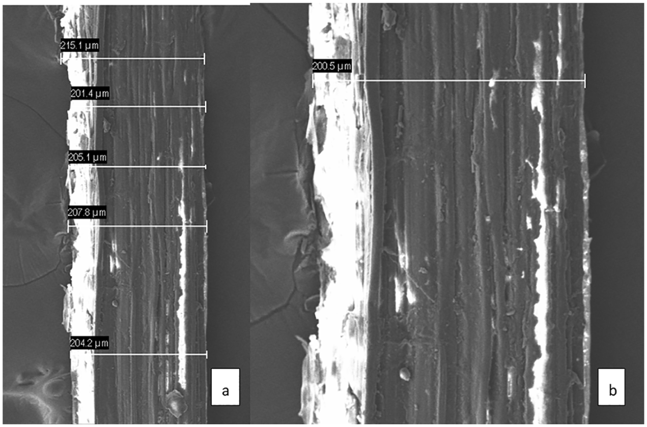
Figures 6 to 10 illustrate the SEM images of the mechanically extracted fibres at different magnifications. It indicates the cloudy, unclear surfaces of fibre due to the presence of entangled and broken single fibres, which is a result of the decortication technique. This exhibits the fibre damages resulting due to mechanical extraction, and confirms that the lower tenacity of mechanical-extracted fibre resulted in fibre damage due to the decortication process. This is despite the higher binder content available in the mechanical extracted fibres, which can contribute to the strength. However, binders occupy the spaces in the fibre and result in a higher diameter, leading to lower tenacity values.

SEM images of mechanically extracted Atamuru (ABB) fibre with magnification factor (a) 300× and (b) 600×.

SEM images of mechanically extracted Ambul (AAB) fibre with magnification factor (a) 300× and (b) 600×.
SEM images of mechanically extracted Puwalu (AB) fibre with magnification factor (a) 300× and (b) 600×.

SEM images of mechanically extracted Ambun (AAA) fibre with magnification factor (a) 300× and (b) 600×.

SEM images of mechanically extracted Kolikuttu (AB) fibre with magnification factor (a) 300× and (b) 600×.
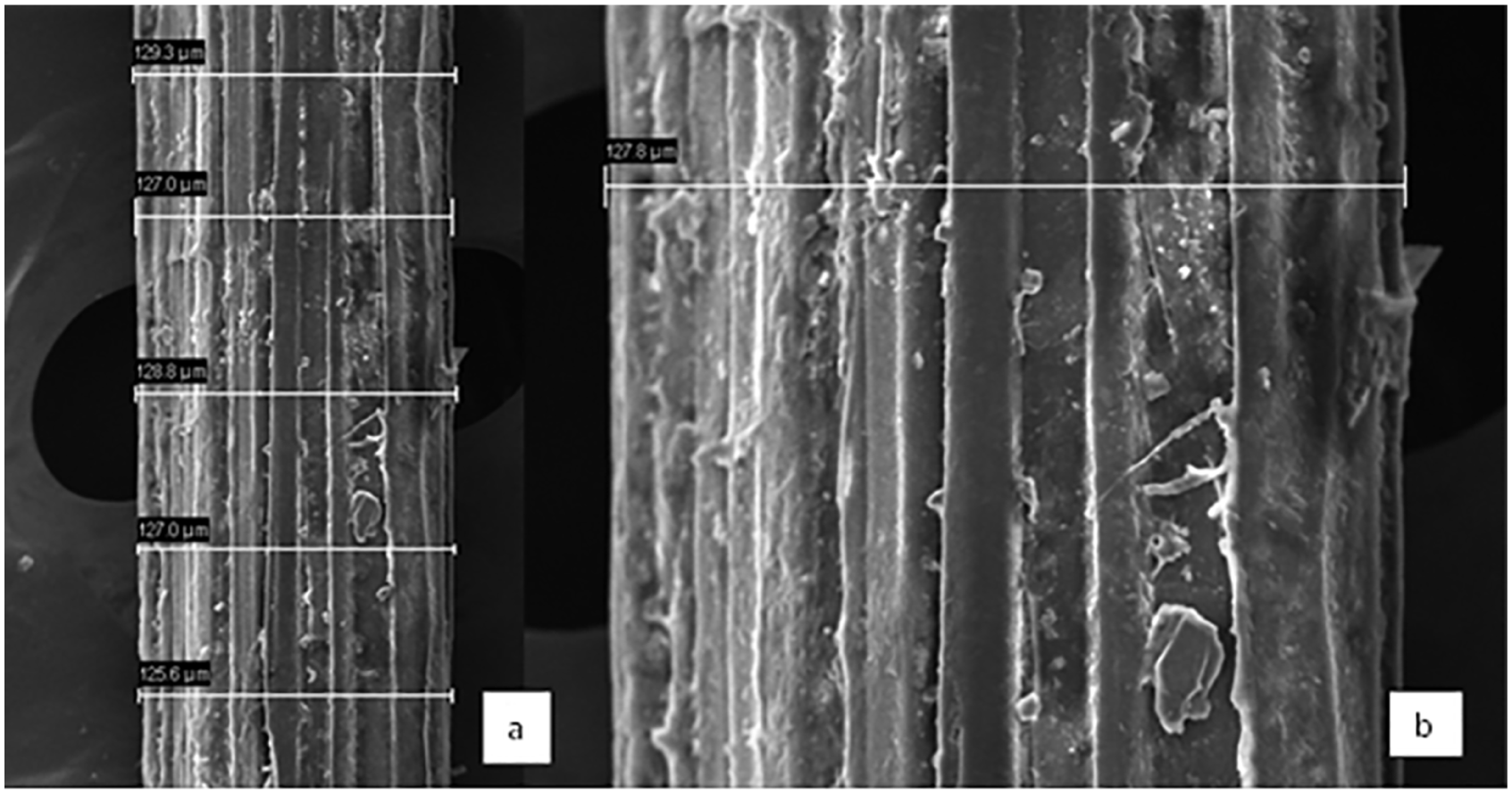
Figures 11 to 15 illustrate the SEM images of chemically treated fibres at different magnifications. The parallel orientation of fibres in the bundle and the cementing natural binder material is noticeable. The separation of binder, which was in between the fibrils, and the presence of partially removed binder material from the surface are evident. This indicates the partial removal of binder material. Hence, it is possible that chemical extraction has an effect on removing and separating the binder within the fibrils. Therefore, a factor leading to the reduction of fibre diameter is the separation of binder from the main bundle. However, a thorough cleaning process is essential to clean the partially removed binder materials that are held on the surface.

SEM images of chemically extracted Atamuru (ABB) fibre with magnification factor (a) 600× and (b) 1500×.
SEM images of chemically extracted Ambul (AAB) fibre with magnification factor (a) 600× and (b) 1500×.

SEM images of chemically extracted Puwalu (AB) fibre with magnification factor (a) 600× and (b) 1500×.

SEM images of chemically extracted Ambun (AAA) fibre with magnification factor (a) 600× and (b) 1500×.

SEM images of chemically extracted Kolikuttu (AB) fibre with magnification factor (a) 600× and (b) 1500×.
Figures 16 to 20 illustrate the SEM images of the biologically treated fibres and indicate the similar separation as of chemical extraction and partial removal of binder material. Furthermore, they illustrate the removal of binder material from the inter-fibre surface, but the removed binders stagnate on top of the outer surface of the fibre. Githinji et al. 18 state that the partial removal of non-cellulosic binder material facilitates easy rearrangement of fibrils in the direction of fibre axis, and hence enable better load sharing, which results in increase of fibre tenacity. SEM analysis confirms the better removal of binder material, without damaging the fibrils. Hence it is a factor leading to a smaller diameter and increased tenacity.

SEM images of biologically extracted Atamuru (ABB) fibre with magnification factor (a) 600× and (b) 1500×.

SEM images of biologically extracted Ambul (AAB) fibre with magnification factor (a) 600× and (b) 1500×.

SEM images of biologically extracted Puwalu (AB) fibre with magnification factor (a) 600× and (b) 1500×.

SEM images of biologically extracted Ambun (AAA) fibre with magnification factor (a) 600× and (b) 1500×.

SEM images of biologically extracted Kolikuttu (AB) fibre with magnification factor (a) 600× and (b) 1500×.
FTIR analysis
Figure 21 illustrates the FTIR spectroscopy of the different extraction methods. The diagnostic region above 1500 cm−1 exhibits the presence of C=C double bond and Csp3–H bond having sp3 hybridized carbon in the structure. The presence of lignin is evident in the mechanically extracted fibre through strong band intensity and asymmetric stretching of C=C double bond by the peaks at 1605 and 1637 cm−1. The band intensity becomes weak in the range of 1605–1637 cm−1 in biologically (enzyme) extracted fibre and insignificant in chemically extracted fibre. 19 Hence, this indicates the reduction of lignin through biological and chemical extraction methods.

Combined FTIR spectroscopy graph of mechanical (blue), chemical (red) and biological (green) extraction methods.
Furthermore, broadening of the peak around 3400 cm−1 indicates O-H stretching, which exhibits the presence of absorbed water, aliphatic primary and secondary alcohols found in cellulose, hemicellulose and lignin. 20 , 21 The broadening of band increases and the intensity reduces in the order of mechanical, chemical and biological extraction. This may be due to reduction in the total content and opening the avenue to cross-linking through removal of cementing substances in the inter-fibre space. Jagadeesh et al. 22 state that the increased breadth of the band of hydroxyl group stretching indicates the increased cellulose percentage in fibre and the resulting higher strength.
In the fingerprint region below 1500 cm−1, two asymmetric peaks can be seen at about 1024 and 1318 cm−1, which represent, respectively, the presence of C–O–C asymmetric stretching and OH in-plane deformation. Lower band intensity in biological extraction at 1318 cm−1 emphasizes the reduction of lignin and increase of crystallinity. Reduction of band intensity at about 1024 cm−1 explains the reduction of carbohydrate functional groups in chemical extraction. 19 Therefore, FTIR analysis further strengthens the results obtained through physical tests.
A summary of the FTIR results is shown in Table 1.
Summary of FTIR analysis.

FTIR: Fourier transform infrared spectroscopy.
Conclusion
In conclusion, the results of this study provide some interesting factors on fibre behaviours with different extraction methods. It is evident that enzymatic biological extraction results in higher tenacity and strength values, whereas chemical extraction results in the lowest diameter values. Comparatively, while the mechanical extraction method resulted in coarser fibres, chemical extraction resulted in a 30% reduction in the fibre diameter. Biologically extracted fibre showed a 40% increase in the fibre strength compared with mechanically extracted fibres. SEM analysis revealed the presence of damaged fibre in mechanical extraction and removal of binder material in chemical and biological extraction. Furthermore, FTIR analysis proved the removal of lignin in the chemical and biological extraction methods. Different cultivars presented different behaviours in extensibility, which is common in natural fibres. The overall results of this study open up new avenues for investigation related to banana fibre extraction and suggest that further more focused studies need to be conducted in relation to biological extraction in order to obtain fine fibre that could be used for the production of textile and apparel.
Footnotes
Declaration of conflicting interests
The author(s) declared no potential conflicts of interest with respect to the research, authorship and/or publication of this article.
Funding
The author(s) disclosed receipt of the following financial support for the research, authorship and/or publication of this article: This work was supported by Sri Lanka Institute of Textile and Apparel, Ministry of Industry and Commerce, Sri Lanka, under national project funding.