
Abstract
Diabetes-induced erectile dysfunction (DIED) is a type of refractory erectile dysfunction which can be clinically treated using the traditional Chinese medicine leech whose main ingredient is hirudin. Oxidative stress can damage vascular endothelial cells, affect blood circulation, and induce fibrosis of smooth muscle cells. This study assessed the efficacy of hirudin in treating DIED before exploring its potential mechanism of action. DIED was induced in rats using streptozotocin, while experimental apomorphine was used to screen for erectile dysfunction models. The rats were then divided into four groups: a blank control group (NC group), a model group (M group), a hirudin group (H group), and an inhibitor group (YC group). After 2 weeks, the serum levels of malondialdehyde (MDA), superoxide dismutase (SOD), and nitric oxide (NO) were determined. The histological features and HIF-1α/RhoA/ROCK signaling pathway-related proteins of the penile corpus cavernosum were detected. Erectile function improved in the H and YC groups without significantly affecting body weight and blood glucose levels, with histopathological analysis also showing improvement in penile structure in these groups. In addition, the expression of HIF-1α/RhoA/ROCK signaling pathway-related proteins was lower in the penile cavernous tissue of rats in the H and YC groups (p < .05), with the serum levels of NO and SOD also being higher in these groups (p < .05). The serum level of MDA decreased in the YC and H groups (p < .05). In this study, only animal experiments were conducted to investigate the regulation of Rho/ROCK pathway by HIF-1α. Cellular studies of the underlying mechanisms are lacking.
Introduction
Erectile dysfunction (ED) is a common sexual dysfunction characterized by the inability to consistently achieve and maintain a sufficient penile erection for satisfactory sexual activity (Defeudis et al., 2022). Studies have shown that ED affects approximately 52% of men between the ages of 40 and 70 years, and it serves as an important indicator of overall health (Saffati et al., 2024). Diabetes-induced erectile dysfunction (DIED), a prevalent urological condition among men, is a form of ED triggered by diabetes mellitus (DM), and has a reported prevalence of 52.5%, which is 3.5 times higher than in nondiabetic individuals (Kouidrat et al., 2017). Managing DIED is more challenging than ED alone due to impaired immune regulation, oxidative stress, and heightened inflammation in diabetic patients. In addition, DIED is associated with various factors such as pathological changes in the penile vascular endothelium and smooth muscle as a result of high glucose toxicity, sensory nerve dysfunction, hormonal imbalances, and psychological factors (Kwon et al., 2024). DIED is currently treated with oral phosphodiesterase type 5 inhibitors (PDE5i), dopamine agonists, and surgical methods, but these options often prove insufficient to fully address patient needs (Kim et al., 2021). In traditional Chinese medicine (TCM), DIED is classified under “thirst-quenching and impotence,” and previous studies have suggested that TCM may help to regulate oxidative stress, apoptosis, atherosclerosis, and endothelial function through multiple targets and signaling pathways, thereby improving symptoms, reducing PDE5i dosage, and minimizing side effects (Feng et al., 2021).
Previous research indicated that the TCM leech can alleviate vascular endothelial injury, inhibit abnormal platelet activation, and treat DIED by inactivating the CaSR/PLC/PKC signaling pathway, which is responsible for reducing endothelial cells (ECs) apoptosis and decreasing the mRNA and protein expression of intercellular cell adhesion molecule-1 (ICAM-1), nuclear factor kappa B (NF-κB), and protein kinase C β (PKCβ) (Ma et al., 2021). Hirudin, the main active component in leeches, has been shown to possess anticoagulant activity, protect ECs, increase blood flow, and modulate NOD-like receptor thermal protein domain-associated protein 3 (NLRP3) to regulate inflammatory responses (Montinari & Minelli, 2022). The current team previously demonstrated that hirudin could improve vascular endothelial damage, regulate smooth muscle contraction, and inhibit smooth muscle fibrosis, thereby improving erectile function. Hirudin may be a potential agent for the treatment of DIED and may be helpful in improving the efficacy of DIED.
The RhoA/Rho kinase pathway is crucial for cellular morphology, smooth muscle contraction, and other biological functions (Y. Zhang et al., 2018). While studying DIED rats, Li et al. (2011) found RhoA/ROCK expression to be up-regulated in their penile tissues. Finally, aberrant activation of the RhoA/Rho kinase signaling pathway, induced by HIF-1α under hypoxic conditions, was also shown to be involved in tissue fibrosis (Sodhi et al., 2011). Building on the above findings, the current study established a rat model of DIED to further investigate hirudin’s mechanism of action in treating DIED. By comparing the penile tissue samples and the expression levels of Rho and ROCK across samples, this study aimed to provide a basis for hirudin’s therapeutic potential in managing DIED.
Materials and Methods
Main Reagents
The research-grade freeze-dried hirudin powder of research grade (batch no. 230501) was purchased from Wuhan Shengtianyu Technology Co. (China), while HY-14927 and STZ (batch no. S8050) were obtained from MCE (China) and Lamblade (China), respectively. In addition, the antibodies ACTIN (GB15001), Endothelial Nitric Oxide Synthases (eNOSs) (GB115277-100), and HIF-1α (GB111339-100) were purchased from Servicebio (China), while those of RHOA (10749-1-AP), ROCK1 (21850-1-AP), and ROCK2 (21645-1-AP) were obtained from Wuhan Three Eagles Biotechnology Co. (China). Finally, the antibody P-MYPT1 (AP0835) was purchased from MCE (China).
Modeling and Grouping
Thirty-six 8-week-old male Sprague-Dawley (SD) rats (SPF-grade of mean weight 200 ± 20 g) were obtained from Beijing HuafuKang Biotechnology Co. Ltd [China; Animal License No.: SCXK (Jing) 2014-0004]. All animals, housed in a clean animal laboratory at the Experimental Animal Centre of Dongzhimen Hospital, Beijing University of Chinese Medicine, were initially subjected to mating tests to confirm their normal sexual function. The rats were then given free access to solid feed and deionized water for 1 week under controlled conditions: a temperature of 18–25°C, a room humidity of 55%–80%, and exposure to 10–12 h of light per day. All experimental procedures were performed according to guidelines approved by the Animal Ethics Committee of Beijing University of Chinese Medicine (Authorization Number: BUCM-2023052402-2198).
DM Rat Model
Thirty-six 8-week-old SD rats were provided regular feed for 1 week to allow for adaptation. We numbered and randomly assigned the rats using the SPSS 22.0 software random number method. Of these, six animals, randomly selected according to the random number table method, were used as the control group and maintained on the regular feed. The remaining ones were then switched to a high-fat and high-sugar diet for 4 weeks, after which they were intraperitoneally injected with a 1% streptozotocin solution (55 mg/kg of body weight). For the control group, an equal volume of sodium citrate buffer was injected. On the third day postinjection, blood samples were collected from the tail vein using the tail cutting method to determine fasting blood glucose levels. In this case, diabetes was considered to be successfully induced in the rats when their fasting blood glucose levels exceeded 16.7 mmol/L for two consecutive measurements. Eventually, 18 rats met the above criteria and were included as DM rat models in further experiments.
DIED Rat Model
Apomorphine (APO) was administered subcutaneously into the neck of DM rat models at a dose of 100 μg/kg before recording any penile erection response occurring within 30 min. Animals in which penile erection was not observed were classified as DIED rats and included in subsequent experiments.
Grouping and Drug Administration
In addition to the blank control group (n = 6, hereafter referred to as the NC group), 18 rats from the successfully established animal models were randomly divided into the following three drug intervention groups: the model group (n = 6, hereafter referred to as M group), the hirudin group (n = 6, hereafter referred to as H group), and the inhibitor group (n = 6, hereafter referred to as YC group).
The intervention methods were as follows: all experimental rats were regularly fed and treated once daily at 10 a.m. The NC and M groups then received a subcutaneous injection of saline in the abdomen. In the current team’s preliminary studies, a dose of 6 IU/100 g-d of hirudin was found to be more effective for enhancing erectile function in rats. For the H group, hirudin was dissolved in saline and administered subcutaneously at this dose, while for the YC group, YC-1 was dissolved in dimethyl sulfoxide (DMSO) and corn oil before being injected subcutaneously. The dose of inhibitor 0.1 mg/100 g-d was based on the literature (Komsuoglu Celikyurt et al., 2014). After 2 weeks of drug intervention, the rats in each group were weighed, and APO-induced erection was assessed, with penile erection recorded as in the model screening stage.
Tissue Collection
At the end of the experiment, all rats were fasted for 12 h before being anesthetized with 1% pentobarbital sodium (30 mg/kg body weight). The sampling time was determined to be 1 h after the last treatment. A 5-mL blood sample was then collected from the abdominal aorta, with serum subsequently obtained by low-temperature centrifugation at 3,000 r/min for 20 min for analyzing the superoxide dismutase (SOD), malondialdehyde (MDA), and testosterone (T) levels. In addition, penile cavernous tissues were collected for paraffin embedding, hematoxylin and eosin (H&E) staining, and Masson staining before being observed using transmission electron microscopy.
Evaluation of Erectile Function
After administering the drug for 2 weeks, the rats from each group were placed in transparent observation cages with a quiet environment and dim light to allow optimal observation. APO injections, at a dose of 100 μg/kg body weight, were then administered into the neck of the animals before observing and recording the number of penile erections occurring within 30 min.
H&E Staining of Penile Corpus Cavernosum Tissues
The tissue sections were processed by being sequentially immersed in environmental-friendly dewaxing solution I for 20 min, followed by environmental-friendly dewaxing solution II for 20 min, anhydrous ethanol I for 5 min, anhydrous ethanol II for 5 min, and eventually 75% alcohol for 5 min, after which they were washed using tap water. This was followed by a 4-min staining with hematoxylin solution, and after being washed with tap water, the slices were differentiated in differentiation solution, washed using tap water, and returned to blue in a bluing solution before being eventually rinsed under running water. Sequential dehydration was then performed by immersing the sections in 85% and 95% gradient alcohol (5 min each), and after a 5-min staining in eosin solution, they were cleared using three rounds of anhydrous ethanol (referred to as anhydrous ethanol I, anhydrous ethanol II, and anhydrous ethanol III) for 5 min each before being immersed in dimethyl I for 5 min, followed by dimethylbenzene II for 5 min. The sections were eventually sealed with neutral tree gum and examined under a microscope to collect and analyze the images.
Measurement of Serum Levels of Cytokines Associated With Oxidative Stress and Endothelial Injury
NO is one of the important markers of vascular damage (Bondonno et al., 2016). SOD and MDA are markers of oxidative stress. SOD can protect cells from oxidative damage. MDA reflects oxidative damage to cell membrane lipids (Hua et al., 2023) (red font in the paper). For each group of rats, the collected serum was centrifuged before detecting the MDA, SOD, and NO levels using enzyme-linked immunosorbent assay (ELISA). The kit was first equilibrated to room temperature before use, and the reagents, samples, and standard solutions were then prepared according to the provided instructions. Briefly, the assay steps involved dilution, incubation, washing of plates, addition of substrates A and B, incubation in the dark, reaction termination by adding the stop solution, and recording OD values at 450 nm. The OD values were then plotted against standard concentrations to obtain a standard curve from which the serum concentration of the different components was determined based on their OD values.
Western Blotting for the Detection of HIF/RhoA/ROCK Pathway-Related Proteins
Penile corpus cavernosum tissues were rapidly ground in liquid nitrogen, with the resulting powder subsequently transferred into pre-cooled EP tubes. To each tube, 50 μL of cell lysis buffer was then added, and after a 20-min incubation on ice, centrifugation was performed for 10 min at 10,000 r/min and 4°C. The supernatants were then transferred to clean pre-cooled EP tubes to determine the protein concentration according to the bicinchoninic acid (BCA) protein quantification method. Following protein separation by sodium dodecyl sulfate-polyacrylamide gel electrophoresis (SDS-PAGE) gel electrophoresis, the protein bands were transferred to polyvinylidene fluoride (PVDF) membranes, which were then blocked for 60 min in a blocking solution. The membranes were subsequently incubated overnight at 4°C with primary antibodies that had been diluted in the blocking solution. This was followed by a 60-min incubation with the secondary antibody (diluted 300 times with 1×Tris buffered saline Tween (TBST)) before eventually determining the relative protein expression by analyzing the gray scale values of the protein bands.
Statistical Analyses
All results were analyzed with GraphPad Prism 8 and SPSS 22.0 software. A variance test was used for data with normal distribution, and a nonparametric test was used for data without normal distribution. Normally distributed data were first presented as mean ± standard deviation (x ± s), and those with homogeneous variance were then assessed using one-way analysis of variance (ANOVA), with intergroup pairwise comparisons subsequently performed with the least significant difference test. In addition, non-normal distributions were analyzed with nonparametric rank-sum tests, while unordered categorical data were analyzed using chi-square tests. Results were considered statistically significant at p < .05. In addition, to provide a more nuanced understanding of the findings, 95% confidence intervals (CIs) for the critical comparisons are reported.
Results
Hirudin’s Effects on the Number of Erections, Body Weight, and Blood Glucose Level
After the drug intervention, the number of erections in rats from M group decreased significantly compared to those of NC group, while YC and H groups showed notable improvement in the number of erections compared with M group (Figure 1A). However, the body weight did not change significantly for M group compared with NC group or for H and YC groups in comparison with M group (Figure 1B). Similarly, the blood glucose levels were not significantly different between the NC and M groups or when comparing the H and YC groups with the M group (Figure 1C).
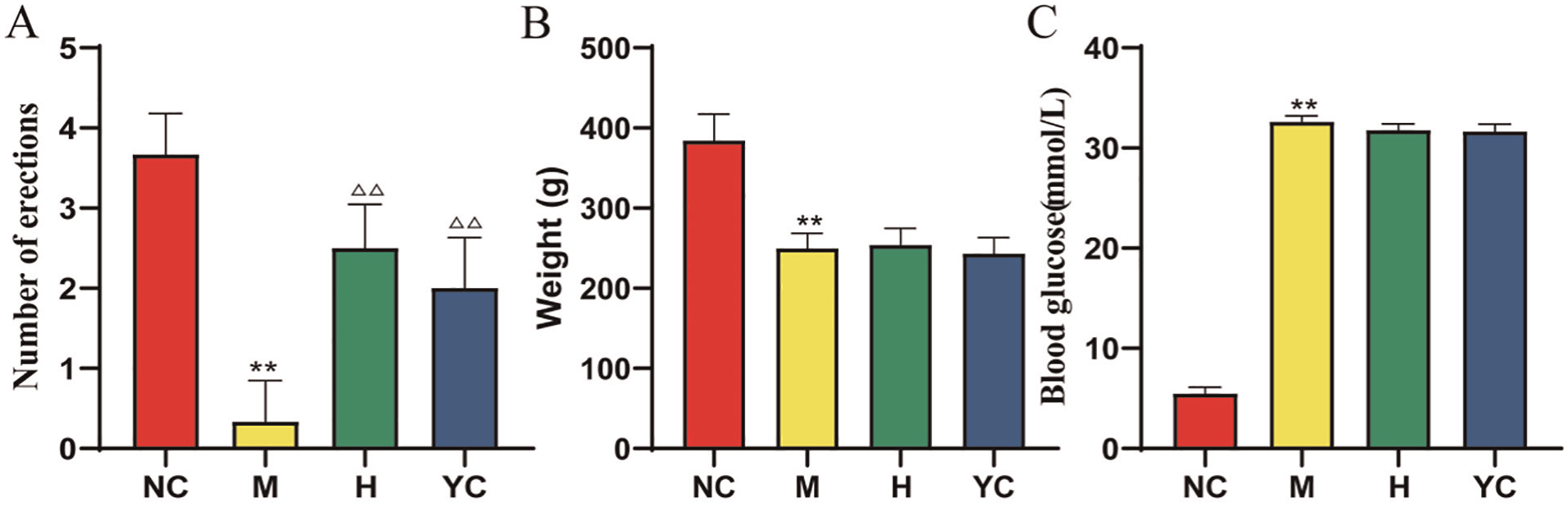
Hirudin’s Effects on the Number of Erections, Body Weight, and Blood Glucose Level in Rats From Different Group, **p < .05 in Comparison With the NC Group, △△ p < .05 in Comparison With the M Group. (A) Effect of Hirudin on Number of Erections of Rats in Each Group, (B) Effect of Hirudin on Body Weight of Rats in Each Group, and (C) Effect of Hirudin on Blood Glucose Levels of Rats in Each Group
Hirudin’s Effects on the Penile Structure of Rats
In the NC group, the corpus cavernosum displayed a well-organized structure, with regularly arranged trabeculae and evenly distributed blood-filled sinuses containing a few red blood cells. The trabeculae contained mainly smooth muscle, with few collagen fibers and small regularly shaped blood vessels, and showed no signs of interstitial hyperplasia. In contrast, the M group exhibited reduced cavernous sinus size, fewer small blood vessels, increased collagen fiber content, and significantly lower smooth muscle cell density compared with the control. Furthermore, H and YC groups showed a more regular distribution of blood sinusoids, increased smooth muscle cell density, and a reduction in collagen fibers within the blood sinusoids. A few erythrocytes were also visible in the blood sinusoids, with an increase in the smooth muscle cell density (Figure 2).
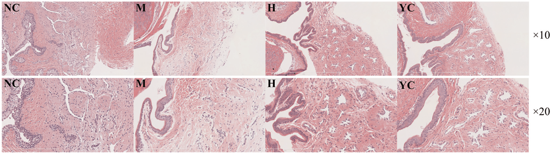
Hirudin’s Effects on the Different Histological Structures of the Corpus Cavernosum of Rats in Each Group (HE Staining, ×10 Magnification, Scale: 200 μm; ×20 Magnification, Scale: 100 μm)
Effects of Hirudin on the Level of Oxidative Stress
Serum levels of NO and SOD were significantly lower in M group compared with NC group but significantly higher in YC and H groups compared with M group (Figure 3A and C). However, serum levels of MDA were significantly higher in M group compared with NC group but significantly lower in YC and H groups compared with M group (Figure 3B).
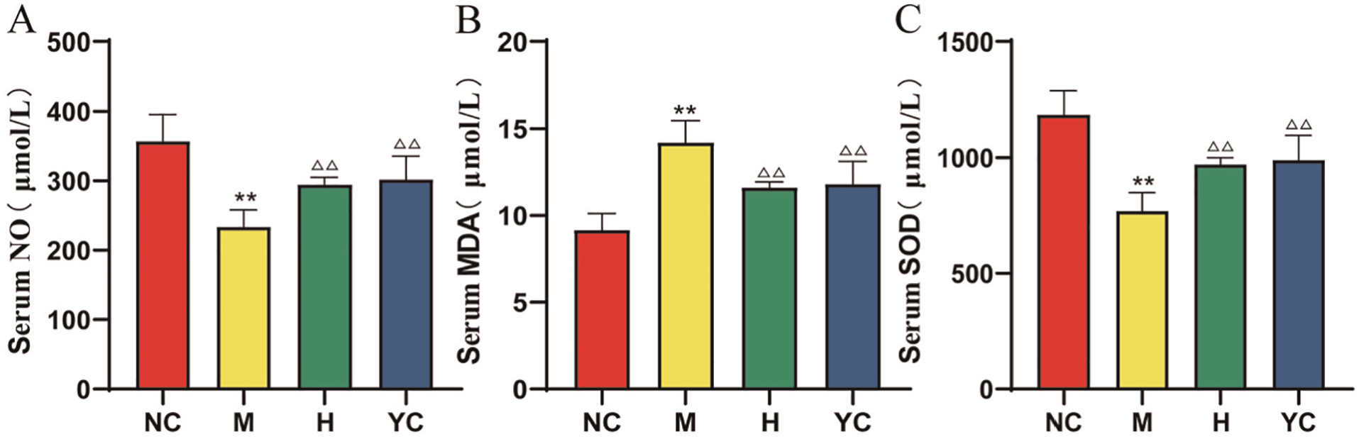
Hirudin’s Effects on the Serum Levels of NO, MDA, and SOD in Rats From Different Group, ** p < .05 in Comparison With the NC Group, △△ p < .05 in Comparison With the M Group. (A) Effect of Hirudin on Serum Level of NO of Rats in Each Group, (B) Effect of Hirudin on Serum Level of MDA in Each Group, and (C) Effect of Hirudin on Serum Level of SOD in Each Group
Effects of Hirudin on the Protein Expression of the HIF-1α/RhoA/ROCK Pathway
The relative expressions of HIF-1α, RhoA, ROCK1, ROCK2, and P-MYPT1 in the penile corpus cavernosum of M group were significantly higher compared with NC group, while those of H and YC groups were significantly lower compared with M group (Figure 4A–F).
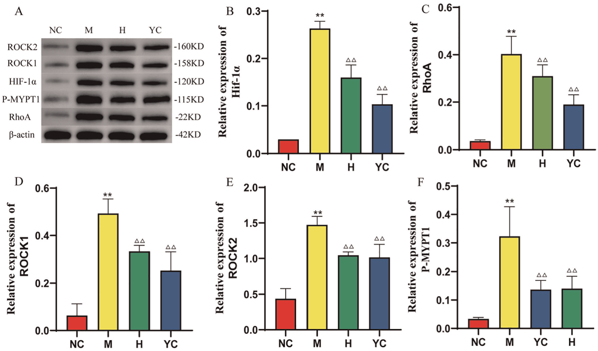
Hirudin’s Effects on the Expression of HIF-1α/RhoA/ROCK Pathway-Related Proteins in the Different Groups of Rats, ** p < .05 in Comparison With the NC Group, ΔΔ p < .05 in Comparison With the M Group. (A) Protein Immunoblotting in Penile Corpus Cavernosum Tissues of Rats in Each Group, (B) Relative Expression Levels of HIF-1α in Penile Corpus Cavernosum Tissues of Rats in Each Group, (C) Relative Expression Levels of RhoA in Penile Corpus Cavernosum Tissues of Rats in Each Group, (D) Relative Expression Levels of ROCK1 in Penile Corpus Cavernosum Tissues of Rats in Each Group, (E) Relative Expression Levels of ROCK2 in Penile Corpus Cavernosum Tissues of Rats in Each Group, and (F) Relative Expression Levels of P-MYPT1 in Penile Corpus Cavernosum Tissues of Rats in Each Group
Discussion
Recent research has shown that DIED’s pathogenesis is closely linked to neuropathy, vasculopathy, endothelial dysfunction, reduced NOS expression in penile tissues, and a lower smooth muscle to collagen ratio, with impaired diastolic function of penile tissues being a key mechanism (Irwin, 2019). Leech, a TCM commonly for DIED treatment, contains hirudin as its main ingredient. In this study, hirudin was found to increase the frequency of erections in DIED rats without significant changes in their body weight and blood glucose levels, hence suggesting that its therapeutic effects could be due to the regulation of oxidative stress, altered smooth muscle diastole, and improved vascular conditions rather than blood glucose control. Hirudin may provide a supplement for the treatment of DIED in the future.
Oxidative stress triggers a series of intracellular cellular responses, especially through HIF-α which regulates multiple factors in an oxygen-dependent manner. For instance, eNOS, which is transcriptionally regulated by HIF-1α during hypoxia, catalyzes the production NO from L-arginine, with the latter being a key regulator of vasodilatation that facilitates penile cavernous vasodilatation and increases intracavernous pressure to enable erection (Leslie & Sooriyamoorthy, 2024; Serocki et al., 2018). Although transient activation of HIF-1 during hypoxia supports tissue repair, the newly formed tissues may only partially restore the original structure and functions. However, in chronic hypoxia or injury, prolonged activation of the hypoxic pathway can drive fibrosis, leading to excessive scarring and compromised organ function (Darby & Hewitson, 2016). Under sustained hyperglycemia and hypoxia, continuously high HIF-1 expression promotes Vascular Endothlial Growth Factor (VEGF) expression which increases the levels of Interleukin–1β (IL-1β) and adhesion factors (e.g., Intercellular Cell Adhesion Molecule-1 (ICAM-1) and Vascular Cell Adhesion Molecule 1 (VCAM-1)) and results in abnormal proliferation of vascular ECs that induces ECs expansion and enlarges cell gaps to further damage the vascular microenvironment (Lou et al., 2019). Persistent high expression of HIF-1 also upregulates platelet-derived growth factor (PDGF) which binds to its receptor (PDGFR) to promote pericyte proliferation, migration, and differentiation into myofibroblasts. This transformation eventually results in extracellular matrix deposition, fibrosis, and further damage to the penile cavernous vascular microenvironment (Y. J. Zhang et al., 2014b).
The oxidative stress induced by DM is a key factor contributing to DIED. Indeed, oxidative stress can disrupt NO signaling and promote fibrosis in the penile cavernous smooth muscle (Chen et al., 2024; Zhang et al., 2024a, 2024b). Therefore, reducing oxidative stress may lead to improved ED (Y. Zhang et al., 2018). NO acts as a signaling molecule and a major mediator of penile erection, with its levels being closely related to erectile function. However, diabetes can induce oxidative stress in the body, resulting in the abnormal activation of enzymes, such as NADPH oxidase, xanthine oxidase, and eNOS, which scavenge NO and reduce its levels. This leads to impaired relaxation of penile cavernous smooth muscle, thereby contributing to ED (Kaltsas et al., 2024). In this context, Yang et al. (2021) found that hirudin could inhibit Myeloperoxidase (MPO) enzyme activity and regulate oxidative stress levels to treat diabetic ED. Similarly, in this study, it was observed that hirudin could reduce MDA concentrations, increase the serum levels of NO and the antioxidant enzyme SOD as well as improve oxidative stress in penile corpus cavernosum (Kaltsas et al., 2024).
The RhoA/Rho kinase pathway plays a crucial role in maintaining cellular morphology, smooth muscle contraction, and other biological functions. RhoA, a member of the Ras-related Rho family, acts as a molecular switch by binding to small Guanosine Triphosphate (GTP) and Guanosine diphosphate (GDP) molecules (Aida et al., 2024). During cavernous smooth muscle contraction, nerve endings and ECs in the corpus cavernosum initially release substances, such as endothelin (ET) and angiotensin II (Ang II), which convert Rho A-GDP to Rho A-GTP, and transfer RhoA to the cell membrane to activate its targets, ROCK1 and ROCK2. ROCK then phosphorylates MYPT-1, the light-chain-binding subunit 1 of myosin, resulting in the inactivation of myosin light chain phosphatase (MLCP) and the contraction of the cavernous smooth muscle (Innico et al., 2021). RhoA influences ED by reducing NO production through the inhibition of eNOS activity, thereby down-regulating the NO/cGMP pathway (Ulker et al., 2019). In addition, HIF-1α can promote the aberrant activation of the RhoA/Rho kinase signaling pathway which contributes to tissue fibrosis. Similarly, angiopoietin-like 4 (ANGPTL4), which is regulated by HIF-1α and has increased expression in diabetic models, binds directly to neurofibrillary protein 1 (NRP1) and NRP2 on ECs. This further activates the RhoA/ROCK signaling pathway, resulting in the transformation of vascular smooth muscle cells into myofibroblasts and leading to the fibrosis of penile cavernous smooth muscle (Vogel et al., 2010; Wei et al., 2024). In this study, hirudin was found to inhibit the fibrosis of corpus cavernosum smooth muscle and improve its relaxation by suppressing HIF-α expression and regulating the RhoA/ROCK pathway, thereby treating DIED. Hirudin may provide a supplement for the treatment of DIED in the future. This study provides a basis for the subsequent research and development of hirudin drugs. More cell and animal studies and clinical trials need to be carried out.
Our study has some limitations. First, we did not verify whether the known targets regulated by hirudin ECs and smooth muscle cells. More cell and animal studies and clinical trials need to be carried out. Second, we could not evaluate whether repression of HIF-1α/Rho/ROCK pathway-related protein expression by hirudin had a direct effect on erection in DIED. Studies are needed to evaluate the detailed mechanism and function of HIF-1α/Rho/ROCK pathway-related proteins in ED and other vascular and/or neurological disorders. The effect of hirudin on DIED patients is still unclear, and further clinical trials are needed for evaluation.
Conclusion
In this study, hirudin was found to improve the oxidative stress state, inhibit smooth muscle fibrosis, reduce vascular injury, and enhance erectile function in DIED rats by modulating HIF-α to regulate the RhoA/ROCK pathway. This study provides a certain research basis for hirudin in the future treatment of DIED. In the future, further cell experiments and clinical trials are needed to clarify its efficacy.
Footnotes
Acknowledgements
All the authors of the manuscript are immensely grateful to the foundations for their valuable support.
Declaration of Conflicting Interests
The author(s) declared no potential conflicts of interest with respect to the research, authorship, and/or publication of this article.
Funding
The author(s) disclosed receipt of the following financial support for the research, authorship, and/or publication of this article: This research was supported by the Pilot Project for Enhancing Clinical Research and Translational Capacity at Dongzhimen Hospital, funded by the Clinical Research and Business Fee Support Program for High-Level Traditional Chinese Medicine Hospitals from the Central Government (DZMG-MLZY-23006): Construction of the Diagnostic and Treatment System for Male Diseases Based on Professor Li Yueqing’s Academic Experience about the Theory of Deficiency and Blood Stasis, the Talent Training Programme Project, the Clinical Research Operating Expenses of Centralised High-level Chinese Medicine Hospitals, and the Beijing University of Chinese Medicine 2024 New Teacher Project (2024-JYB-XJSJJJ-041), and the Wu Jieping Medical Foundation Clinical Research Special Funding Fund (320.6750.2022-25-1).
Consent to Participate Statement
Patient consent was not required in accordance with local or national guidelines.
Data Availability Statement
The data sets analyzed during the current study available from the corresponding author on reasonable request.