
Abstract
Holothurian glycosaminoglycan (hGAG) is extracted from the body wall of the sea cucumber, and previous studies have shown many unique bioactivities of hGAG, including antitumor, anti-angiogenesis, anti coagulation, anti thrombosis, anti-inflammation, antidiabetic effect, antivirus, and immune regulation. The effects of 3W and 5W molecular weights hGAG with hematoporphyrin derivative-photodynamic therapy (HPD-PDT) on lung cancer were investigated. Human lung adenocarcinoma A549 cells were divided into 6 groups: control group, 3W molecular weight hGAG group, 5W molecular weight hGAG group, HPD-PDT group, 3W molecular weight hGAG + HPD-PDT group, and 5W molecular weight hGAG + HPD-PDT group. Cell morphology was observed under inverted phase contrast microscope. Cell proliferative activity was detected by CCK8 and cell apoptosis was assayed by Hoechst33258 staining and flow cytometry. The results showed that two different molecular weights hGAG could inhibit proliferation, promote apoptosis rates of A549 cells, and enhance the sensitivity of A549 cells to HPD-PDT. The combined use of hGAG and HPD-PDT has synergistic inhibitory effects on A549 cells, and the effects of 3W molecular weight hGAG are better than that of 5W molecular weight hGAG.
Introduction
Lung cancer, the second most commonly malignant tumors, caused 2.20 million new diagnoses and 1.79 million tumor-related deaths in 2020, and the latter accounts for about 18% of total cancer mortality. 1 The survival rate of patients with lung cancer after 5 years was only 10% to 20% in most countries, including China.2-4 In China, it was estimated that there were 0.81 million new lung cancer cases in 2020, 0.54 million in men, accounting for 38% of global new lung cancer cases in males, and 0.27 million in women, accounting for 36% of global new lung cancer cases in females. 5 In histopathological classification, lung cancer consists of 2 main types, non-small cell lung cancer (NSCLC) and small cell lung cancer (SCLC), and the proportion of the former is more than 85%.6,7 In NSCLC, adenocarcinoma is the most prevalent subtype, accounting for approximately 40% of lung cancer’s new diagnoses. 8
Sea cucumber is of high pharmacological value, and there are many kinds of active components existing in the sea cucumber. Sea cucumber polysaccharide is a kind of acidic mucopolysaccharide, which is extracted from the body wall of the sea cucumber and includes 2 main subtypes: one is holothurian glycosaminoglycan (hGAG) and the other is sea cucumber fucoidan. 9 With deep analysis of the structure and bioactivities of hGAG, the content of sulfate groups, the position of sulfation and molecular weight may predominantly determine the bioactivities of hGAG.10,11 Nowadays, many unique biological activities of hGAG have been discovered, such as antitumor, anti-angiogenesis, 12 anti coagulation,11,13 anti thrombosis, 14 anti inflammation, 15 antidiabetic effect, 16 antivirus, 17 and immune regulation. 18 Moreover, a previous study of our research group has confirmed that hGAG can strengthen the sensitivities of tumor cells to chemotherapy. 19
Photodynamic therapy (PDT) is also called photoradiation therapy or photochemical therapy, and now is becoming a new form of tumor treatment. It encompasses 3 important parts: photosensitizer, laser with appropriate wavelength and oxygen in tumor cells. 20 The process of PDT for cancer treatment is as follows: tumor cells selectively ingest photosensitizers, and then laser irradiation triggers the photochemical reaction that leads to tumor cell apoptosis and necrosis via reactive oxygen species (ROS), while normal tissues or organs are less affected. Nowadays, the most prevalent kind of photosensitizers is hematoporphyrin in clinical practice. 21 Previous studies indicated that PDT combined with chemotherapy, target therapy, and immune therapy could gain better curative effectiveness in patients with tumor.22-24 However, it is still not known that whether hGAG could enhance the sensitivity of tumor cells to HPD-PDT or not. Based on the previous research results, this study was performed to explore the combined effects of 3W and 5W molecular weights hGAG and photodynamic therapy on inhibiting proliferation and promoting apoptosis in A549 cells, and explore whether there were differences in antitumor effects of sea cucumber polysaccharides with 3W and 5W molecular weights.
Material and Methods
Substances
Herein, 3W and 5W molecular weights sea cucumber polysaccharide was isolated and purified by Ocean University of China. The structure of the monosaccharide is as shown in Figure 1, and 3W and 5W molecular weights hGAG are polymers consisting of different amounts of monosaccharides.

Chemical structure of hGAG monosaccharide.
Cell Culture
The human lung adeno carcinoma cell line A549 was provided by the Central Laboratory of the Affiliated Hospital of Qingdao University. The cells were cultured in complete medium which consisted of 10% heat-inactivated fetal bovine serum (HyClone), 1% penicillin-streptomycin (100 U/ml penicillin and 100 U/ml streptomycin; Servicebio), and 89% DMEM high-glucose medium (HyClone) at 37°C in humidified 5% CO2. All experiments were performed when cells grew to 80% to 90% confluence.
CCK8 Detection
Cell viability was assessed by CCK8 (Beyotime Institute of Biotechnology, Shanghai, China). A549 cells were seeded in 96-well plates at a density of 1 × 10⁴ cells/well. A proper amount of complete culture medium was added into each well, and cells were incubated for 24 hours. As for the concentration of hGAG, we referred to our previous study, 19 in which it was found that concentration at 100 µg/ml of 3W and 5W hGAG did not show any cytotoxicity to the cancer cells and showed the most obvious effect compared with other concentrations. Cells were divided into 6 groups: control group; 3W molecular weight hGAG therapy group (the concentration of hGAG was 100 μg/ml); 5W molecular weight hGAG group (the concentration of hGAG was 100 μg/ml); HPD-PDT group (hematoporphyrin derivative concentration was 8 μg/ml and cells were irradiated by a 630 nm laser; in the process, the laser energy density was 50 mW/cm2, the spot diameter was about 9 cm, the irradiation time was 2 minutes and the vertical distance between the emitter and the 96-well plate was 9 cm.); 3W molecular weight hGAG combined with photodynamic therapy group (the concentration of hGAG was 100 μg/ml; after incubated for 24 hours and washed with PBS, the remaining treatment was the same as HPD-PDT group); 5W molecular weight hGAG combined with photodynamic therapy group (the concentration of hGAG was 100 μg/ml and the treatment was the same as 3W hGAG + HPD-PDT group). Each group included 3 wells, washed with 100 µl phosphate-buffered saline (PBS) and observed by inverted phase contrast microscope (Olympus, China). We added 10 µl CCK8 + 90 µl complete medium into each group and cultured A549 cells at 37°C. A microplate reader was used to measure the optical density (OD) at 450 nm after 30 minutes. The cell viability ratio was calculated as follows: Cell survival ratio (%) = (OD2 − OD0)/(OD1 − OD0) × 100%. OD2 is the absorbance of the experimental group, OD1 is the absorbance of the control group, and OD0 is the absorbance of the blank control group. The coefficient of drug interaction (CDI) was utilized to evaluate the inhibitory effects of the combinations. The formula to calculate CDI is as follows: CDI = AB/(A × B). According to the absorbance of each group, AB is the ratio of the combination group to the control group, and A or B is the ratio of the non-combination group to the control group. According to CDI, if the CDI value is <1, it indicates there is synergism between hGAG and HPD-PDT; especially if the CDI value is <0.7, hGAG and HPD-PDT are significantly synergistic; if the CDI value is =1, hGAG and HPD-PDT are additive; if the CDI value is >1, there is antagonism between hGAG and HPD-PDT.
Hoechst33258 Staining
A549 cells were inoculated in 24-well plates, divided into 6 groups and treated with various concentrations of agents as described above. The culture medium was removed after 48 hours treatment. 0.5 ml cell fixative was added to each well and cells were fixed for 15 minutes. After washing twice using PBS, 0.5 ml Hoechst staining solution (Beyotime Institute of Biotechnology, Shanghai, China) was added into each well. After 5 minutes of staining, we removed the staining solution and washed again twice with PBS. An inverted fluorescence microscope (Olympus, China) was used for observation.
Flow Cytometric Analysis of Apoptosis
Apoptotic cells in the early stage and the late stage can bond with Annexin V-FITC and propidium iodide (PI), respectively, and Annexin V-FITC/PI double-staining could be used to distinguish early and late apoptotic cells. After being treated for 48 hours and washed with PBS, A549 cells were centrifuged for 5 minutes (1000 rpm) and washed twice with PBS, and then mixed with 5 µl Annexin V-FITC and 10 µl PI. After incubation at room temperature for 10 minutes, a flow cytometer (BD Biosciences, Franklin Lakes, NJ) was used to analyze the apoptosis of A549 cells.
Statistical Analysis
All experiments were repeated at least 3 times. SPSS statistical software, version 26.0 (SPSS Inc, IBM, Chicago, IL) was used for data analysis, and the numerical variables were expressed as mean ± standard deviation (X ± S). All groups were tested for homogeneity of variance before comparison. One-way analysis of variance was used to compare the mean between groups. LSD-t (least significant different-t) test was used for pairwise comparison, with the level of the test α = .05, that is, when the value of P is < .05, the difference was statistically significant.
Results
Morphology of A549 Cells Line
The morphological changes of the A549 cells line were observed under inverted phase contrast microscope. It showed that A549 cells in the control group were in a good state and arranged closely, with a large number and small intercellular space. Cells were flat and grew adherently. There were clear nucleoli, full cell body, and transparent cytoplasm. In contrast, after hGAG and HPD-PDT treatment, the cell count in the culture medium decreased; the cells were sparse and less dense; the cell body protrusions got shorter; the cellular volume became smaller, and some necrotic cells were visible, among which the 2 combination groups were the most obvious. Compared with 5W hGAG group, the morphological changes of the cells in 3W hGAG group were more obvious (Figure 2).
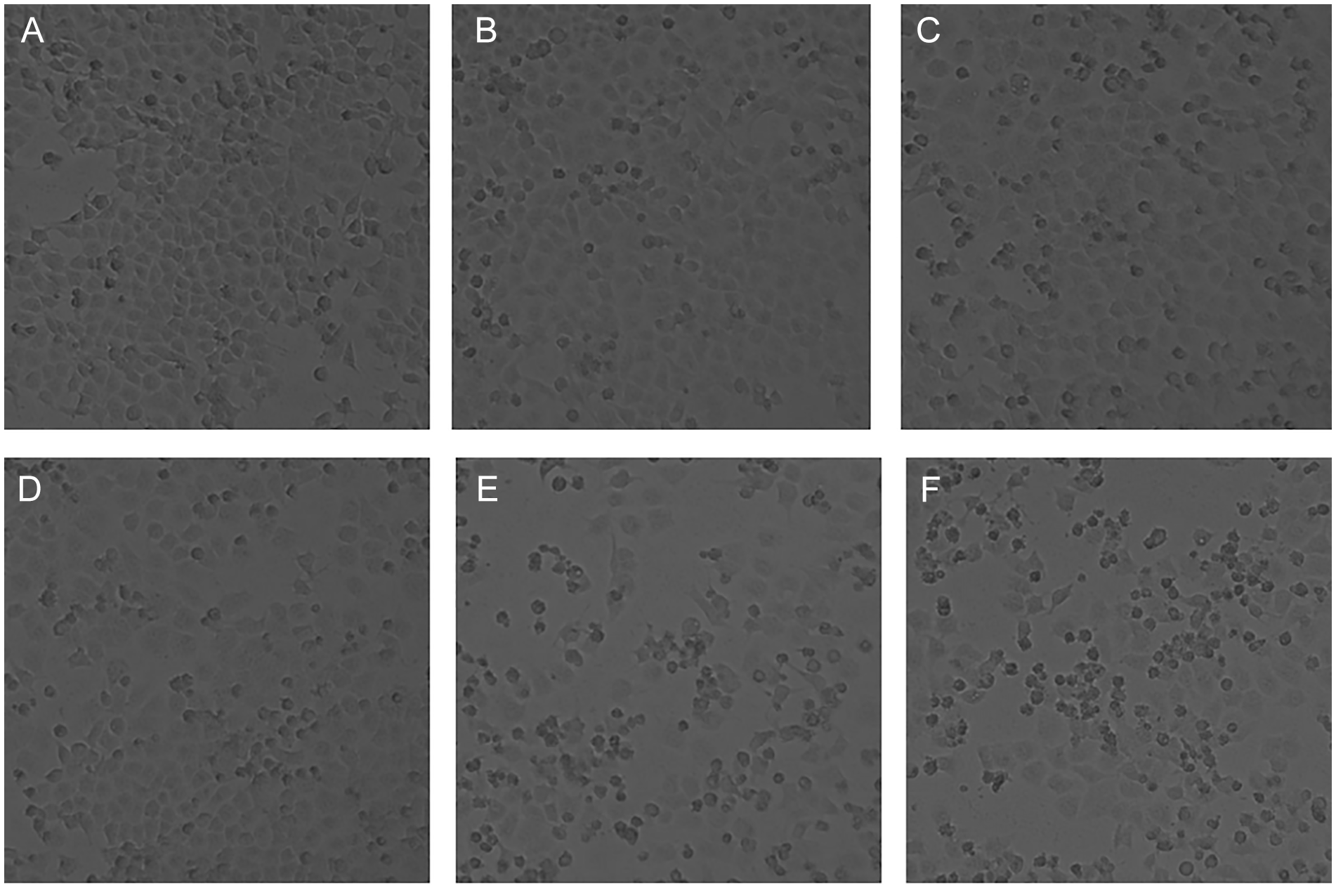
Observation of A549 cells showed sparse and less dense cells, decreased cell count, short cell body protrusions, small cellular volume, and some necrotic cells in experimental groups compared with the control group. (×10) (A) Control group. (B) 3 W hGAG group. (C) 5 W hGAG group. (D) HPD-PDT group. (E) 3 W hGAG + HPD-PDT group. (F) 5 W hGAG + HPD-PDT group.
The Inhibition Effects of hGAG and HPD-PDT on A549 Cell Line
To reveal the effects of hGAG and HPD-PDT on cell viability, CCK8 assay was performed. The OD value and proliferative activity of A549 cells were measured (Table 1). It was observed that 3W and 5W molecular weights hGAG and HPD-PDT exhibited inhibitory effects on the proliferation of A549 cells (P < .05). And hGAG could promote the proliferation inhibition of A549 cells by HPD-PDT and enhance the sensitivity of HPD-PDT (P < .05). Especially the inhibitory effect of 3W molecular weight hGAG was more clear than that of 5W molecular weight hGAG (P < .05). CDI values of the 2 molecular weights of hGAG and photodynamic combination were 0.894 (3W) and 0.933 (5W), respectively, which showed that synergistic effects could be obtained when hGAG and HPD-PDT are applied together.
The OD Value and Proliferative Activities in Different Groups (Mean ± SD, n = 3).
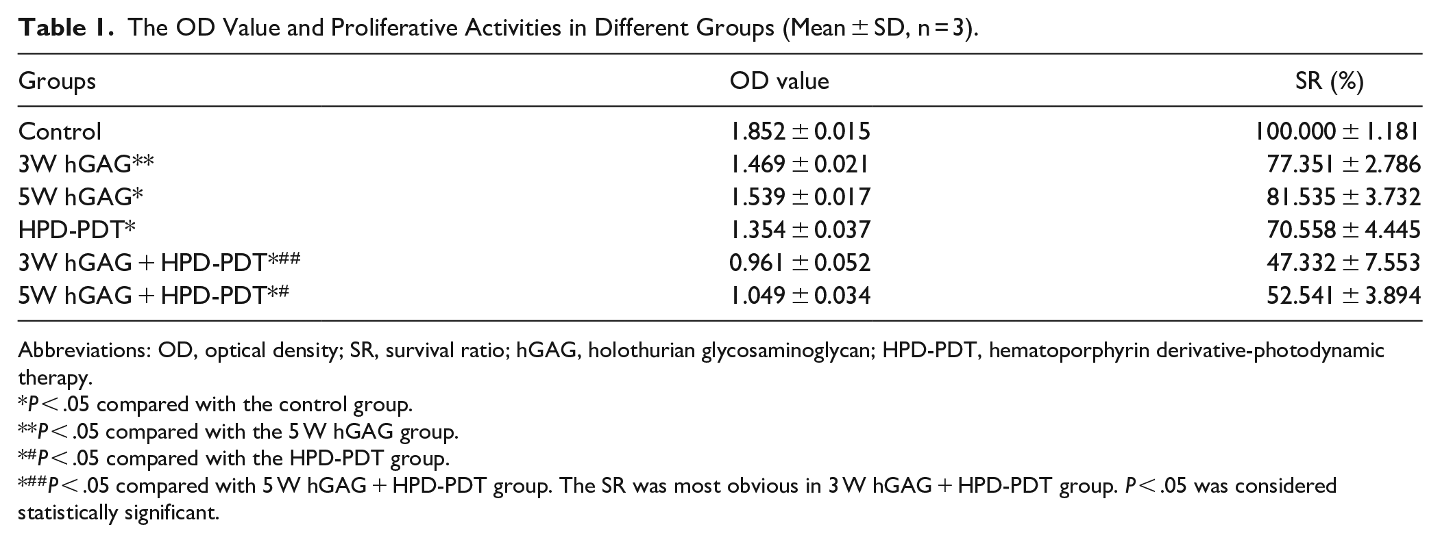
Abbreviations: OD, optical density; SR, survival ratio; hGAG, holothurian glycosaminoglycan; HPD-PDT, hematoporphyrin derivative-photodynamic therapy.
P < .05 compared with the control group.
P < .05 compared with the 5 W hGAG group.
#P < .05 compared with the HPD-PDT group.
##P < .05 compared with 5 W hGAG + HPD-PDT group. The SR was most obvious in 3 W hGAG + HPD-PDT group. P < .05 was considered statistically significant.
The Apoptosis Effects of hGAG and HPD-PDT on A549 Cells
Apoptotic A549 cells were detected by Hoechst33258 staining. There were no obvious apoptotic cells in the control group, while shrunken, hyperchromatic, and pyknotic cells and nuclear fragmentation could be observed under microscope in experimental groups. (Figure 3). And we observed lower cell count and less cell density and more obvious apoptotic cells in the 3W molecular weight hGAG group than in the 5W molecular weight hGAG group. The 2 combination groups were the most obvious with the lowest cell count and the least cell density. The results of total apoptosis rates of A549 cells from flow cytometric analysis also showed that apoptosis rates in experimental groups significantly increased compared with the control group and the apoptosis rates in each combination group were the highest compared with the single drug group (P < .05). It indicated that 3W and 5W molecular weights hGAG and HPD-PDT could induce the apoptosis of A549 cells and hGAG increased the apoptosis rates induced by HPD-PDT (P < .05). The apoptosis effect of 3W molecular weight hGAG was more obvious than that of 5W molecular weight hGAG (P < 0.05; Table 2 and Figures 3 and 4).
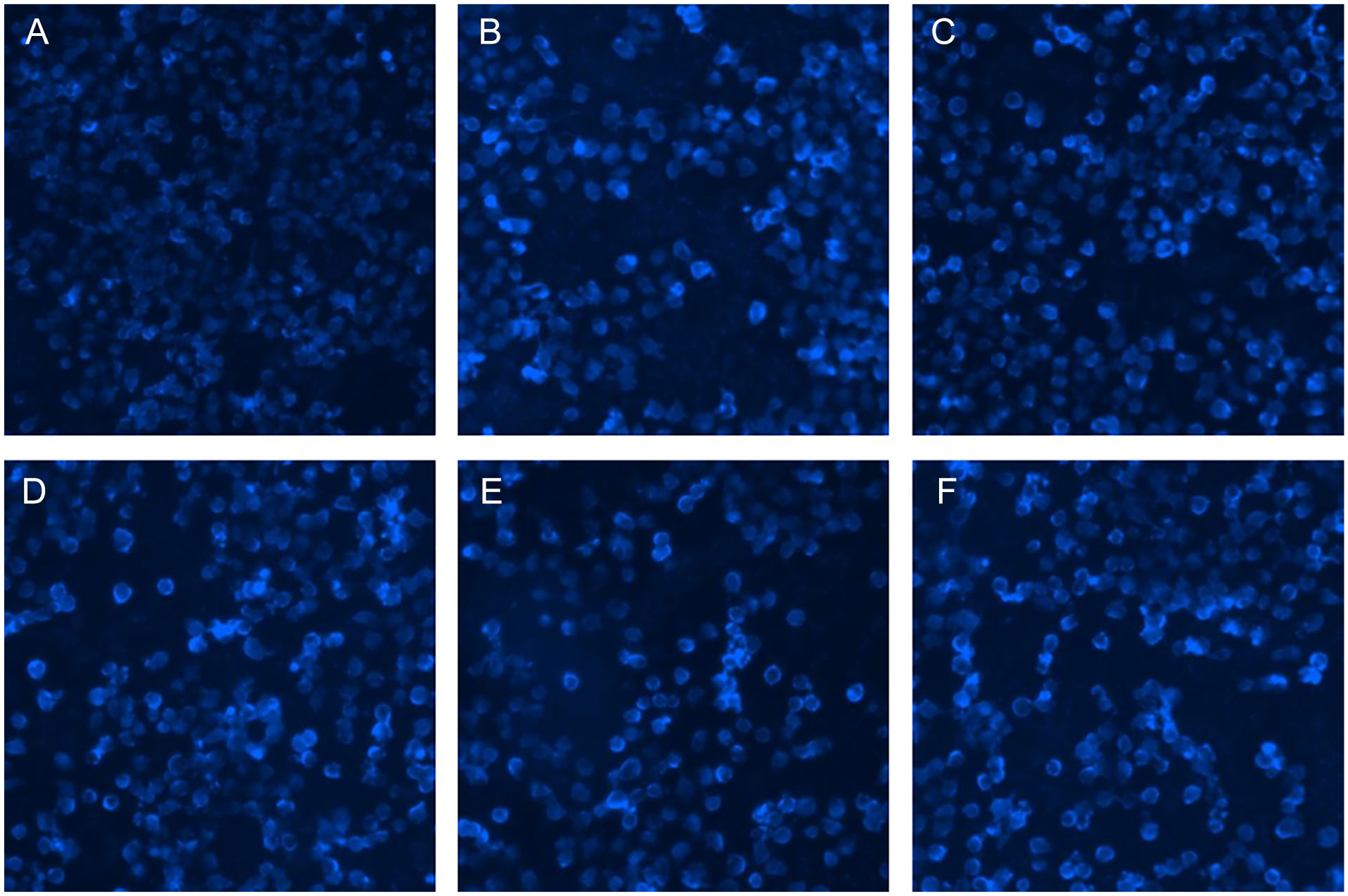
A549 cells were treated with different methods for 48 hours. The cells were stained with Hoechst33258 and images were captured by using an inverted fluorescence microscope. There were shrunken, hyperchromatic, and pyknotic cells and fragmented nuclei in experimental groups. (×10) (A) Control group. (B) 3W hGAG group. (C) 5W hGAG group. (D) HPD-PDT group. (E) 3W hGAG + HPD-PDT group. (F) 5W hGAG + HPD-PDT group.
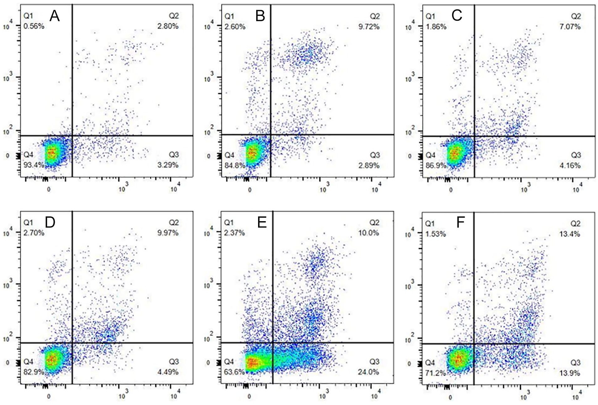
A549 cells were treated with different methods for 48 hours. AnnexinV-FITC/PI double staining was used to distinguish early and late apoptotic cells. The proportion of apoptotic cells was measured by flow cytometry. (A) Control group. (B) 3W hGAG group. (C) 5W hGAG group. (D) HPD-PDT group. (E) 3W hGAG + HPD-PDT group. (F) 5W hGAG + HPD-PDT group.
The Apoptosis Rates of A549 Cells in Different Groups (Mean ± SD, n = 3).

Abbreviations: A, early apoptosis rate; B, late apoptosis rate; C, total apoptosis rate; hGAG, holothurian glycosaminoglycan; HPD-PDT, hemoporphyrin derivative-photodynamic therapy.
P < .05 compared with the control group.
P < .05 compared with the 5W hGAG group.
#P < .05 compared with the HPD-PDT group.
##P < .05 compared with 5W hGAG + HPD-PDT group. The total apoptotic rate was most obvious in 3W hGAG + HPD-PDT group. P < .05 was considered statistically significant.
Discussion
Worldwide, the morbidity and mortality of lung cancer rank first in males, while in females cancer deaths rank third, after breast cancer and colorectal cancer and new cases rank second, after breast cancer. 1 Lung cancer has become a heavy burden for China. 25 With the implementation of low-dose CT screening, more and more patients at early stages have been discovered and a significant decrease has been gained in mortality. 26 In the meantime, with the development of chemotherapy, target therapy, and immune therapy, the overall survival and disease-free survival have greatly progressed. 27 However, the limitations of these treatments are also assignable. Radiochemotherapy can better improve the prognosis of patients at present. But its side effects also need to be paid attention to, such as damage to the immune system and destruction of normal organ functions. 28 In recent years, target therapy and immune therapy have better effects on some patients with particular oncogenic drivers and PD-L1 expression, but drug resistance limits their application in lung cancer treatment. 29 Therefore, exploring new treatments and new active components with antitumor effects is essential.
HGAG is an active component extracted from the body wall of sea cucumber in recent years, and cannot be synthesized artificially at present. In previous studies, hGAG has been shown to have antitumor effects, the mechanism for which includes anti-angiogenesis, heparanase activity inhibition, suppressing cell adhesion to platelet-coated surface, regulation of immunoreactivities, cell cycle inhibition, and induction of apoptosis,12,30-32 and hGAG can strengthen the sensitivity of tumor cells to chemotherapy. 19 Nowadays, more kinds of low molecular weight hGAG are obtained and their structure-bioactivities are becoming a focus of current studies. There is a close correlation between bioactivities and molecular weight. Some studies have proved that low molecular weight hGAG, as a depolymerized product, demonstrated the same bioactivities as naïve hGAG, such as anti coagulation and hematopoiesis-stimulation.11,33 Furthermore, lower molecular weight hGAG can dramatically improve the immune regulation activity. 10 In this study, low molecular weight sea cucumber polysaccharide was obtained by chemical degradation of high molecular weight sea cucumber polysaccharide. Through the assay of cell proliferative activity and apoptosis rates, the results indicated that the inhibitory effects on A549 cells of 3W molecular weight hGAG was superior to 5W molecular weight hGAG, which also pointed out the direction for further exploring the anti tumor effects of lower molecular weight.
In recent years, as a potential and non-invasive treatment, photodynamic therapy (PDT) is gradually used for the treatment of lung carcinoma. The antitumor mechanisms of PDT include inducing tumor cell necrosis, apoptosis and autophagy, regulating immune response, and destroying tumor nutrient vessels.34-39 Because of the capability of normal tissues to clear or eliminate the photosensitizers, the photochemical reaction almost occurs in pathological tissues, and there are almost no phototoxic effects in surrounding normal tissues. 37 Compared with conventional treatments, PDT has the advantages of less trauma, less toxicity, repeatability, and high selectivity. With the development of endoscopic technology, PDT is broadly applied in many kinds of tumors, including benign tumors and malignant tumors. Moreover, combination with some novel technologies, such as nanotechnology, liposome and lipoprotein, and electroporation provides the possibility for improving the efficacy of PDT. 40 Besides the above aspects, finding out some new components to maximize the efficacy of PDT as far as possible is also a promising direction for lung cancer treatment.
However, due to the limitation on the experimental conditions, this study was only performed on A549 human lung adenocarcinoma cells. It is needed to further investigate the effects of the 2 different molecular weights hGAG on other cell lines and animals. Furthermore, based on our previous study, the mechanism of the sensitization enhancement of hGAG to chemotherapy may correlate with low expression of survivin and Bcl-2 mRNA and protein and high expression of Bax and caspase-3 mRNA and protein. 19 The signaling pathway of the anti tumor effects and immunostimulatory effects is possibly related to suppression of the activation of the ERK1/2/p38 and MAPK/NF-κB pathway.12,18 The mechanism and signaling pathway of the sensitization enhancement of hGAG to HPD-PDT need further elucidation. We speculate that the mechanism of hGAG combined with HPD-PDT on apoptosis may be also associated with the above mechanism. And we will explore protein and gene expressions as well as signaling pathways to validate our speculation in our following experiments. Because of the limitations of separation and purification techniques, only 3W and 5W molecular weights hGAG can be obtained by us, and it is also essential to separate and purify lower molecular weight hGAG, and reveal the inhibitory effects of more kinds of lower molecular weights hGAG combined with HPD-PDT to find appropriate molecular weight hGAG for improvement in HPD-PDT. It is believed that in the future hGAG will become a promising candidate in lung cancer treatment and contribute to the clinical application of HPD-PDT, which brings more benefits to clinicians and lung cancer patients.
Conclusion
In conclusion, 3W and 5W molecular weights hGAG, whether in combination with HPD-PDT or not can cause proliferation inhibition of A549 cells and promote apoptosis rates of A549 cells, which can enhance the sensitivity of A549 cells to HPD-PDT. The combined use of hGAG and HPD-PDT has synergistic effects and the inhibitory effect of 3W molecular weight hGAG is better than that of 5W molecular weight hGAG, which indicates that this synergistic effect may be associated with the molecular weight of hGAG.
Footnotes
Declaration of Conflicting Interests
The author(s) declared no potential conflicts of interest with respect to the research, authorship, and/or publication of this article.
Funding
The author(s) disclosed receipt of the following financial support for the research, authorship, and/or publication of this article: This project was supported by the Key Laboratory of Marine Drug, Ministry of Education (KLMDOUC201307).