
Abstract
Thermotherapy and thermochemotherapy have been used in clinics to treat patients with malignant diseases, including colon cancer, and their efficacy has been well proved. Heat shock proteins (HSPs), especially Hsp70, play important roles in neutralizing their efficacy. It has been reported that quercetin can suppress cancer by inhibiting the intratumoral expression of Hsp70. This study was designed to investigate whether quercetin could enhance sensitivity to thermotherapy and thermochemotherapy. Soluble quercetin liposome was used in this study. The effects of quercetin were investigated in vitro and in mouse colon cancer models of subcutaneous tumor and peritoneal carcinomatosis. The results showed that quercetin liposome inhibited the upregulation of Hsp70 and enhanced apoptosis induced by hyperthermia and thermochemotherapy. Systemic administration of quercetin liposome can sensitize CT26 cells to thermotherapy and chemothermotherapy. This study suggests that quercetin liposome might be potentially applied for clinical cancer therapy.
Introduction
Thermotherapy and thermochemotherapy are proven effective approaches in cancer therapy. Hyperthermia thermotherapy is usually applied to increase tumor temperature to the range of 40°C to 43°C. 1 The mechanisms of thermotherapy in cancer consist of direct cell cytotoxicity, heat-induced alterations of tumor microenvironment, induction of apoptosis, and sensitizing tumor to chemotherapeutic drugs or other treatments. 2 Thermotherapeutic perfusion, one approach of thermotherapy, is often used in peritoneal cavities of patients with colon cancer or ovarian cancer.
Hyperthermia exposure also induces synthesis and overexpression of heat shock proteins (HSPs), which attenuates the effects of thermotherapy and thermochemotherapy, and has been linked to cancer resistance to stress-mediated apoptotic signals. Targeting HSPs has proven to be an effective strategy to reverse the thermotolerance of cancer cells. Hsp70 is the largest and the most important member to offer protection for cells or organs. 3 It plays a key role in promoting nascent protein folding, refolding of misfolded protein, and hydrolyzing the aggregation of denatured proteins.4,5 In tumor cells, Hsp70 has been reported to be expressed at higher levels than in normal cells3,4 and to be induced at a high level during hyperthermia, which enables the tumor to overwhelm the effect of hyperthermia. 5 Therefore, Hsp70 can be a potential target in strategies of cancer thermotherapy.
Quercetin, a ubiquitous bioactive flavonoid, can inhibit proliferation and induce apoptosis in a variety of cancer cells.6-10 Quercetin inhibits the growth of cancer cells through various mechanisms: inhibition of glycolysis, macromolecule synthesis, and enzymes; freezing cell cycle; and interaction with estrogen II binding sites.9-15 In addition, it was reported that quercetin could inhibit the induction of Hsp70 and thermotolerance without affecting the synthesis of other proteins.16,17 We assumed that combined therapy of quercetin with hyperthermia would enhance the suppression of tumor growth, through quercetin-mediated downregulation of Hsp70expression.
In this study, we tested the efficacy of quercetin liposome, a solution as described previously by our laboratory,9,10,18 in sensitizing tumor cells to hyperthermia and hypothermic peritoneal perfusion in mice. The results showed that quercetin liposome could inhibit expression of Hsp70 and enhance sensitivity of tumor to thermotherapy and thermochemotherapy.
Materials and Methods
Reagents, Cells, and Mice
CT26 colon cancer cells were obtained from the American Type Culture Collection and cultured at 37°C with 5% CO2 in 75 cm2 square flask or 6-well plate in RPMI 1640 medium (Life Technologies, Bedford, MA) supplemented with 10% fetal bovine serum and 100 U/mL mycillin. Quercetin (Sigma-Aldrich Co, St Louis, MO) liposome was prepared as described previously. 18 Female BALB/c mice aged 5 weeks were purchased from the Laboratory Animal Center, Sichuan University. All animal experiments were carried out according to the protocol approved by the Ethics Committees on Animal Experimentation of Sichuan University.
Flow Cytometry
CT26 cells were seeded into 6-well plates at 2 × 10⁵/well. When cells grew to 50% confluence, cells were treated as follows: added quercetin liposome at 150 µM, heat shocking of cells at 42°C for 2 hours or added quercetin at 150 µM followed by heat shocking of cells at 42°C for 2 hours, then continued incubation at 37°C for 4 hours. 9 The cells were trypsinized for harvesting and washed with phosphate buffered saline (PBS). Ice-cold ethanol (75%) was added to the cells before vortexing, and then PBS with PI-stain and RNase A were added. The percentage of apoptotic cells was analyzed using an EpicsXLMCL flow cytometry (Beckman Coulter, Brea, CA) as described previously. 19
Western Blotting Analysis
The liposome quercetin was added into the culture medium at final concentrations of 0, 50, 100, 150, and 200 µM, when cells were 70% confluent. Cells were heat shocked at 42°C for 2 hours followed by incubation at 37°C for 4 hours, and then Western blot was performed as described. 20 Briefly, the cells were harvested and lysed with radioimmunoprecipitation assay buffer. Cell lysates were separated by SDS/PAGE and electroblotted using Sartoblot (Sartorius, Goettingen, Germany) onto a polyvinylidene fluoride membrane. The membrane was blocked at 4°C with 5% nonfat dry milk and probed with an Hsp70 antibody (Santa Cruz Biotechnology Inc, Santa Cruz, CA) at 1:500 dilutions. Biotinylated goat anti-mouse IgG (Vector Laboratories, Burlingame, CA) was used as the secondary antibody. Staining signals were visualized with the use of the Vectastain ABC kit (Vector Laboratories).
Thermotherapy
The mouse model of colon tumor was established by subcutaneously inoculating 3 × 10⁵ viable CT26 tumor cells into the right hind legs of BALB/c mice. 21 When tumors were palpable, 20 BALB/c mice were randomly divided into 4 groups (n = 5): the first group was administered normal saline intravenously (IV) alone; the second, thermotherapy at 42°C for 1 hour; the third, quercetin liposome IV at a dosage of 10 mg/kg; and the fourth, liposome quercetin IV followed by thermotherapy at 42°C for 1 hour, every 3 days for a total of 5 times. Thermotherapy was performed as described. 21 Briefly, the tumor-bearing legs were immersed in water bath at 42°C for 1 hour. Every 3 days tumor volume was measured and calculated according to the formula, V (mm3) = 0.52 × a × b2, where a is the largest superficial diameter, b is the smallest superficial diameter, and 0.52 is approximately equal to π/6. The mice were sacrificed on the 18th day after the first treatment, tumor tissues excised, and fixed in 10% formalin. 18
Perfusion Thermochemotherapy
The tumor-bearing mouse model of hyperthermic peritoneal perfusion was established. Briefly, 5 × 10⁵ viable CT26 tumor cells were injected into the abdominal cavity of each BABL/c mouse. Five days after inoculation, 24 BABL/c mice were randomized into 4 groups with 6 mice in each group for further administration as follows: (1) control without hyperthermic peritoneal perfusion, (2) hyperthermic peritoneal perfusion with quercetin liposome at 10 mg/kg/mouse, (3) hyperthermic peritoneal perfusion with 5-fluorouracil (5-FU) at 50 mg/kg/mouse, and (4) hyperthermic perfusion with both quercetin liposome and 5-FU. The treatment was initialized according to the protocol, where 1 mL of agent solution at 43°C should be perfused into the abdominal cavity as soon as possible. Treatment was performed every 3 days for 5 times. The mice were sacrificed on day 18 after the first treatment; then tumor nodus was calculated and volume of the intraperitoneal tumor was measured.
Immunohistochemistry Analysis
The excised tumors, fixed in 10% formalin, from the thermotherapy experiment were embedded in paraffin and sectioned at 4-µm thickness. The sections were used for analysis of Hsp70 expression by immunohistochemistry using an anti-Hsp70 monoclonal antibody.
TUNEL Assay
The sections, the same as that used for immunochemistry analysis, were used for in situ analysis of apoptosis by TUNEL (terminal deoxynucleotidyl transferase dUTP nick end labeling). TUNEL staining was performed with an in situ apoptotic cell detection kit (Boehringer, Mannheim, Germany) following the manufacturer’s protocol.
Statistical Analysis
The data were analyzed by analysis of variance and presented as the mean ± standard deviation. A value of P < .05 was considered to be statistically significant.
Results
Hsp70 Upregulation by Hyperthermia and Downregulation by Quercetin
CT26 cells were treated with different concentration of quercetin liposome and hyperthermia at 42°C, and CT26 cells incubated at 37°C and that were not subjected to any treatment served as control, and the level of Hsp70 was examined by Western blotting. The results indicated that Hsp70 was largely induced by hyperthermia at 42°C (Figure 1A), and quercetin liposome could down regulate Hsp70 during hyperthermia in a dose-dependent manner starting at 50 µM (P < .05). When exposed to 150 µM—compared with 50µM—quercetin liposome the levels of Hsp70 were remarkably decreased (P < .05), and Hsp70 levels were lowest when exposed to 200 µM, compared with cells treated at 42°C alone (Figure 1B).
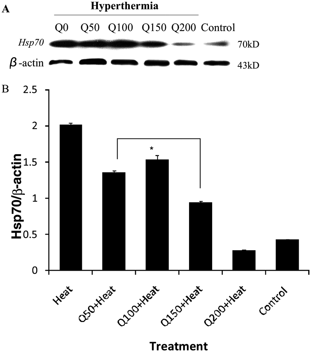
Quercetin inhibits the hyperthemia-induced Hsp70 expression: (A) Western blot bands for Hsp70 at varying quercetin concentrations and (B) density of the Hsp70 bands CT26 cancer cells were administered quercetin liposome at different concentrations for 30 minutes, then heat treated at 42°C for 2 hours. The cells were cultured at 37°C for 5 hours and total protein was harvested for Western blotting. Downregulation of Hsp70 was initiated at a concentration of 50 µM, was evident at a concentration of 150 mM, and reached a low at a concentration of 200 µM. *P < .05 (each treatment vs control or Q50 vs Q150).
Quercetin Enhances Apoptosis In Vitro
CT26 cells were treated by hyperthermia, quercetin, or the combination treatment of hyperthermia and quercetin, and flow cytometry was performed to detect cell apoptosis. The quantitative assessment of apoptotic cells was achieved by estimating the rate of apoptotic cells via flow cytometry (Figure 2A and B). The results indicate that quercetin liposome could obviously enhance the efficacy of apoptosis induced by hyperthermia.
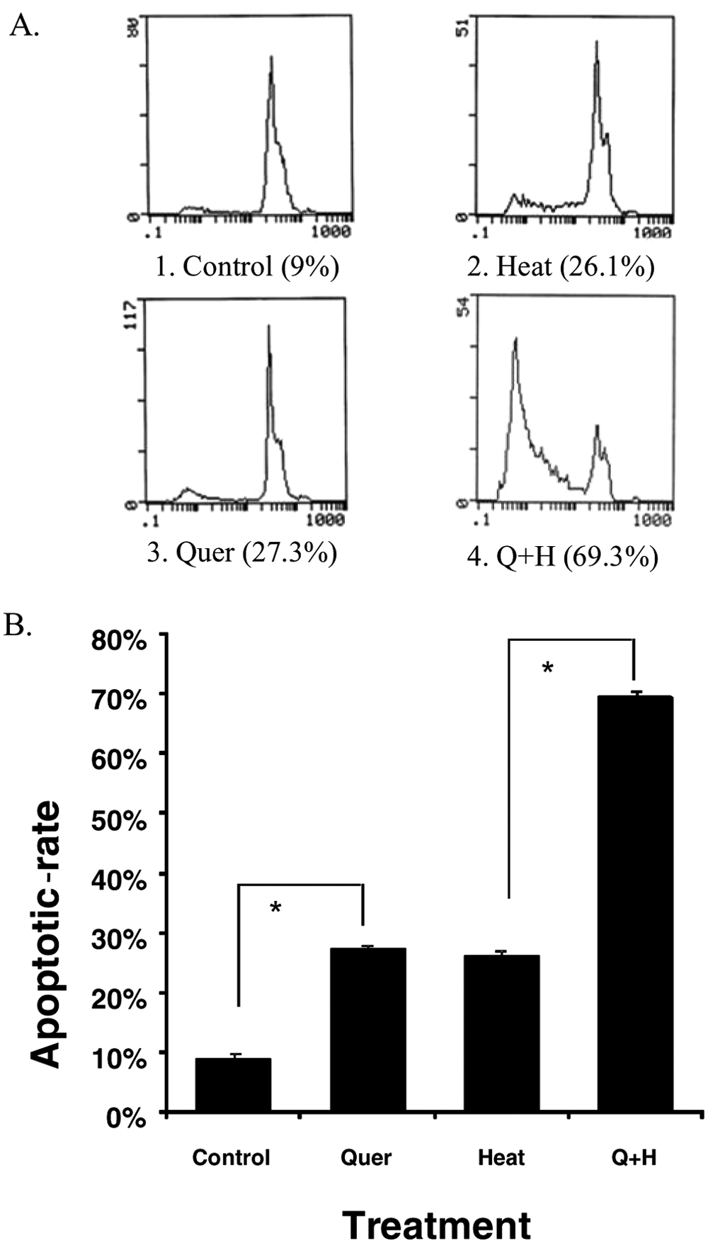
Quercetin increased hyperthermia-induced apoptosis in vitro: (A) Flow cytometry analysis and (B) apoptosis rate
Increased Tumor Inhibition by Quercetin Liposome Plus Thermotherapy In Vivo
Tumor models were established with CT26 cells. Tumor-bearing mice were subjected to different treatment for 5 times. The tumor volume was measured every 3 days, and the results were displayed (Figure 3). The results revealed that the tumor growth was obviously inhibited by thermotherapy and quercetin liposome. In the cohort treated with quercetin liposome followed by thermotherapy, tumor volume was measured and calculated as 1/20 of that in control. The in vivo observations demonstrated that the most effective suppression of tumor growth is ascribed to a combination of thermotherapy and quercetin liposome, which is also more effective than thermotherapy alone (P < .05).

Quercetin liposome increased antitumor effects of heat treatment in vivo
Quercetin Liposome Plus Thermotherapy Induced Hsp70 Downregulation and Increased Apoptosis
The expression of Hsp70 was examined by immunohistochemistry, and apoptosis of excised tumors by TUNEL (Figure 4A-C). Hsp70 expression was observed obviously within tumor tissue from control (Figure 4A-a), whereas hyperthermia treatment resulted in significant Hsp70 up regulation (Figure 4A-b). Intratumoral expression of Hsp70 in mice treated with quercetin liposome alone was scarcely observed, which indicated that systemic administration of quercetin liposome can inhibit the baseline expression of Hsp70 (Figure 4A-c). The expression of Hsp70 in mice treated with quercetin liposome followed by hyperthermia was much less than that in mice treated with hyperthermia alone (Figure 4A-d). These findings suggested that systemic administration of quercetin liposome can effectively inhibit the upregulation of Hsp70 induced by hyperthermia. Furthermore, the TUNEL assay showed that the combined treatment with quercetin liposome and hyperthermia could increase the induction of apoptosis significantly than that of other groups (P < .05; Figure 4B and C).

Immunohistochemistry for Hsp70 expression, and TUNEL analysis under a fluorescence microscope (×200): (A) Immunohistochemistry staining, (B) TUNEL analysis, and (C) apoptosis rate
Hyperthermic Peritoneal Perfusion With 5-FU and Quercetin Liposome Decreases Tumor Burden
Tumor models with peritoneal metastases tumors were established with CT26 cells. Tumor-bearing mice were subjected to different treatment, namely, 5-FU, quercetin liposome, and a combination of quercetin liposome and 5-FU. The number of tumor nodules in abdominal cavity was counted and their volume was measured. The results showed that hyperthermic peritoneal perfusion with 5-FU or with both 5-FU and quercetin liposome can obviously reduce number and volume of abdominal tumors compared with the control (P < .05; Figure 5A-C); the number of nodules in 5-FU plus quercetin liposome group was notably less than that in only 5-FU group (P < .05; Figure 5B). These results revealed that quercetin liposome notably enhanced the antitumor effect of hyperthermic peritoneal perfusion with 5-FU (P < .05)

Quercetin liposome enhanced the effects of intraperitoneal perfusion thermochemotherapy
Discussion
It has been reported that the upregulation of HSP70 compromises the effects of thermotherapy, whereas targeting HSP70 has proven to be an effective strategy to reverse the thermotolerance of cancer cells.22,23 Previous investigations have shown that quercetin inhibits the expression of Hsp70 and induces cancer cell apoptosis. 9 Here, we used water-soluble quercetin liposome to investigate the sensitizing effects on thermotherapy and thermochemotherapy by intravenous administration. Our data clearly show that quercetin liposome can inhibit expression of Hsp70 and enhance sensitivity of hyperthermia and thermochemotherapy by enhancing cancer cell apoptosis. In vitro, the suppression of Hsp70 by quercetin liposome was proved by Western blotting; the apoptosis of colon cancer cells was shown by flow cytometry. In vivo Hsp70 suppression by quercetin was shown by immunohistochemistry; the treatment of colon cancer solid tumors with quercetin plus hyperthermia showed much greater inhibition than that treated with hyperthermia alone. Our data suggest that the systemic administration of quercetin liposome may be used as a new strategy to enhance the therapeutic effects of cancer thermotherapy or thermochemotherapy.
Heat shock proteins not only offer common cells and organisms resistance against apoptosis induced by exogenous and endogenous damage but also offer protection against drugs or other treatment to tumor cells. Therefore almost all kinds of cancer possess tolerance against drugs and other treatment. 8 However, it is not Hsp90 or Hsp60, but Hsp70, 19 which is demonstrated to be the major Hsp responsible for anti-apoptosis and resistance to drugs or other treatment to tumor cells. 24 It was reported that quercetin could down regulate expression of Hsp70 in several types of tumor cells at an ordinary temperature. Cell apoptosis was significantly increased as a result.9,10 However, the studies on whether quercetin could significantly inhibit Hsp70 induction at higher temperature and increase tumors sensitivity to thermotherapy and chemotherapy in animal model have been seldom reported. To this end, this study was designed, and results revealed that quercetin could significantly inhibit the expression of Hsp70 at high temperature of 42°C in a dose-dependent manner, and could enhance apoptosis when combined with hyperthermia or hyperthermic chemotherapy both in vivo and in vitro.
5-Fluorouracil is the most common chemotherapeutic agent in the treatment of colon carcinoma.21,25-27 Although the efficacy of 5-FU is desirable, the intrinsic or acquired drug resistance remains an obstacle and is difficult to deal with. 28 Hyperthermia is a very effective method to overcome drug resistance and to intensify its efficacy. 1 Hyperthermic peritoneal perfusion is a special type of hyperthermia. Colon carcinoma is characterized by a high rate of morbidity and fatality, as well as peritoneal metastases and nonperitoneal metastases. Therefore, hyperthermia and hyperthermic peritoneal chemotherapy were introduced to treat patients with colon cancer for very effective treatment outcomes. 29 One of the most important reasons for performing peritoneal chemotherapy is that local administration results in a 20- to 40-fold higher concentration of the drug in the abdominal cavity than in blood. 30 However, HSPs can be released because of hyperthermia exposure, and it was reported that Hsp70 is associated with the resistance to 5-FU. 31 Therefore, it is very intriguing to reveal the correlation among Hsp70, hyperthermia, and resistance to 5-FU. So we mainly studied the role of inhibiting Hsp70 with quercetin during hyperthermia or hyperthermic peritoneal chemotherapy in this study. Our results indicated that quercetin could enhance the suppression of tumor by inhibiting Hsp70.
The clinical application of quercetin was limited by its poor water solubility. And liposome is an effective way to solve the problem of water solubility. So quercetin was embedded within liposomes. Its stability and activity were confirmed by our laboratory previously. In present study, although the quercetin dose used was as low as 150 to 200 µM, the effects of tumor suppression and apoptosis in tumor cells induced by it were significant.
In conclusion, the data presented in this study strongly indicate that quercetin can play an important role in sensitizing chemotherapy and thermotherapy by inhibiting the expression of Hsp70. These findings may be of importance to explore further clinical applications of quercetin liposome.
Footnotes
Author’s Note
Authors Bing He and Xin Wang contributed equally to this article.
Declaration of Conflicting Interests
The authors declared no potential conflicts of interest with respect to the research, authorship, and/or publication of this article.
Funding
The authors disclosed receipt of the following financial support for the research, authorship, and/or publication of this article: This work received research grants from the National Major Project 2009ZX09503-005.