
Abstract
Aged garlic has been extensively studied and has been shown to have a number of medicinal properties, including immunomodulatory, hepatoprotective, antimutagenic, anticarcinogenic, and antioxidant effects. The objective of this study was to investigate the mechanisms of the cardioprotective effect of aged garlic extract (AGE), a widely used herbal medicine with potent antioxidant activity, against doxorubicin-induced cardiotoxicity. Moreover, the study investigated if the cardioprotective effect of AGE might be at the expense of the antitumor effect of the anticancer drug doxorubicin (DOX). Primary cultured neonatal rat cardiac myocytes were treated with DOX, AGE, and their combination for 24 hours. DOX increased p53 and caspase 3 activity–induced apoptotic cell death, whereas AGE pretreatment suppressed the action of DOX. AGE pretreatment did not interfere with the cytotoxic activity of DOX, but it increased the DOX uptake into tumor cells and increased the long term survivors of tumor-bearing mice from 30% to 70%. In conclusion, DOX impairs viability of cardiac myocytes, at least partially by activating the p53-mediated apoptotic signaling. AGE can effectively and extensively counteract this action of DOX and may potentially protect the heart from severe toxicity of DOX. At the same time, AGE did not interfere with antitumor activity of DOX.
Introduction
Doxorubicin (DOX) was introduced in cancer therapy in the late 1960s. It has emerged as one of the most potent broad-spectrum antitumor anthracycline antibiotics. DOX can be administered as a single agent or in combination with other chemotherapeutic agents. It is widely used to treat a variety of cancers, including leukemias, lymphomas, soft-tissue sarcomas, and solid tumors. Its cytotoxic effects on malignant cells, however, are complicated by an increase in the risk of cardiotoxicity.1,2
Because of the increasing worldwide prevalence and health burden of DOX-induced cardiotoxicity, it has become increasingly important to find pharmacological remedies to protect against this serious side effect. Garlic has been extensively studied and has been shown to have a number of medicinal properties, including immunomodulatory, hepatoprotective, antimutagenic, anticarcinogenic, and antioxidant effects.3-6 Various garlic preparations and components have also been shown to have antioxidant activity, including the ability to lower reactive oxygen species in vivo.7-9
We recently reported that using a model of DOX-induced heart damage in rats, pretreatment with aged garlic extract (AGE), a strong antioxidant, offered protection against DOX-induced myocardial cell damage. 4 The present study was undertaken to investigate how AGE could protect the rat heart against DOX-induced cardiotoxicity and whether this interaction could be at the expense of antitumor activity of DOX.
Materials and Methods
Reagents
Doxorubicin hydrochloride was purchased from Sigma–Aldrich Co (St. Louis, MO). AGE (Kyolic) was kindly provided by Wakunaga of America (Mission Viejo, CA). It is prepared by soaking sliced raw garlic (Allium sativum) in 15% to 20% aqueous ethanol for at least 10 months at room temperature. The extract is then filtered and concentrated under reduced pressure at low temperature. The content of water-soluble compounds is relatively high whereas that of oil-soluble compounds is low. The AGE used in these experiments contained 28.6% extracted solids (286 mg/mL), and S-allyl cysteine, the most abundant water-soluble compound in AGE, was present at 1.47 mg/mL.
Animals and Tumor
Female Swiss albino mice (8 weeks old, 20-25 g body weight) were obtained from King Fahd Medical Research Center, King Abdulaziz University, Jeddah, Saudi Arabia. The animals were conditioned for 1 week at room temperature. A commercial balanced diet and tap water, ad libitum, were provided throughout the experiment. A line of Ehrlich ascites carcinoma cells (EAC) cells was supplied by Prof Abdel-Moneim and maintained in our laboratory by weekly intraperitoneal (IP) transplantation of 2.5 × 10 6 cells/mouse. This study was approved by the Ethical Committee, Faculty of Medicine, King Abdul-Aziz University, Jeddah, Saudi Arabia.
Experimental Protocol
Cell Culture
Neonatal, ventricular rat cardiac myocytes (Lonza, Cambridge, UK) were maintained in rat cardiac myocyte complete medium (Lonza, Cambridge, UK). They were routinely cultured into T-75 flasks precoated with poly-
Detection of Apoptosis
Propidium iodide uptake by cardiac myocyte
Cardiac myocyte were stained with propidium iodide for detection of morphologic characteristics of apoptosis. Round, glass cover slips were sterilized by heating at 200°C, by autoclaving, or by soaking in 100% ethanol for 20 minutes. With the use of sterile forceps 1 sterile, round, glass cover slip was placed per well of a 24-well plate. Cells (5 × 10 4 to 6 × 10 4 ) were seeded on each cover slip and were grown until they reach 80% to 90% confluence. After a period of 24 hours, 0.25 µM staurosporine and different drug treatments were added in a fresh complete medium for 24 hours. One hour before the end of experiment, 10 µg/mL propidium iodide was added and the plate was incubated in a CO2 incubator protected from light. Afterward, the medium was discarded and the cells washed twice with phosphate-buffered saline (PBS). Then the cells were fixed in 4% paraformaldehyde (50 µL/well) and incubated at room temperature for 20 minutes. Next, the paraformaldehyde was discarded and cells rinsed with PBS. Finally, 1 drop of mounting medium with 4′-6-diamidino-2-phenylindole (DAPI; Vectashield mounting medium for fluorescence with DAPI) was placed on a slide and the cover slip was put upside down on the mounting medium and the slides kept at 4°C. Next day the slides were examined using fluorescent microscope equipped with the appropriate filters. Apoptotic cells were defined on the basis of nuclear changes such as apoptotic body, fragmentation, and chromatin condensation.
Measurement of active p53 levels in rat cardiac myocyte cells
The p53 levels in rat cardiac myocyte were evaluated by enzyme-linked immunosorbent assay (ELISA, Duoset, DYC1355, R&D Systems, Abingdon, UK). The cells were washed with PBS and solubilized with 400 µL of freshly prepared lysis buffer A. Then, in a microcentrifuge, the solubilized cells were centrifuged at 16 000 × g for 5 minutes at 4°C. The cytosolic supernatant was discarded and the nuclear pellet was solubilized with 200 µL freshly prepared lysis buffer B. Then the nuclear extract was vortexed for 10 seconds and incubated on ice for 20 minutes. Finally, samples were centrifuged at 16 000 × g for 5 minutes at 4°C. Further details of ELISA protocol were performed according to manufacturer’s instructions.
Measurement of active caspase 3 in rat cardiac myocyte cells
Active caspase 3 in rat cardiac myocyte was quantified using ELISA kit (R&D Systems, Abingdon, UK) according to instructions from the manufacturer. 10
Measurement of 8-isoprostane in cardiac myocyte culture medium
The level of 8-isoprostane (8-ISP) in rat cardiac myocyte culture medium cells was determined using the 8-ISP ELISA kit (Cayman, cat #516351).11,12 Assay plates were precoated with mouse monoclonal antibody. Following 18 hours incubation with tracer, antiserum, and either standard or sample, the plate was washed and developed with the addition of Ellman’s reagent, representing the acetylcholinesterase (AChE) substrate. The product of the enzymatic reaction absorbs at 412 nm. The intensity of the light detected spectrophotometrically is proportional to the amount of tracer bound to the well, and inversely proportional to the amount of free 8-ISP present in the well during incubation.
Protein determination
Protein concentration was assayed by a Bio-Rad protein assay kit (Bio-Rad Lab, Tokyo, Japan) using bovine serum albumin as the standard. 13
Cytotoxic Activity
Evaluation of antitumor activity
The effect of AGE on the antitumor activity of DOX was evaluated using the regimen of Donenko et al 14 with slight modification. In brief, EAC were inoculated IP into 40 female Swiss albino mice (2.5 × 10 6 cells/mouse). After a period of 24 hours, the mice were equally divided into 4 groups. Group 1: Mice were administered 0.2 mL distilled water p.o. daily for 6 days, and received 0.2 mL saline IP every other day for a total of 3 doses and served as control group. Group 2: Mice were administered AGE 2860 mg/kg p.o. once daily for 6 days and served as the aged garlic group. Group 3: Mice were injected with DOX (2 mg/kg IP) every other day for a total of 3 doses and served as DOX group. Group 4: Mice were administered AGE 2860 mg/kg p.o. once daily for 6 days before DOX injection (2 mg/kg intraperitoneally) every other day for a total of 3 doses and served as DOX-aged garlic group. Average survival days of mice and long-term survivors are defined as the mice surviving to the end of the experiment (90 days) without an apparent evidence of tumor cell growth.
Effect of AGE pretreatment on DOX uptake in EAC cells
EAC cells were inoculated as described above at 2.5 × 10 6 cells/mouse. Twenty-four hours later, AGE (2860 mg/kg p.o.) or an equal volume of saline was administered once per day for 6 days. On the sixth day, DOX was injected IP in a single dose (15 mg/kg). Six hours after DOX therapy, EAC cells were withdrawn from each group, counted, homogenized, and extraction of DOX was performed according to the method of Bachur et al. 15 The concentration of DOX was measured spectrofluorometrically using PerkinElmer Fluorescence Spectrometer LS55 with excitation and emission wave lengths of 470 and 585 nm, respectively.
Tissue distribution of DOX in the presence and absence of AGE
A total of 48 female Swiss albino mice (20-25 g) were inoculated with 0.2 mL of (2.5 × 10 6 ) EAC IP Twenty-four hours later, animals were divided into 2 groups (24 mice each). Group I was injected with DOX (15 mg/kg IP) 10 days after daily administration of 0.2 mL distilled water orally. Group II was injected with DOX (15 mg/kg IP) 10 days after daily administration of AGE 2860 mg/kg orally. At the end, mice were anesthetized and blood samples were collected from ophthalmic artery in the orbit rim prior to sacrifice at 24, 48, 72, and 120 hours after treatment, serum was separated, and the heart of each animal was dissected and used for determination of DOX concentration according to the method of Bachur et al. 15
Statistical Analysis
Results are expressed as mean ± standard error of the mean (mean ± SEM). The experimental data were statistically analyzed using independent Student’s t test and one-way analysis of variance test followed by least significant difference test. The level of significance was set at P ≤ .05.
Results
Detection of Apoptosis in Cardiac Myocyte
Morphological changes of cardiac myocytes exposed to 10 µM DOX for 24 hours, in the presence or absence of 1000 µg AGE, were examined by fluorescence microscopy and propidium iodide staining (Figure 1). Cardiac myocytes treated with DOX showed chromatin condensation and nuclear fragmentation, which is well known as typical apoptosis (Figure 1C). With AGE pretreatment there was a significant (P < .05) decrease in apoptotic cells (Figure 2). Propidium iodide–positive cells appear red when DAPI is used to counterstain all nuclei. Nonapoptotic nuclei remain blue.
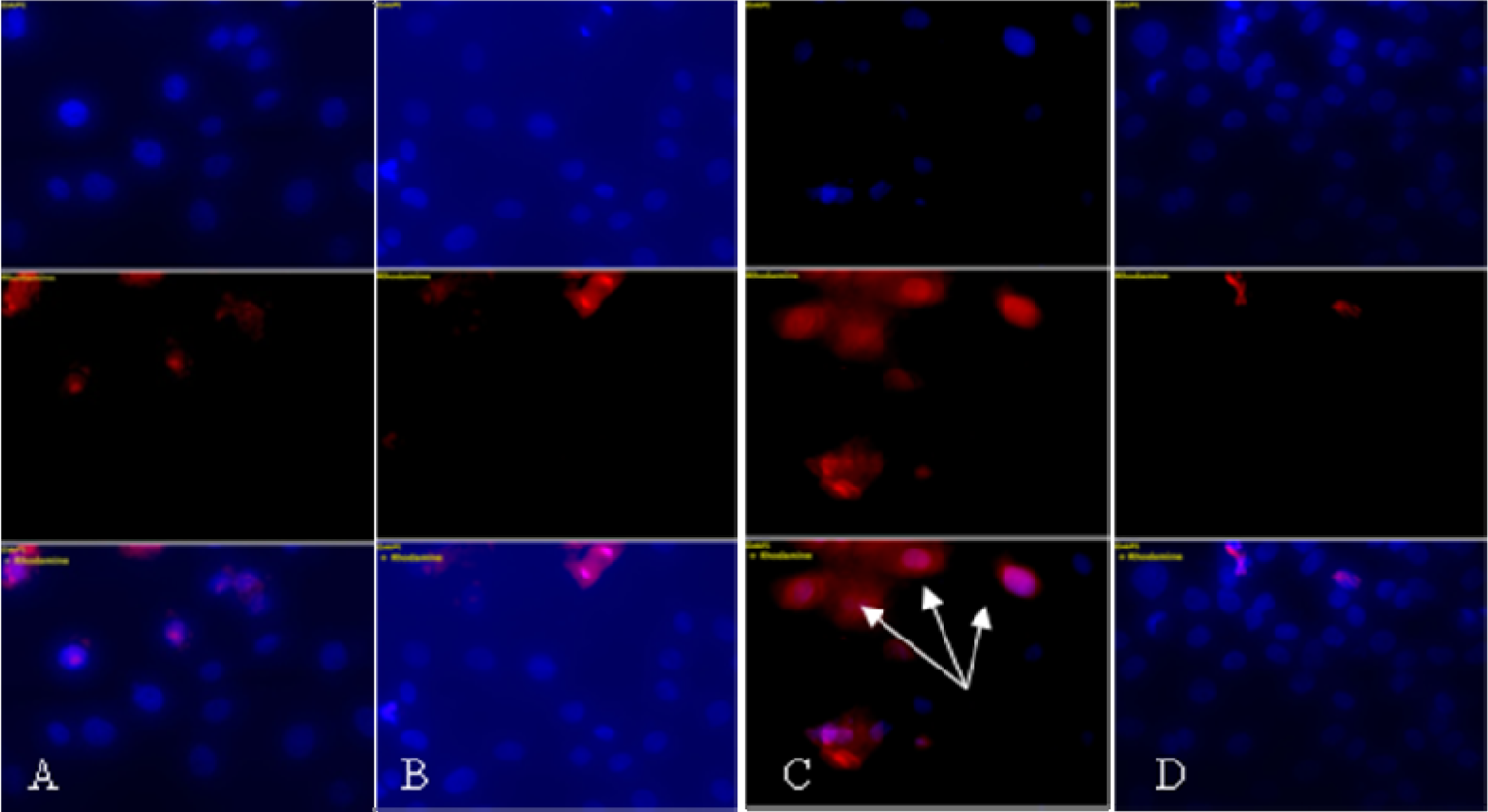
Effect of AGE pretreatment (1000 µg) on the morphology of cardiac myocytes treated with DOX (10 µM). Cardiac myocytes were treated with AGE (1000 µg) and/or DOX (10 µM) for 24 hours. Cells were fixed and stained with PI, and visualized under a fluorescence microscope. (A) Control, (B) AGE 1000 µg, (C) DOX 10 µM, and (D) AGE 1000 µg + DOX 10 µM. Cardiac myocytes are stained by the PI method with DAPI as a counterstain. PI-positive cells appear red when DAPI is used to counterstain all nuclei. Nonapoptotic nuclei remain blue. The arrow indicates a positively stained apoptotic cell
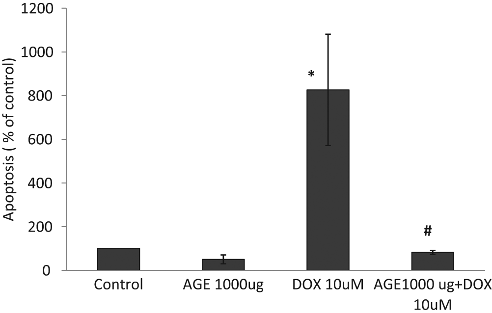
Effect of aged garlic extract (AGE) pretreatment (1000 µg) on the percentage apoptosis in rat cardiac myocytes treated with doxorubicin (DOX; 10 µM). Mean of apoptotic cell number is plotted by percentage of control. Data are mean ± SEM for a minimum of 3 independent experiments for each condition
Levels of Active p53 in Rat Cardiac Myocyte
The change in p53 levels in rat cardiac myocyte after treatment with 10 µM DOX for 24 hours and/or 1000 µg AGE were evaluated. Active p53 was increased in DOX-treated cells by about 1.21-fold compared with control. In the presence of AGE there were no significant changes in p53 activity compared with control (Figure 3).

Effect of aged garlic extract (AGE) pretreatment (1000 µg) on the activity of p53 in rat cardiac myocytes treated with doxorubicin (DOX; 10 µM). Values are expressed as mean ± SEM (n = 3). Similar results were obtained in 3 independent experiments
Active Caspase 3 Levels in Rat Cardiac Myocyte
When cardiac myocytes were treated for 24 hours with 10 µM DOX, there was a 17% increase in active caspase 3 concentrations (P < .05 compared with control). Pretreatment of cells with 1000 µg AGE normalized the caspase 3 level in cardiac myocytes (Figure 4).

Effect of aged garlic extract (AGE) pretreatment (1000 µg) on the activity of caspase 3 in rat cardiac myocyte treated with doxorubicin (DOX; 10 µM). Values are expressed as mean ± SEM (n = 3). Similar results were obtained in 3 independent experiments
8-Isoprostane in Cardiac Myocyte Culture Medium
Treatment of cardiac myocyte with 10 µM DOX for 24 hours resulted in a significant (P < .05) increase in 8-ISP by 37.45% compared with the control untreated cells. However pretreatment of cells with 1000 µg AGE normalized the 8-ISP in cardiac myocytes (Figure 5).
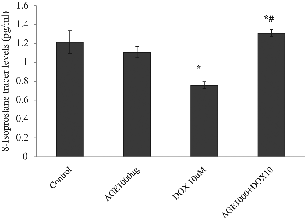
Effect of aged garlic extract (AGE) pretreatment (1000 µg) on the levels of 8-isoprostane tracer competitor (AChE) in cell culture supernatants of cardiac myocyte treated with doxorubicin (DOX; 10 µM). Values are expressed as mean ± SEM (n = 3). Similar results were obtained in 3 independent experiments
Survival of Mice Bearing EAC Tumor
Table 1 and Figure 6 show the effect of AGE pretreatment on the cytotoxic activity of DOX against the growth of EAC cells inoculated IP into female Swiss albino mice. Control tumor-bearing mice showed a mean survival time (MST) of 17 days, whereas, administration of DOX (2 mg/kg) for 3 doses increased the MST to 50 days, with a 30% long-term survivors. AGE pretreatment 2860 mg/kg IP once daily for 6 days increased the MST of tumor-bearing mice treated with DOX to 88 days with 70% long-term survivors.
Effect of AGE Pretreatment on the Antitumor Activity of DOX in Mice Bearing EAC Cells

Abbreviations: AGE, aged garlic extract; DOX, doxorubicin; EAC, Ehrlich ascites carcinoma; MST, mean survival time (= average survival days of mice); LTS, long-term survivors (defined as the mice that survived to the end of experiment [90 days] without an apparent evidence of tumor cell growth.
Indicates significant change from control (P < .05).
Significantly different from DOX (P < .001).

Effect of aged garlic extract (AGE) pretreatment on the antitumor activity of doxorubicin (DOX) in mice bearing Ehrlich ascites carcinoma (EAC) cells. Changes in percentage animal survival expressed as mean ± SEM. Each group consists of 10 animals
Effect of AGE Pretreatment on DOX Uptake in EAC Cells
DOX uptake was increased in EAC cells, 6 hours after administration of a single dose of DOX (15 mg/kg; Table 2). AGE pretreatment (2860 mg/kg for 10 days) significantly increased the cellular level of DOX (144.85 ng/10 8 cells compared with 81.11 ng/10 8 cells after DOX alone).
Effect of AGE Pretreatment (2860 mg/kg) on the Uptake of DOX (Single Dose of 15 mg/kg, IP) in Tumor Cells of EAC-Bearing Mice a
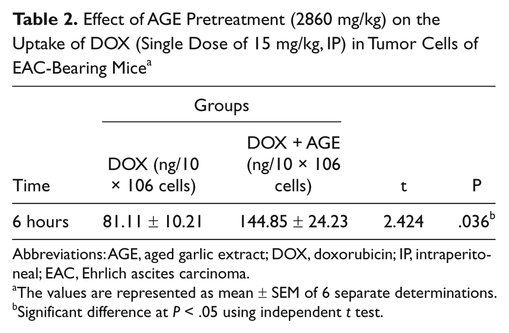
Abbreviations: AGE, aged garlic extract; DOX, doxorubicin; IP, intraperitoneal; EAC, Ehrlich ascites carcinoma.
The values are represented as mean ± SEM of 6 separate determinations.
Significant difference at P < .05 using independent t test.
Effect of AGE Pretreatment on DOX Tissue Distribution
Figures 7 and 8 demonstrate tissue distribution of DOX (15 mg/kg, IP),when injected alone or pretreated with AGE (2860 mg/kg, p.o.) daily for 10 days, in serum and heart of female Swiss albino mice at 24, 48, 72, and 120 hours.
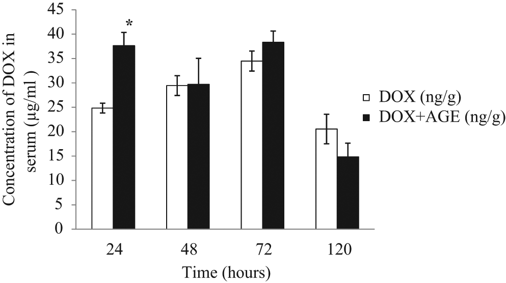
Effect of aged garlic extract (AGE) pretreatment (2860 mg/kg × 10 days) on serum doxorubicin (DOX) level (single dose of 15mg/kg, IP) of mice bearing Ehrlich ascites carcinoma (EAC) cells. The values are represented as mean ± SEM of 6 separate determinations
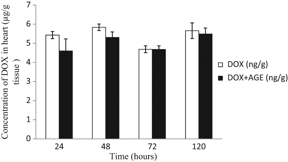
Effect of aged garlic extract (AGE) pretreatment (2860 mg/kg p.o. × 10 days) on the uptake of doxorubicin (DOX) single dose of 15 mg/kg, IP) in heart of mice bearing Ehrlich ascites carcinoma (EAC) cells. The values are represented as mean ± SEM of 6 separate determinations
There was a significant increase in concentration of DOX in the serum of mice pretreated with AGE after 24 hours (P < .001) compared to mice treated with DOX alone. No significant changes in DOX concentration in the heart have been observed after treatment with DOX alone and DOX pretreated with AGE at all time points tested.
Discussion
The usefulness of DOX as an important cytotoxic drug in the treatment of cancer is usually limited by its severe cardiotoxicity. 2
AGE has been proven to have cardioprotective effect against DOX-induced cardiotoxicity. 4 The mechanism of DOX-induced cardiotoxicity has been studied since this complication was first discovered, but the solution to this potentially fatal complication has not been discovered yet. So the present study was designed to investigate how AGE could protect the rat heart against DOX-induced cardiotoxicity. In the heart, many previous investigations have designated apoptosis of cardiac myocyte as the most direct cause of DOX cardiotoxicity. 16
Our study demonstrated that DOX has a profound effect on p53, where it increased p53 levels, suggesting the role of p53 in DOX-induced cardiac myocyte apoptosis. Since stresses that activate p53 may include the DNA damaging signals and free radicals, 17 the cytotoxic and the adverse free radical–generating action of DOX could be the relevant triggers for generating this apoptosis-inducing factor in cardiac myocyte. The current study showed that AGE alleviated DOX-induced cardiac myocyte apoptosis through return of the p53 level and caspase 3 activity to the normal value. This implicates the additional counteracting action of AGE against DOX apoptotic cardiotoxicity at multiple cellular levels. This assumption is supported by recent reports showing that AGE treatment is associated with a significant myocardial protection.4,5,18-21
Since there was recovery of 8-ISP levels in animal pretreated with AGE (Figure 5), it suggests that AGE may have an attenuating effect on DOX-induced oxidative stress.
Morphological changes of cardiac myocyte exposed to 10 µM DOX confirmed the aforementioned results (Figure 1), where cardiac myocyte treated with DOX showed chromatin condensation and nuclear fragmentation, which is well known as typical apoptosis (Figure 1C). On the other hand, AGE pretreatment showed a decrease in apoptotic cells (Figure 2). These molecular results could be illustrated on the basis that AGE treatment significantly decreases the lipid peroxidation and increases endogenous total antioxidants status in rats treated with DOX. 4 Our result agrees with that reported by Demirkaya et al 22 who showed that chronic garlic intake augmented endogenous antioxidants in conditions of oxidant stress–induced injury, which may have important cytoprotective effects on the heart.
A critical question remains: Does AGE that protects the heart against DOX-induced cardiotoxicity 4 interfere with cytotoxic activity of DOX? The current study showed that AGE did not interfere with cytotoxic activity of DOX, but at the same time, it increased its cytotoxic activity, where the mean survival time of tumor-bearing mice was increased from 50 days after DOX treatment compared with 88 days after combination of AGE and DOX. This action may be because of suppression of P-glycoprotein associated energy-dependent efflux of DOX pump, leading to an increased intracellular drug concentration and increased cellular toxicity. 23
In our study, there was a higher serum level of DOX in AGE pretreated group (Figure 7), but there was no increase in DOX concentration in cardiac tissue (Figure 8). Therefore, it seems that AGE prevented more uptake of DOX in cardiac tissue (cardioprotection) but at the same time it increased the concentration of DOX in tumors (Table 2), which correlated well with the increased cytotoxicity against Ehrlich cells (Figure 6), where there was 30% long-term survivors after DOX treatment compared with 70% after combination of AGE and DOX.
In conclusion, all experiments performed consistently show that DOX treatment may impair survival of cardiac muscle cells at least partially through triggering p53-mediated cell apoptosis, whereas AGE ameliorates nearly all of these apoptotic actions of DOX and preserves cell viability via its antioxidant activity of AGE. 4 Our results provide a window for reduction of the serious cardiac complication after addition of AGE. In addition, the results showed that AGE did not interfere with the cytotoxic activity of DOX but increased its activity against tumor cells. Since AGE does not compromise the antitumor effect of DOX, the combined treatment of DOX and AGE holds promise as a safe and effective chemotherapeutic strategy.
Footnotes
Authors’ Note
This work is a part of thesis prepared at King Fahd Medical Research Center, Jeddah, Saudi Arabia and School of Biology, Chemistry and Health Science, Manchester Metropolitan University, MI5GD, Manchester, UK.
Declaration of Conflicting Interests
The authors declared no potential conflicts of interest with respect to the research, authorship, and/or publication of this article.
Funding
The authors disclosed receipt of the following financial support for the research, authorship, and/or publication of this article:
Financial support was obtained from the Scientific Channel Program, King Fahd Medical Research Center, King Abdul-Aziz University.