
Abstract
Introduction
Breast cancer (BC) is a common cancer characterized by a high molecular heterogeneity. Therefore, understanding its biological properties and developing effective treatments for patients with different molecular features is imperative. Calcium-sensing receptor (CaSR) has been implicated in several regulatory functions in various types of human cancers. However, its underlying pathological mechanism in BC progression remains elusive.
Methods
We utilized The Cancer Genome Atlas and Gene Expression Omnibus databases to explore the function of CaSR in the metastasis of BC. Gene ontology analysis, Kyoto Encyclopedia of Genes and Genomes analysis, and Gene Set Enrichment Analysis of biological processes and cell signaling pathways revealed that CaSR could be activated or inhibited. Importantly, quantitative reverse transcriptase-polymerase chain reaction and western blotting were used to verify the gene expression of the CaSR. Wound healing and transwell assays were conducted to assess the effect of CaSR on the migration of BC cells.
Results
We demonstrated that CaSR expression in metastatic BC was higher than that in non-metastatic BC. It is the first time that database information has been used to reveal the biological process and molecular mechanism of CaSR in BC. Moreover, the CaSR expression in normal breast epithelial cells was notably less compared to that in BC cells. The activation of CaSR by Cinacalcet (a CaSR agonist) significantly enhanced the migration of BC cells, whereas NPS-2143 (a CaSR antagonist) treatment dramatically inhibited these effects.
Conclusion and future perspective
Bioinformatics techniques and experiments demonstrated the involvement of CaSR in BC metastasis. Our findings shed new light on the receptor therapy and molecular pathogenesis of BC, and emphasize the crucial function of CaSR, facilitating the metastasis of BC.
Introduction
Breast cancer (BC) is a major health concern that substantially affects women worldwide. The latest data from the International Agency for Research on Cancer show that BC is the most common cancer globally, surpassing lung cancer for the first time1–3. Despite a decline in the mortality rate of BC, effective control of tumor growth by surgery and chemotherapy, and advances in targeted and combined therapies for metastatic disease, death from metastatic BC remains a serious concern 4 . Unfortunately, about 30% of patients with BC succumb to metastatic disease. Although research has made great strides in treating primary BC, little progress has been achieved in the treatment of metastatic BC 5 . Invasion-metastasis cascades involve multiple steps and numerous proteins and signaling molecules, each of which is regulated by changes in gene expression. There is an urgent need to explore detailed molecular mechanisms contributing to BC metastasis 6 .
Calcium-sensing receptor (CaSR) was discovered in 1993 as the first membrane receptor to sense calcium ions in the extracellular environment7,8. CaSR belongs to the C family of G protein-coupled receptors with seven transmembrane domains. In addition to extracellular calcium ions, various other substances such as cations, polyamines, peptides, and antibiotics can activate this receptor. This broad ligand combination triggers complex intracellular signaling networks affecting multifarious physiological and pathological processes, such as hormone secretion, chemotaxis, proliferation, apoptosis, and differentiation.9,10 We have previously reported that the CaSR controls the inflammatory responses of PMNs, and exosomes derived from CaSR-stimulated PMNs can reduce ischemia/reperfusion (I/R) injury in acute myocardial infarction. 11
Recently, the function of CaSR in tumors has been explored with a complex underlying mechanism. CaSR is a multipotent receptor that promotes proliferation and stimulates metastasis in certain cancers, such as breast, lung, prostate, and kidney cancers, whereas it inhibits proliferation in other cancers, such as the parathyroid gland, colon, and pancreas cancers. 12 Cancer tissues, such as the microenvironment of bone metastasis, are characterized by high extracellular calcium concentration that stimulates CaSR. It has been reported that the signaling capabilities of CaSR in certain cancer cells are altered. 13 However, the molecular mechanism of CaSR in BC has remained still elusive. In the present study, we conducted bioinformatic analysis, followed by in vitro studies to explore the implications of CaSR in BC.
Methods
Data Collection
The Gene Expression Omnibus (GEO) database is a globally recognized and data-intensive public platform. The oncology research has greatly benefited from the public sequencing data available in this database. Following an extensive search and evaluation process, three datasets (GSE29431, GSE73540, and GSE102484) with gene expression information were selected, including GSE29431 (primary tumor = 14, metastatic tumor = 25), GSE73540 (primary tumor = 18, metastatic tumor = 10), and GSE102484 (primary tumor = 582, metastatic tumor = 101). The RNA-seq (FPKM) data of 1097 patients with BC were downloaded from The Cancer Genome Atlas (TCGA) database. Clinicopathological features such as gender, age, stage, and prognosis of corresponding patients with BC were simultaneously downloaded from the UCSC Xena website.
Differentially Expressed Genes
We used the R package sva 14 to eliminate the batch effects from GSE102484 and GSE29431 to obtain an integrated GEO dataset. We next analyzed the effect of CaSR gene expression on BC tumor tissues. We divided the tumor samples into high and low expression groups according to the median CaSR gene expression value in the integrated GEO and TCGA datasets, respectively, and R package Limma 15 was used for the differential analysis of groups. We set the genes with logFC > 1 and P < 0.05 as up-regulated genes. Genes with logFC < −1 and P < 0.05 were down-regulated. The intersection of differentially expressed genes (DEGs) in the two datasets was used to obtain differentially expressed CaSR-related genes. Afterward, prognosis-related genes of patients with BC were calculated by univariate Cox analysis and utilizing patient prognostic data from the TCGA database (P < 0.05). Differentially expressed CaSR-related prognostic genes are defined as the intersection of differentially expressed CaSR-related genes and prognostic genes in patients with BC.
Gene ontology and Kyoto Encyclopedia of Genes and Genomes Enrichment Analyses
Gene ontology (GO) is a popular approach to examining the functional enrichment of genes across various dimensions and levels. It is conducted from three levels: biological process (BP), molecular function (MF), and cellular component (CC). 16 Kyoto Encyclopedia of Genes and Genomes (KEGG) is a widely used database for storing information on genomes, diseases, drugs, and biological pathways. 17 The R package “clusterProfiler” 18 was utilized for conducting GO functional annotation and KEGG pathway enrichment analysis for differentially expressed CaSR-related prognostic genes to pinpoint the significantly enriched pathways and biological processes.
Gene Set Enrichment Analysis
The reference gene set “c5.go.v7.4.entrez.gmt” was obtained from the MSigDB database using the gene expression data of patients with BC. 19 We used the Gene Set Enrichment Analysis (GSEA) method included in the R package “clusterProfiler” for enrichment analysis of integrated GEO and TCGA datasets.
Protein-protein Interaction
The protein-protein interaction (PPI) networks consist of individual proteins that interact with each other to play roles in multiple life processes, including biological signal transmission, gene expression regulation, energy metabolism, and cell cycle regulation. STRING is a database to identify the interactions between known proteins and predicted proteins. 20 We used the STRING database to identify genes with a total score exceeding 400 for building the PPI network related to the expression of the CaSR gene and differential prognostic expression. The PPI network model was visualized using Cytoscape (v3.7.0). 21
Cell Culture
Procell (Wuhan, China) supplied BC cell lines MDA-MB-231, BT-549, and MCF-7, as well as the normal cell line MCF-10A. To create the full medium, 10% fetal bovine serum (Gibco, Shanghai, China) and 1% penicillin-streptomycin were mixed into the essential medium (Corning, Shanghai, China). The respective cell cultures were incubated in the full medium at 37 °C with 5% CO2. Following subculturing, the cells were used for western blotting and additional experiments.
Quantitative Reverse Transcriptase-polymerase Chain Reaction
CaSR expression in cells was identified through RT-qPCR analysis. The total RNA was isolated from cells using the Total RNA Kit I (Omega Bio-tek, GA, USA). The RNA was transcribed in reverse using the NovoScript Plus All-in-one 1st Strand cDNA Synthesis SuperMix kit from Novoprotein in Suzhou, China. The relative expression of CaSR was assessed through RT-qPCR using the NovoStart SYBR qPCR SuperMix Plus kit (Novoprotein, Suzhou, China), with glyceraldehyde-3-phosphate dehydrogenase (GAPDH) serving as an internal reference control. The primer sequences for GAPDH and CaSR were as follows:
GAPDH-F: 5′-GACATGCCGCCTGGAGAAAC-3′, GAPDH-R: 5′-AGCCCAGGATGCCCTTTAGT-3′, CaSR-F: 5′-GAAAGACAGAATGTCACCAG-3′, CaSR-R: 5′-CATATGGTATAGGAGGGGTG-3′.
The thermal cycling parameters included an initial denaturation step at 95 °C for 10 min, followed by denaturation at 95 °C for 10 s, annealing and extension at 60 °C for 30 s, repeated for a total of 40 cycles.
Western Blotting
The protein expression of CaSR in BC cell lines was detected using western blotting. Proteintech Group Lnc, USA, provided the anti-CaSR antibody. Total intracellular proteins were obtained using radioimmunoprecipitation analysis lysis buffer. An enhanced bicinchoninic acid protein analysis kit was used to detect protein concentrations. Afterward, proteins were separated by loading onto an SDS-PAGE gel and then transferring to a PVDF membrane. Following this, the PVDF membrane was exposed to primary antibodies (CaSR, β-actin, 1:1000) overnight, rinsed, treated with secondary antibodies (1:5000) for an hour, and ultimately identified using the enhanced chemiluminescence reagent.
Transwell Assay
BC cells were seeded at a density of 1 × 10⁵ cells/well in 200 µL of culture medium without serum into the top compartment of the transwell system (Corning, USA). At the same time, 600 µL of the solution with 20% fetal bovine serum was placed into the bottom section of a 24-well plate. Next, cells were treated with 4% paraformaldehyde for 20 min after 16 h and then stained with 0.1% crystal violet for another 20 min. Afterward, an inverted microscope was used to capture images of five randomly selected regions on the lower membrane, followed by cell counting.
Wound Healing Assay
Exponentially growing BC cells were seeded in 6-well plates at 85% confluency. A pipette (100 µL) was used to create a straight-line scratch in the center of the plate. The cells were rinsed gently with phosphate-buffered saline and then switched to a serum-free medium before imaging the scratch width at the same spot after 0 h and 24 h using a microscope and then analyzing the results with ImageJ software (v1.8.0).
Statistical Analysis
Statistical analysis was performed using the GraphPad Prism 9.0 software. Mean ± standard deviation is shown in the experimental data. A comparison of the average values between the two groups was conducted using a t-test. One-way analysis of variance was utilized for comparing groups with three or more members. *P < 0.05, **P < 0.01, and ***P < 0.001 suggest a statistically significant difference.
Results
Correlation Between CaSR Gene Expression and Breast Cancer Metastasis
Analyzing the correlation between CaSR levels and BC cancer metastasis in patients with BC involved comparing CaSR expression differences in metastatic and non-metastatic BC patients across three datasets. The results demonstrated that patients with metastatic BC had high CaSR gene expression, among which the BC patient data set GSE29431 contained 25 patients with metastasis and 14 patients without metastasis (P = 0.006, Figure 1A), and GSE73540 contained 10 patients with metastasis and 18 patients without metastasis (P = 0.033, Figure 1B). GSE102484 included 101 patients with metastasis and 582 patients without metastasis (P = 0.0021, Figure 1C).

Correlation between CaSR gene expression and breast cancer metastasis. (A) Box plot was created using the CaSR expression from the GSE29431 dataset. (B) Box plot showing CaSR expression in the GSE73540 dataset. (C) A box plot was created using the CaSR expression from the GSE102484 dataset. CaSR, calcium-sensing receptor.
Relationship Between CaSR Expression and Genome-Wide Expression Profile
The relationship between CaSR expression and genome-wide expression profile in patients with BC was analyzed by first integrating the first two significantly dispersed GEO data sets (Figure 2A) and the integrated GEO data set after removing the batch effect (Figure 2B). The GEO and TCGA datasets were integrated, and the patients were divided into a high CaSR expression group and a low CaSR expression group according to the mean CaSR expression. The integrated GEO dataset contained 361 patients in the high CaSR expression group and 361 patients in the low CaSR expression group. A total of 526 patients were present in the high CaSR expression group and 571 patients in the low CaSR expression group in the TCGA dataset. An analysis of gene expression differences between the patient groups identified 1242 genes that were differentially expressed based on a fold change > 1.2 and false discovery rate (FDR) < 0.05 in the integrated GEO dataset (Figure 2C) according to fold change > 1.2 and FDR < 0.05. In total, 3629 DEGs were screened in the TCGA dataset (Figure 2D). Subsequently, a univariate COX analysis was conducted on the genes within the TCGA expression dataset based on the survival data from the TCGA database, revealing that 2440 genes showed a significant correlation with the prognosis of BC patients (P < 0.05). Next, the overlap between DEGs and genes related to prognosis in the two datasets was utilized to identify 56 differentially expressed CaSR-related prognostic genes (Figure 2E, Table S1).

Differentially expressed CaSR-related prognostic genes. (A) PCA analysis before GEO data integration. (B) PCA analysis after GEO data integration. (C) Volcano map of differentially expressed genes between high and low CaSR expression groups in the integrated GEO dataset. (D) Volcano map of differentially expressed genes between high and low CaSR expression groups in the TCGA dataset. (E) Venn diagram illustrating genes that are expressed differently and genes that are related to prognosis. CaSR, calcium-sensing receptor; TCGA, The Cancer Genome Atlas; GEO, Gene Expression Omnibus.
Enrichment Analysis of Differentially Expressed CaSR-Related Prognostic Genes
We further investigated the relationship between CaSR-related prognostic genes and cell components, biological processes, molecular functions, and biological pathways using the functional enrichment analysis (Figure 3A, Table S2). DEGs were largely enriched in hemidesmosome assembly, cell chemotaxis, positive regulation of neutrophil chemotaxis, epidermis development, and chemokine-mediated signaling pathway biological processes (Figure 3B). Those enriched in cellular components included extracellular region, proteinaceous extracellular matrix, extracellular space, extracellular exosome, keratin filament (Figure 3C), and those enriched in chemokine activity, heparin-binding, CXC chemokine receptor (CXCR) binding, structural molecule activity, serine-type endopeptidase activity, structural constituent of cytoskeleton, and high mobility group box domain binding-related molecular functions (Figure 3D). Next, an analysis of pathway enrichment was conducted on DEGs. The results demonstrated that DEGs were enriched in the cytokine-cytokine receptor interaction and chemokine signaling pathway, hsa04060: cytokine-cytokine receptor interaction (Figure 3E) and hsa04062: Chemokine signaling pathway (Figure 3F).
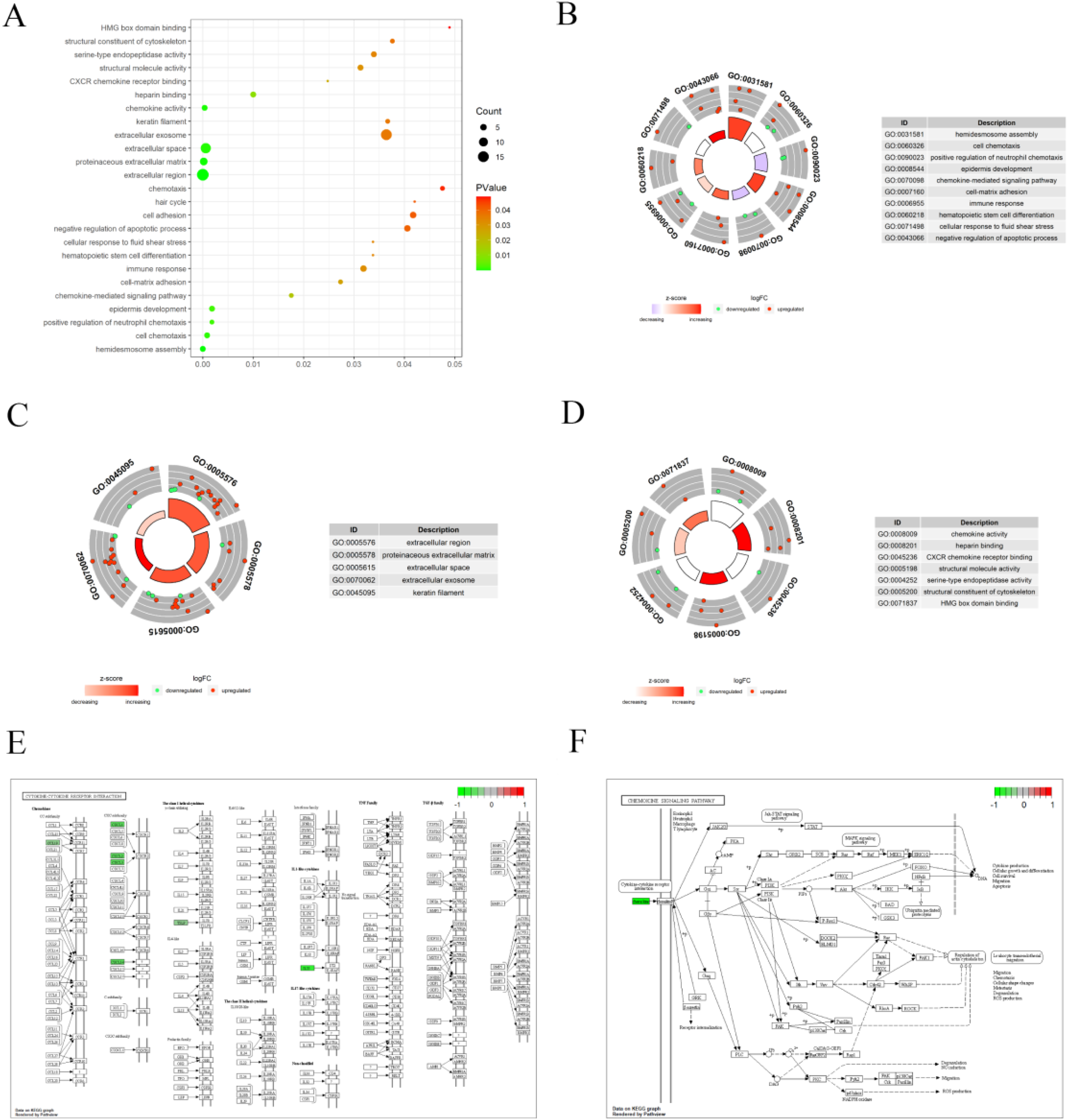
Go functional annotation and KEGG pathway enrichment analysis of CaSR-related prognostic genes. (A) Go functional annotation. (B) Biological process, BP (hemidesmosome assembly, cell chemotaxis, etc). (C) Cellular component, CC (extracellular region, proteinaceous extracellular matrix, etc). (D) Molecular function, MF (chemokine activity, heparin-binding, etc). (E,F) KEGG pathway enrichment analysis (cytokine-cytokine receptor interaction, chemokine signaling pathway). CaSR, calcium-sensing receptor; KEGG, Kyoto Encyclopedia of Genes and Genomes.
GSEA of CaSR in BRCA
The impact of CaSR gene expression on BC was verified by studying the relationship between the genes in the CaSR high expression group and the CaSR low expression group from the two databases. The results demonstrated that the biological functions affected by genes in the integrated GEO database were highly consistent with those in the TCGA database, among which 222 biological processes were significantly activated and 1832 biological processes were inhibited in the integrated GEO data (Figure 4A,B, Table S3). In the TCGA data, 206 biological processes were activated, whereas 1738 biological processes were inhibited (Figure 4C,D, Table S4). The intersection of activated or inhibited biological processes in the two datasets found that 205 biological processes were co-activated (Table S5) and 1716 biological processes were simultaneously inhibited (Table S6).

GSEA between high and low CaSR expression groups. (A,B) GSEA enrichment analysis of integration GEO data. (C,D) GSEA enrichment analysis of TCGA data. CaSR, calcium-sensing receptor; TCGA, The Cancer Genome Atlas; GEO, Gene Expression Omnibus; GSEA, Gene Set Enrichment Analysis.
PPI Network and Prognosis Analysis of CaSR-Related Prognostic Genes
We constructed the PPI networks among 56 differentially expressed CaSR-related prognostic genes (Figure 5A). These genes were primarily enriched in chemokine activity, cornification, hemidesmosome assembly, type I hemidesmosome assembly, and BP230 keratin K5/K14 and interleukin-10 signaling functions (Figure 5B,C). We obtained 11 hub genes using MCODE (Figure 5D), and the results demonstrated that the expression of 11 genes was highly correlated in both the TCGA-BC and the integrated GEO datasets. The correlation between NRT5 and KRT14 in the TCGA dataset reached 0.82 (P < 0.05, Figure 5E), and the correlation between NRT5 and KRT6B in the integrated GEO dataset reached 0.87 (P < 0.05, Figure 5F). Moreover, the survival analysis demonstrated that the expression of eight hub genes, namely COL17A1, CXCL1, CXCL2, TSLP, IL33, KRT14, CCL19, and KRT6B, significantly impacted the prognosis of patients with BC (P < 0.05, Figure 5G).

PPI network and prognosis analysis of differentially expressed CaSR-related prognostic genes. (A) PPI network of differentially expressed CaSR-related prognostic genes. (B,C) PPI network function analysis of differentially expressed CaSR-related prognostic genes. (D) Hub genes of CaSR-related prognostic genes in the PPI network. (E) The correlation of hub genes expression in integrated GEO datasets. (F) The correlation of hub genes expression in TCGA datasets. (G) The relationship between the expression of hub genes and the prognosis of patients with BC. BC, breast cancer; CaSR, calcium-sensing receptor; TCGA, The Cancer Genome Atlas; GEO, Gene Expression Omnibus, PPI, protein-protein interaction.
Effects of CaSR on the Migration of BC Cells
Leveraging big data analysis, we confirmed through quantitative reverse transcriptase-polymerase chain reaction and western blotting that the level of CaSR expression in normal breast epithelial cells (MCF-10A) was notably less compared to that in BC cells (MCF-7, MDA-MB-231, and BT-549). In addition, the CaSR expression was the most pronounced in MDA-MB-231 cells exhibiting the most robust metastatic potential (Figure 6A,B).

The expression of CaSR in normal breast cells and BC cells. (A) Experimental findings from qRT-PCR demonstrated that CaSR expression was elevated in BC cells compared to normal breast cells. (B) Western blotting revealed that the level of CaSR was elevated in BC cells compared to normal breast cells (*P < 0.05, **P < 0.01, ***P < 0.001). BC, breast cancer; CaSR, calcium-sensing receptor; qRT-PCR, quantitative reverse transcriptase-polymerase chain reaction.
Subsequently, transwell and wound healing experiments were further conducted to appraise the impact of CaSR activation on BC cell migration. Two cell lines were treated with Cinacalcet (CIN, a CaSR agonist, 100 nM) and NPS-2143 (NPS, a CaSR antagonist, 100 nM). As shown in Figure 7A,B, CaSR activation induced by CIN markedly facilitated cell migration in BC cells, whereas NPS treatment dramatically suppressed the above effects.

Effects of CaSR on the migration of BC cells. (A) Transwell assay results of BC cells treated with CIN or NPS. Migrating cells were quantified after 16 h (x100). (B) Results from the wound healing experiment of BC cells treated with either CIN or NPS. The rate of wound healing was assessed at 0 h and 24 h (x100, *P < 0.05, **P < 0.01, ***P < 0.001). BC, breast cancer; CaSR, calcium-sensing receptor.
Discussion
BC is one of the most common malignant tumors globally. Despite the significant advancements made in multimodal treatment, the intricate molecular subtypes and extensive heterogeneity continue to present significant obstacles in clinical scenarios, including issues with drug resistance, recurrence, and metastasis. The 5-year survival rate for metastatic BC remains unsatisfactory. 22 Individuals with metastatic BC face multiple challenges and disparities in obtaining optimal care. Enhanced comprehension of causal molecular abnormalities can identify new biomarkers for the early diagnosis, targeted therapy, and prognosis evaluation of metastatic BC.
Although the expression of CaSR is stringently controlled in normal physiological conditions, it becomes disrupted in cancer. The CaSR activity, as a crucial participant in the intricate calcium signaling network, is associated with the growth, differentiation, and apoptosis of various cancer cells. Previous research has indicated that CaSR could function as either a tumor suppressor or an oncogene in the advancement of various malignant tumors, depending on the type of tumor. It acts as a cancer inhibitor in the colon, 23 parathyroid, 24 and neuroblastoma, 25 whereas it behaves as an oncogene in the ovary 26 and kidney 27 cancers. These two significantly different functions could be attributed to the diverse expression of CaSR and different downstream signaling pathways in multiple tumors. The current understanding of the molecular mechanisms of CaSR in cancer is still in its infancy, and further basic experiments are required to investigate more defined axes of signaling regulation. This is essential to better understand the therapeutic application and clinical translation of CaSR. Currently, the treatment revolving around the CaSR gene is mainly focused on parathyroid and bone tumors. Combined with the current hot areas of cancer research, it is imperative to further study the role of CaSR in treatment and prognosis.
We first used the database to demonstrate that CaSR expression in metastatic BC was higher than that in non-metastatic BC. Next, we screened 56 differentially expressed CaSR-related prognostic genes and further performed a series of bioinformatics analyses. It was the first time that database information was used to reveal the biological process and molecular mechanism underlying the function of CaSR in BC. Advancing the knowledge of CaSR biology in BC is crucial. We proved that CaSR was significantly upregulated in BC cells and was the highest expressed in MDA-MB-231 cells. Similarly, Liu reported that the expression of CaSR in lung cancer with bone metastases was notably elevated, promoting the differentiation and maturation of osteoclasts. 28 A recent study indicated that the level of CaSR was down-regulated in pancreatic cancer tissues, which was associated with tumor differentiation, tumor size, and decreased patient survival rates. 29
The CaSR can be activated by different types of endogenous ligands, as well as by synthetic modulators such as CIN, which is clinically used to treat secondary hyperparathyroidism in patients with chronic kidney diseases. In our study, we found that CaSR is activated by CIN, promoting the migration of BC cells. Conversely, CaSR is inhibited by NPS, dramatically suppressing the above effects. CaSR regulates the downstream cell signaling via the G protein pathways, including the increase in intracellular calcium concentration, either releasing it from the internal storage or extracellular space. The activation of downstream intracellular calcium effector molecules further conducts different cell cycle processes and cell metastasis. 30 Aggarwal et al revealed that the expression of CaSR was negatively correlated with the markers of epithelial-mesenchymal transformation in colorectal cancer. 31 Increased concentrations of calcium are associated with protecting against colorectal cancer. The CaSR gene polymorphism is correlated with a higher risk of developing colorectal cancer and survival of patients.
Bioinformatics technology was utilized to predict and analyze the function of CaSR in BC metastasis and its possible mechanisms, and CaSR-related prognostic genes were derived, whereas this research used simple cellular experiments to verify. Importantly, our study is also valuable in providing predictive data as well as ideas for CaSR in BC. We plan to expand the clinical samples along with conducting large-scale cellular and animal experiments for verification. Moreover, we will further explore the biological value of CaSR in the diagnosis and treatment of BC.
Collectively, the past decade has witnessed significant advances in treatment modalities for BC, with the introduction of targeted and immunotherapy-based therapies. The increasing link between CaSR and cancer demonstrates that CaSR could function as biomarkers or therapeutic targets for BC.
Conclusion and Future Perspective
Bioinformatics techniques and experiments demonstrated that CaSR was involved in BC metastasis. Our findings shed new light on the molecular pathogenesis of BC as well as receptor therapy and emphasize the crucial function of CaSR in mediating the metastasis of BC. Future studies should explore the basis of CaSR as a potential anticancer drug target, develop new CaSR-specific receptor blockers, and strengthen clinical trials to provide novel ideas for gene targets of CaSR in cancer therapy.
Supplemental Material
sj-doc-1-tct-10.1177_15330338241254219 - Supplemental material for Calcium-sensing Receptor, a Potential Biomarker Revealed by Large-scale Public Databases and Experimental Verification in Metastatic Breast Cancer
Supplemental material, sj-doc-1-tct-10.1177_15330338241254219 for Calcium-sensing Receptor, a Potential Biomarker Revealed by Large-scale Public Databases and Experimental Verification in Metastatic Breast Cancer by Wanlin Xie, Huimin Xu, Yangyang Cheng, Xin Lin, Jingya Zeng and Yihua Sun in Technology in Cancer Research & Treatment
Supplemental Material
sj-doc-2-tct-10.1177_15330338241254219 - Supplemental material for Calcium-sensing Receptor, a Potential Biomarker Revealed by Large-scale Public Databases and Experimental Verification in Metastatic Breast Cancer
Supplemental material, sj-doc-2-tct-10.1177_15330338241254219 for Calcium-sensing Receptor, a Potential Biomarker Revealed by Large-scale Public Databases and Experimental Verification in Metastatic Breast Cancer by Wanlin Xie, Huimin Xu, Yangyang Cheng, Xin Lin, Jingya Zeng and Yihua Sun in Technology in Cancer Research & Treatment
Supplemental Material
sj-doc-3-tct-10.1177_15330338241254219 - Supplemental material for Calcium-sensing Receptor, a Potential Biomarker Revealed by Large-scale Public Databases and Experimental Verification in Metastatic Breast Cancer
Supplemental material, sj-doc-3-tct-10.1177_15330338241254219 for Calcium-sensing Receptor, a Potential Biomarker Revealed by Large-scale Public Databases and Experimental Verification in Metastatic Breast Cancer by Wanlin Xie, Huimin Xu, Yangyang Cheng, Xin Lin, Jingya Zeng and Yihua Sun in Technology in Cancer Research & Treatment
Supplemental Material
sj-docx-4-tct-10.1177_15330338241254219 - Supplemental material for Calcium-sensing Receptor, a Potential Biomarker Revealed by Large-scale Public Databases and Experimental Verification in Metastatic Breast Cancer
Supplemental material, sj-docx-4-tct-10.1177_15330338241254219 for Calcium-sensing Receptor, a Potential Biomarker Revealed by Large-scale Public Databases and Experimental Verification in Metastatic Breast Cancer by Wanlin Xie, Huimin Xu, Yangyang Cheng, Xin Lin, Jingya Zeng and Yihua Sun in Technology in Cancer Research & Treatment
Footnotes
Abbreviations
Acknowledgements
We acknowledge TCGA and GEO database for providing their platforms and contributors for uploading their meaningful datasets.
Declaration of Conflicting Interests
The authors declared no potential conflicts of interest with respect to the research, authorship, and/or publication of this article.
Ethical Approval
Our study did not require an ethical board approval because it did not contain human or animal trials.
Funding
The author(s) disclosed receipt of the following financial support for the research, authorship, and/or publication of this article: This research was funded by the Natural Science Foundtion of Heilongjiang Province, grant number 2021H082.
Supplemental Material
Supplemental material for this article is available online.
References
Supplementary Material
Please find the following supplemental material available below.
For Open Access articles published under a Creative Commons License, all supplemental material carries the same license as the article it is associated with.
For non-Open Access articles published, all supplemental material carries a non-exclusive license, and permission requests for re-use of supplemental material or any part of supplemental material shall be sent directly to the copyright owner as specified in the copyright notice associated with the article.