
Abstract
Keywords
Introduction
Breast cancer is the leading cause of cancer-related death among females worldwide. 1 However, systematic treatment has limited influence on survival of breast carcinoma patients owing to heterogeneity of disease. Thus, the search for novel prognostic biomarkers to improve patient prognosis has great clinical significance.
Notch pathway is activated by the interaction of Notch receptors (Notch1, 2, 3, or 4) with different types of ligands (Jagged1, Jagged2, DLL1, DLL3, or DLL4). The role of aberrant Notch3 expression in promoting breast tumor self-renewal, invasiveness, and poor outcome is corroborated, which is involved in the sustainable development of breast cancer.2,3 High intensity of Dll4 expression in breast tumor is a statistically significant adverse prognostic factor, and enhances cancer stem cell self-renewal ability.4–6 The interplay between Notch3 and DLL4 may trigger tumor growth and survival in T-cell acute lymphoblastic leukemia. 7 DLL4 activating Notch3 regulates MUSASHI-1 expression, which adjusts colorectal cancer cell metastasis. 8 Gastric cancer patients with positive lymphangion invasion and distal metastasis have significantly higher expression rates of Notch3 and DLL4. 9 This is consistent with the role of Notch3 and DLL4 as oncogenes in pancreatic ductal adenocarcinoma. 10 However, little is known about the role of the interaction between Notch3 and DLL4 in breast carcinogenesis and development, particularly in prognostic values.
In this study, we examined the mRNA and protein expression of Notch3 and DLL4 by quantitative real-time polymerase chain reaction (qRT-PCR), western blot, and immunohistochemical staining. Then, the clinical significance of Notch3 and DLL4 in breast cancer patients, and its prognostic value were determined. Taken together, these results suggest that the expression of Notch3 and DLL4 are dramatically correlated with poor prognosis in breast cancer, which may present as potential prognostic markers in mammary cancer.
Methods
Patients and Samples
The specimens of breast cancer were obtained from (conceal). The 90 cases of breast carcinoma tissues and 60 adjacent normal breast tissues from patients, who underwent initial surgery without neoadjuvant therapy between June 2001 and January 2008, were used in the retrospective study. The clinicopathological parameters including lymph node metastasis, clinical stage, histological grade, estrogen receptor (ER), progesterone receptor (PR), and human epidermal growth factor receptor 2 (HER2) status were obtained from patients’ medical records. The overall survival (OS) data was collected from the date of surgery to date of death or to the end of follow-up. This study was approved by the Ethics Committee of the First Affiliated Hospital of Shihezi University School of Medicine Medical Ethics Committee (approval no. KJ2020-144-01) and written informed consent was obtained from each participant.
Immunohistochemical Staining and Evaluation
Formalin-fixed paraffin-embedded tissue samples were xylene dewaxed, alcohol rehydrated, and antigen retrieval. Immunohistochemistry staining was performed in strict accordance with the manufacturer's instructions. Primary antibodies, including anti-Notch3 (1:300, sc-515825; Santa Cruz Biotechnology) and anti-DLL4 (1:500, sc-365429; Santa Cruz Biotechnology), were incubated followed by wash and incubation with secondary antibodies (Dako Cytomation EnVision System) for 30 min. Subsequently to visualization, staining was displayed with 3,3-diaminobenzidine (DAB) (Dako) solution and sections were lightly counterstained with hematoxylin. Finally, the protein expression level was evaluated by immunohistochemical staining results according to the cell staining intensity and percentage of positive areas. The staining intensity was scored from 0 to 3 as follows: 0 (negative), 1 (buff), 2 (yellow), and 3 (brown). The percentage of positively stained cells was scored on a scale of 0 to 4 as follows: 0 (<1%, absent), 1 (1%-24%, sporadic), 2 (25%-49%, local), 3 (50%-74%, majority), and 4 (75%-100%, vast majority). The scores for percentages of positive cells and staining intensities were then multiplied to generate an immunoreactivity score (IS) for each case. The IS ranged from 0 to 12 (0, 1, 2, 3, 4, 6, 8, 9, and 12). The scores of < 4 were defined as low expression, and the scores of ≥ 4 were designated as high expression.
Quantitative Real-Time Polymerase Chain Reaction
Total RNA was isolated from 30 breast cancer tissues and 20 cases of adjacent nontumorous tissues by the TRIzol method and reverse-transcribed into cDNA. The UltraSYBR Mixture (CWBIO) were used for qPCR assays. Results were calculated using the 2−ΔΔCt method and normalized to the reference housekeeping gene β-actin. The primers used were as follows: Notch3 forward primer 5′-TGGCGACC TCACTTACGACT-3′, reverse primer 5′-CACTGGCAGTTATAGGTGTTGAC-3′; DLL4 forward primer 5′-ACAGGCCGAAAGACAGATAGG-3′, reverse primer 5′-GTGGGTCAGAACTGGTTA TTG-3′; β-actin forward primer 5′-CTCCATCCTGGCCTCGCTGT-3′, reverse primer 5′-GCTGTCAC CTTCACCGTTCC-3′.
Western Blot Analysis
Total protein was extracted from the mammary cancer tissues and adjacent normal tissues using radioimmunoprecipitation assay (RIPA) buffer. Protein samples were separated by SDSPAGE and transferred to the polyvinylidene difluoride transfer membrane that was blocked with 5% skimmed milk for 2 h at room temperature. Then, the membrane was incubated overnight at 4 °C using the primary antibodies. Subsequently, the membranes were incubated with the horseradish peroxidase-conjugated secondary antibody. Finally, protein bands were detected with the enhanced chemiluminescence reagent.
Oncomine Database Analysis
Oncomine (https://www.oncomine.org) 11 was applied to analyze the mRNA levels of Notch3 and DLL4 according to the fold changes using the filters of differential analysis (cancer vs normal), cancer type (breast cancer), sample type (clinical specimen), data type (mRNA), and gene (Notch3 or DLL4). The study was used paired Student's t-test and selected threshold as P value ≤ .05.
UALCAN Database Analysis
Prognostic values of Notch3 and DLL4 mRNA expression in breast cancer patients were assessed by using UALCAN database (http://ualcan.path.uab.edu/). 12 Briefly, in the breast cancer database, we filled in the gene name of Notch3 or DLL4 and chose the OS with keeping all default settings, then obtained the Kaplan–Meier curve. P < .05 was considered as a threshold of statistical significance.
Statistical Analysis
The data obtained were all statistically analyzed by SPSS software (Version 22.0 SPSS). Student's t-test was performed to determine the difference expressions between 2 groups. Spearman's rank correlation test was used to assess the interaction between the 2 proteins’ expressions. The relationship between protein expression and clinicopathological features were carried out using Chi-squared test or Fisher's exact test. Kaplan–Meier univariate and Cox's multivariate survival analyses were performed to identify the independent prognostic factors in breast cancer patients. A P value of less than 0.05 was considered to indicate statistical significance.
Results
The Expression of Notch3 and DLL4 in Breast Tissues
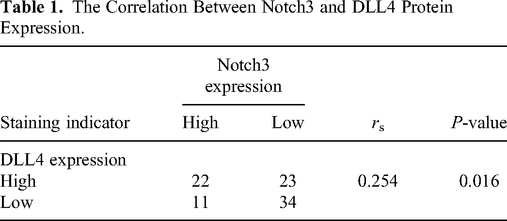
We investigated the clinical importance of Notch3 and DLL4 protein expression in breast samples by immunohistochemistry staining. The both proteins expression was noticeably higher in tumor than in normal tissues (Figure 1, P < 0.001). Besides, we analyzed the relationship of Notch3 and DLL4 expression in breast cancer. Spearman's rank correlation analysis showed Notch3 was obviously positive association with DLL4 (Table 1).

Immunohistochemical analysis of Notch3 and DLL4 protein in normal breast and carcinoma tissues (original magnification × 200). (A) Notch3 expression in normal breast tissues. (B) Notch3 expression in breast cancer. (C) Expression of DLL4 in normal breast tissues. (D) Expression of DLL4 in breast cancer. (E) Notch3 and DLL4 expressions scores in breast cancer and normal tissues, respectively (***P < 0.001).
The Correlation Between Notch3 and DLL4 Protein Expression.
We further verified the increased expression of Notch3 and DLL4 protein in patients with breast tumor by western blotting (P < 0.0001, P = 0.0017, Figure 2). Subsequently, mRNA expression of Notch3 and DLL4 were examined using qRT-PCR. Both genes mRNA expression was raised in breast cancer (P < 0.0001, P = 0.0005, Figure 3). Similar results were observed in TCGA breast, Karnoub breast and Zhao breast microarray datasets from the Oncomine database (P < 0.05, Figure 4).

Notch3 and DLL4 protein expression level was measured in breast cancer and adjacent normal tissues by western blot (N1, 2, 3: Normal 1, 2, 3; C1, 2, 3: Cancer 1, 2, 3. **P < 0.01, **** P < 0.0001).

Increased Notch3 and DLL4 mRNA expression in breast cancer. (A) Notch3 mRNA expression in tumor tissue and adjacent tissue (****P < 0.0001). (B) DLL4 mRNA expression in breast tissues (***P < 0.001).

Notch3 and DLL4 mRNA expression in breast cancer based on Oncomine database (P < 0.05).
Relationship of Notch3 and DLL4 Protein With Clinicopathological Characteristics
The association between Notch3 or DLL4 protein expression and clinicopathological parameters was detected, respectively. We found Notch3 high expression was significantly accompanied with clinical IV stage, histological 3 grade and negative Her2 status. DLL4 protein increased level markedly correlated with lymph node metastasis, advanced stage, high grade, negative ER status and negative Her2 status (Table 2).
Association of Notch3 and DLL4 Expression With Clinicopathological Parameters.

Abbreviations: ER, estrogen receptor; HER2, human epidermal growth factor receptor 2; PR, progesterone receptor.
Survival Analysis
The presence of poor prognosis was significantly associated with Notch3 high expression (P = 0.0098, Figure 5A). The raised level of DLL4 protein leaded to shorter OS (P = 0.015, Figure 5B). Moreover, the co-overexpressing of Notch3 and DLL4 (Notch3/DLL4 high/high) resulted in worse outcome than no co-overexpressing of Notch3 and DLL4 (P = .071, Figure 5C). Furthermore, the group of Notch3/DLL4 high/high had decreased OS time comparing to groups of increased levels of either Notch3 or DLL4 (Notch3/DLL4 high/low or Notch3/DLL4 low/high) or low expression of both Notch3 and DLL4 (Notch3/DLL4 low/low) (P = 0.0242, Figure 5D). UALCAN database was used to evaluate the relationship between Notch3 and DLL4 mRNA and survival of breast carcinoma patients. As results, patients with higher mRNA levels of Notch3 or DLL4 were found to have lower OS (Figure 6A and B), which was consistent with our findings.

Prognostic value of Notch3 and DLL4 expression in breast cancer patients. (A) Association of Notch3 expression and overall survival (OS). (B) Association of DLL4 expression and OS. (C) Association of Notch3/DLL4 high/high and other groups with OS. (D) Association of Notch3/DLL4 high/high comparing to Notch3/DLL4 high/low, Notch3/DLL4 low/ high and Notch3/DLL4 low/low with OS.

Kaplan–Meier curves of overall survival (OS) of breast cancer patients based on expression of Notch3 and DLL4. (A) The relationship between Notch3 mRNA expression and OS. (B) The relationship between DLL4 mRNA level and OS.
The Kaplan–Meier univariate survival analysis demonstrated that poor prognosis of tumor patients was significantly influenced by the lymph node metastasis, clinical IV stage, negative Her2 status, Notch3 and DLL4 (Table 3). In addition, the multivariate Cox's regression revealed that clinical IV stage and Notch3 overexpression were independent predictors of unfavorable prognosis for breast cancer patients (Table 3).
Univariate and Multivariate Survival Analysis for OS.

Abbreviations: ER, estrogen receptor; HER2, human epidermal growth factor receptor 2; OS, overall survival; PR, progesterone receptor.
Discussion
Our studies showed that mRNA and protein levels were increased in breast cancer, which indicated that both proteins might promote tumorigenesis of breast carcinoma. Consisted with our results, Notch313–15 or DLL416–18 overexpressed in mammary cancer and its role in promoting tumor progression have been reported. Akil et al 19 declare that suppression of DLL4 inhibits neoplasia and decreases breast cancer stem cell. Moreover, The Notch3 receptor was positively associated with DLL4 ligand expression and implied their positive synergism in the breast cancerous progression. In ovarian tumor, the expression of DLL4 is found to be elevated in ovarian cancer tissues. 20 Minuzzo 7 reveals Notch3 stimulated by its ligand DLL4 to coordinate escape of tumor cells from quiescence leading to progressive tumor growth in a xenograft model of T-cell acute lymphoblastic leukemia and in colorectal cancer models. It is possible that receptor Notch3 and ligand DLL4 may be interacted with each other and increased the tumorigenic potential.
The relationship between the 2 proteins and clinical characterizes of patients demonstrated Notch3 or DLL4 elevated level was significantly associated with advanced stage, high grade and negative Her2 status. Previous studies illustrate that high TNM stage and tumor grade is related to Notch3 overexpression, 13 and Notch3 signaling appears to have a unique cell proliferative and antiapoptotic role in HER2− but not in HER2+ breast cancer. 21 The Notch3 high expression promotes the migration and invasion of breast carcinoma cells. 22 Xiao et al 4 report that the DLL4 level is increased following stage progression of the disease. In addition, DLL4 was obviously correlated with lymph node metastasis and negative ER status. Similar to reports, DLL4 is markedly linked with nodal, distant metastasis 4 and ER expression. 23 Compelling evidence is emerging that Notch3 and DLL4 higher expression expedites progression of breast cancer.
We found the high expression of protein and mRNA to Notch3 or DLL4 resulted in worse prognostic to breast carcinoma patients. Furthermore, the co-overexpression of Notch3 and DLL4 resulted in worst outcomes in comparison to single high expression of Notch3 or DLL4, respectively. In agreement with a previous report, the Notch3 or DLL4 protein has recently emerged as a critical regulator of breast tumor prognosis.2–4 Tumors with elevated expression of Notch3 and DLL4 provide more opportunities for breast cancer patients to recurrence. 10 These clarified that Notch3 or DLL4 seemed to act as a biomarker to prognosis, and indicated the important synergistic effect of both Notch3 and DLL4 obviously reduced survival time of patients. The results discovered the Notch3 and tumor stages were independent predictors. Somnay 24 identifies that the Notch3 is also an independent predictor for thyroid cancer patients. These evidences suggest that Notch3 besides stage is 1 high risk independent factor for unfavorable prognosis of breast cancer patients.
In summary, our study emphasized the prognostic value of Notch3 and DLL4 in breast cancer. However, the current study still lacks of further research of the molecular mechanism about the affection of Notch3 and DLL4 protein on prognosis, and these will be the focus of our future research.
Conclusion
Our study indicated that Notch3 and DLL4 protein probably promoted carcinogenesis, progression and short survival of patients with breast carcinoma, and Notch3 might be one of independent risk predictors for prognosis of patients.
Footnotes
Acknowledgments
The authors thank all the participants who took part in this study.
Declaration of Conflicting Interests
The authors declared no potential conflicts of interest with respect to the research, authorship, and/or publication of this article.
Funding
The authors disclosed receipt of the following financial support for the research, authorship, and/or publication of this article: This work was supported by the National Natural Science Foundation of China (grant numbers 81560433, 81860498).
Ethical Approval
Our study was approved by The First Affiliated Hospital of Shihezi University School of Medicine Medical Ethics Committee (approval no. KJ2020-144-01).
Informed Consent
All patients provided written informed consent prior to enrollment in the study.