
Abstract
Keywords
Introduction
Globally, lung cancer is the leading cause of cancer-related mortality. The widespread use of low-dose computed tomography (CT) has contributed to the detection of peripheral pulmonary lesions (PPLs) and led to a reduction in lung cancer mortality rates.1–3 Bronchoscopy has been the main diagnostic procedure for PPLs. Moreover, in the past several decades, advanced bronchoscopy with radial endobronchial ultrasound (R-EBUS) and navigation systems have improved the diagnostic yield compared with conventional transbronchial biopsy.4–11
The factors affecting successful diagnostic bronchoscopy when using endobronchial ultrasonography with guide sheath (EBUS-GS) were reported to correlate with the probe position within the lesion.5,11,12 Moreover, regarding the diagnostic yield of small peripheral lung cancer, a previous study reported diagnostic yields of approximately 80% and 70% when the probe was located within or adjacent to the lesion, respectively, compared with the yield of approximately 20% when the probe was located outside the lesion. 12 Therefore, bronchoscopists often try to collect samples from a position where the probe is located within or adjacent to the lesion.
Yamada et al reported that when EBUS-GS was performed on small PPLs (≤30 mm), the optimum number of biopsy specimens was a minimum of 5. 13 Moreover, previous reports have shown that the number of biopsy specimens obtained using multimodality systems such as ultrathin bronchoscopy, R-EBUS, and navigation system was 10 for diagnosing small PPLs (≤30 mm).14,15 Therefore, for the diagnosis of small PPLs with small biopsy forceps, physicians indicated that the number of biopsy specimens ranged from 5 to 10. Although bronchoscopists try to perform a biopsy for at least five times when the probe position is located within the lesion, the diagnostic yield of small PPLs (≤30 mm) with EBUS-GS is reported to be 70%, which was unsatisfactory compared with CT-guided needle aspiration biopsy (CT-NAB) or surgical lung biopsy (SLB).4,7 Moreover, a previous study reported that using EBUS-GS resulted in a diagnostic yield of approximately 40% for small PPLs (≤15 mm). 16
Rapid on-site evaluation (ROSE) during EBUS-GS improves the diagnostic yield of small PPLs including small peripheral lung cancer.17,18 Moreover, the accuracy of ROSE with both histological diagnosis using EBUS-GS and final pathological findings was approximately 80% to 90% for diagnosing such lesions.19–21 Therefore, if ROSE during EBUS-GS for small peripheral lung cancer did not include malignant cells when the probe was located either within or adjacent to the lesion, the bronchoscopic diagnosis had a high possibility of failure. Identification of factors that affected the results of ROSE during EBUS-GS in such a probe position might contribute toward performing additional devices during EBUS-GS, which might lead to overcome the lower diagnostic yield. However, there were few reports associated with the diagnostic accuracy of small peripheral lung cancer and factors affecting the results of ROSE during EBUS-GS when the probe was located either within or adjacent to the lesion. Moreover, when the results of ROSE during EBUS-GS did not include malignant cells in such a probe position, the best technique that could overcome the lower diagnostic yield remains unknown. We hypothesized that when the findings of ROSE during EBUS-GS did not include malignant cells after performing a biopsy five times in the position in which the probe was located either within or adjacent to the lesion, additional conventional transbronchial biopsy (TBB) might help overcome the lower diagnostic yield. Therefore, we retrospectively evaluated factors affecting the positive results of ROSE during EBUS-GS in such a probe position, along with the diagnostic accuracy of ROSE for small peripheral lung cancer. Additionally, we investigated the effectiveness of conventional TBB in addition to EBUS-GS, when the results of ROSE by EBUS-GS were consistently negative after performing a biopsy five times in the same probe position.
Materials and Methods
Patient Enrolmentent Enrolment
We performed a retrospective analysis of consecutive patients who underwent EBUS-GS for PPLs in Handa City Hospital between April 1, 2017 and March 31, 2019. PPLs were defined as lesions surrounded by normal lung parenchyma that were not visualized through bronchoscopy. We included small peripheral lung cancer (≤30 mm) diagnosed by EBUS-GS combined with ROSE. In our study, lesions in which the probe was located outside the lesion and pure ground-glass nodules were excluded. This study was approved by the Handa City Hospital Institutional Review Board (No. 2020-018). Written informed consent for bronchoscopy was obtained from all patients. When using clinical data in this study, if it is difficult to obtain informed consent, the information of this study was disclosed and the study subjects were given the opportunity to refuse.
The Procedure of ROSE and EBUS-GS
All patients were locally anesthetized with a 2% lidocaine spray, and an intravenous bolus of midazolam with or without fentanyl was administered. R-EBUS (bronchoscope: P-260F; Olympus Corporation, Tokyo) was performed using an endoscopic system (EU-ME; Olympus, Tokyo, Japan) equipped with 20-MHz mechanical radial probe (UM-S20-17S; Olympus, Tokyo, Japan) measuring 1.4 mm in diameter. We classified the position of the R-EBUS probe into two groups as follows: (a) within, when the probe was located inside the lesion; (b) adjacent to, when the probe was located at the periphery of the lesion (Figure 1A and B). We performed biopsy at least five times using GS (K-201, external diameter of 1.95 mm; Olympus) in the position in which the probe was located within or adjacent to the lesion, as followed by the Kurimoto method. 5 To perform ROSE, two cytotechnologists evaluated the cell material of forceps biopsy on-site using a quick staining method (Diff-Quik; Kokusaishiyaku, Kobe, Japan). The remaining biopsy specimens were fixed with 10% formalin for histological diagnosis using hematoxylin and eosin staining. If the results of ROSE during EBUS-GS did not include malignant cells consistently after performing biopsy for five times in the probe position of “within” or “adjacent to,” additional conventional transbronchial biopsy (TBB) was performed two or three times at the physician's discretion using conventional biopsy forceps (FB-231D; Olympus). A virtual bronchoscopic navigation system was not used owing to the lack of such a system in our institution.

A. The probe is located within the lesion; B. the probe is located at the periphery of the lesion.
Variables
We investigated the following clinical data of all patients: age, sex, size, lesion lobe, lesion location, structure, bronchus sign on CT, visibility on chest X-ray, probe position on the lesion, ROSE result, bronchoscopic and final diagnosis. The lesion location from the hilum was classified into two groups, namely: inner, for lesions in the inner and middle third ellipses, and outer, for lesions in the outer third ellipse. 22 We classified them into two groups according to the results of ROSE: the ROSE positive group (the group in which the findings of ROSE included malignant cells) and the ROSE negative group (the group in which the findings of ROSE did not include malignant cells). We confirmed the final diagnosis based on the pathologic findings of the biopsy, including bronchoscopy, CT-NAB, or SLB. The diagnosis of inoperable lesions was confirmed by histopathological findings obtained using EBUS-GS. A successful diagnosis obtained using bronchoscopy was defined as malignant lesions, as determined based on histopathology. In contrast, a failed diagnosis was defined when the sample was inadequate (eg, peripheral lung tissue or peribronchial tissue).
Statistical Analysis
Data were presented as median and range. Mann–Whitney U tests and Pearson's Chi-squared tests were used for the statistical analyses of continuous and categorical variables. Univariate and multivariate logistic regression analyses were performed to investigate the significant predictors of the negative results of ROSE during EBUS-GS. The variables analyzed in relation to the results of ROSE during EBUS-GS were as follows: (a) age (<70 years or ≥70 years); (b) sex (male or female); (c) size of the largest diameter (≤15 mm or >15 mm); (d) lobe (upper or others); (e) structure (solid nodule or part-solid nodule); (f) bronchus sign on CT (positive or negative); (g) visibility on chest X-ray (visible or invisible); and (h) R-EBUS image (within or adjacent to). Statistical significance was set at P<.05. All analyses were performed using IBM SPSS Statistics (version 26 Stata Corp, College Station, USA).
Results
Patients Characteristics Between the ROSE Positive and Negative Groups
As shown in Figure 2, we analyzed 67 lesions which were diagnosed as small peripheral lung cancer (≤30 mm) in the probe position being “within” or “adjacent to” using EBUS-GS combined with ROSE. Positive and negative ROSE results were observed in 43 and 24 patients, respectively. The lesion size in the ROSE positive group was significant as compared with that in the ROSE negative group (23.6 mm vs 18.7 mm, P = .008) (Table 1). When the results of ROSE were positive, the diagnostic yield for small peripheral lung cancer (≤30 mm) was 95.3% (41/43). On the other hand, when the results of ROSE were negative, the diagnostic yield of EBUS-GS alone was 16.7% (4/24) (P<.001) (Figure 3).

Among 228 lesions examined using EBUS-GS, we analyzed 67 small peripheral lung cancer subjected to ROSE during EBUS-GS in the position whereby the probe was located within or adjacent to the lesion.

The diagnostic process according to the results of ROSE.
Clinical Baseline Characteristics.

Abbreviations: ROSE, rapid on-site evaluation; CT, computed tomography.
The Correlation Between the Results of ROSE and Histological Findings Using EBUS-GS
The sensitivity and specificity of ROSE were 91.1% and 90.9%, respectively. The positive predictive value and negative predictive value of ROSE were 95.3% and 83.3%, respectively, while the accuracy of ROSE was 91.0% (Table 2).
Comparison of the Results of ROSE With Pathological Findings by EBUS-GS.

Abbreviation: ROSE, rapid on-site evaluation; EBUS-GS, endobronchial ultrasonography with guide sheath.
Factors Affecting the Positive Results of ROSE During EBUS-GS TBB
Multivariate logistic analysis revealed that significant factor affecting positive ROSE results during EBUS-GS was lesion size (>15 mm) (odds ratio [OR], 9.901; 95% confidence interval [CI], 1.916–52.632; P = .006) (Table 3).
Multivariate Logistic Regression Analyses of Factors Affecting the Positive Results of ROSE During EBUS-GS.

Abbreviations: ROSE, rapid on-site evaluation; EBUS-GS, endobronchial ultrasonography with guide sheath, OR, odds ratio; CI, confidence interval; CT, computed tomography; EBUS, endobronchial ultrasonography.
The Effect of Additional Conventional TBB to EBUS-GS
In the ROSE negative group, additional conventional TBB was performed for 12 of 24 cases. The diagnostic yield of additional conventional TBB to EBUS-GS was significantly higher than that of EBUS-GS alone (75.0% vs 33.3%, P = .041) (Table 4).
Effectivity of Additional TBB to EBUS-GS on Diagnostic Yield During Negative Results of ROSE.
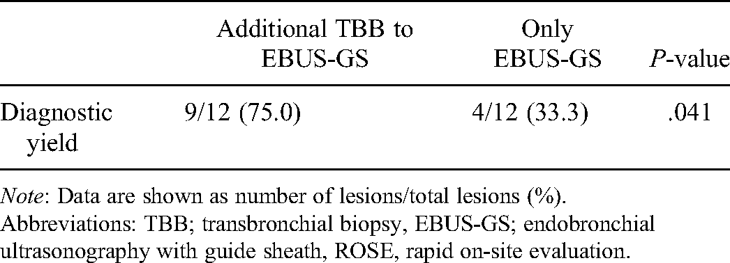
Note: Data are shown as number of lesions/total lesions (%).
Abbreviations: TBB; transbronchial biopsy, EBUS-GS; endobronchial ultrasonography with guide sheath, ROSE, rapid on-site evaluation.
Histological Findings Obtained Using EBUS-GS
The histological findings obtained using EBUS-GS were shown in Table 5. The most frequent histological finding of lung cancer diagnosed with EBUS-GS was adenocarcinoma in the two groups.
Case Diagnoses.

Abbreviations: ROSE, rapid on-site evaluation; EBUS-GS, endobronchial ultrasonography with guide sheath.
Discussion
Our results revealed that the diagnostic accuracy of ROSE during EBUS-GS for diagnosing small peripheral lung cancer was comparable with that reported in a previous study for diagnosing small PPLs. The factor that significantly affected the positive results of ROSE during EBUS-GS was lesion size (>15 mm). When the results of ROSE during EBUS-GS were consistently negative with the probe position being “within” or “adjacent to,” conventional TBB in addition to EBUS-GS improved this lower diagnostic yield, compared with EBUS-GS performed alone.
In previous reports, the introduction of ROSE with bronchoscopy was an entry to perform endobronchial ultrasonography transbronchial needle aspiration (EBUS-TBNA). 23 The diagnostic yield of EBUS-TBNA with ROSE for diagnosing mediastinal and hilar lymphadenopathy was not superior to that of EBUS-TBNA without ROSE. However, EBUS-TBNA with ROSE was reported to reduce the puncture number. 24 On the other hand, EBUS-GS with ROSE was reported to improve the diagnostic yield along with reducing the biopsy number when compared with that without ROSE.17,25 A diagnostic yield of 90% has been reported for instances where EBUS-GS is performed in the position where the probe is located within the lesion. 5 However, EBUS-GS is unsuccessful for 10% of the cases. Nishii et al reported that the reasons for unsuccessful diagnosis of pulmonary malignant lesions are related to the heterogeneity of the EBUS findings and necrosis on histological findings. 26 To overcome these problems, while using EBUS-GS to diagnose small peripheral lung cancer, the use of ROSE may be helpful because we confirmed the presence or absence of malignant cells in the biopsied samples in real time. Our study indicated that when the results of ROSE were positive, the performance of ROSE provided physicians and/or cytopathologists with high diagnostic accuracy and improved the diagnostic yield for small peripheral lung cancer.
The interpretation of ROSE has several problems. First, it is difficult to interpret and distinguish between malignant and benign specimens on ROSE. Second, the discrepancy is usually observed between the individual who reads the findings of ROSE and the person who evaluates the histological findings. Third, there is an additional characteristic observed in the lesions called ground-glass opacity—that usually contains cells that are in the early stages of differentiation—which makes it difficult for pneumonologists or cytologists to confirm atypia. In our study, the characteristics of the lesions were small lesions (≤30 mm) in addition to mainly solid nodule and malignant lesions. In such small lesions, the values of sensitivity, specificity, and accuracy were comparable with those in previous reports.18,20
Lesion size (>15 mm) was a significant factor affecting positive ROSE results during EBUS-GS in the probe positions of “within” or “adjacent to” the lesion. These results indicated that even if the probe was located within or adjacent to the lesion, diagnosing lesions with small PPLs (≤15 mm) using EBUS-GS combined with ROSE may lead to failure. The reason is that collecting sufficient materials from small PPLs (≤15 mm) with a short distance between the peripheral pulmonary lesion (PPL) and EBUS probe is difficult using small biopsy forceps. Previous reports showed the effectiveness of small PPLs (≤15 mm) without a guide sheath compared to with one.27,28 In our study, when the results of ROSE were consistently negative owing to reasons (eg, small PPLs of ≤15 mm) even if the probe was located within or adjacent to the lesion, additional conventional TBB might overcome the lower diagnostic yield compared with EBUS-GS alone by obtaining larger samples (Figure 4).

A case of lung adenocarcinoma in which the results of ROSE were negative and undiagnosed by EBUS-GS, but diagnosed by additional conventional TBB: A. a small nodule in the largest diameter of 11 mm is located on the left S1 + 2; B. the specimen obtained by EBUS-GS did not show malignant findings (hematoxylin and eosin staining); and C. high-power view of the hematoxylin and eosin staining of the specimen obtained by additional conventional TBB showed adenocarcinoma.
The previous report did not demonstrate the usefulness for diagnosing small PPLs, including malignant and benign lesions, by combining ROSE with EBUS-GS and the navigation system. However, the report stated that this combination of techniques might be useful for diagnosing lung cancer. 21 The diagnostic yield of small peripheral lung cancer via EBUS-GS and the navigation system was not always satisfactory compared with CT-NAB or SLB.4,7 Therefore, the technique of adding ROSE to EBUS-GS and the navigation system might be useful for improving the diagnostic yield of small peripheral lung cancer because it is possible to confirm the presence of malignant cells in ROSE in real time. Moreover, the ROSE system might be useful for the operator, as it could help determine changing the sample-collecting procedures when the results of ROSE are consistently negative in a position whereby the probe is located within or adjacent to the lesion using EBUS and the navigation system.
Furthermore, ROSE requires the participation of cytopathologists in our institution. In the present situation, the daily workload of pathologists is heavy; therefore, it is difficult for every institution to dispatch specialized staff to participate in the on-site cytology. Previous reports revealed that additional conventional TBB after performing EBUS-GS without ROSE contributed to improving the diagnostic yield of the small PPLs (≤15 mm) and ground-glass opacity in a facility without ROSE.27,28 Therefore, according to previous reports, we considered that additional conventional TBB might be effective in improving such lesions in a facility without ROSE. Moreover, if we changed to conventional biopsy forceps, we considered taking a biopsy two to three times, as mentioned in a previous report in the facility without ROSE. 27
Our study had certain limitations. First, this was a retrospective and small cohort study. Second, when the results of ROSE using EBUS-GS were consistently negative, we did not fully evaluate the techniques other than conventional TBB (eg, peripheral transbronchial needle aspiration, transbronchial cryobiopsy). Third, we did not thoroughly investigate the change of results of ROSE by additional conventional TBB. The use of ROSE after additional conventional TBB might improve the diagnostic accuracy by confirming the change in the negative results of ROSE to the positive results of ROSE. Furthermore, if the results of ROSE by additional conventional TBB are consistently negative, the addition of techniques other than additional conventional TBB should be considered. Larger prospective studies are needed to overcome these limitations.
In conclusion, our study found that the positive result of ROSE during EBUS-GS was significantly influenced by the lesion size (>15 mm). When the results of ROSE during EBUS-GS were consistently negative after performing a biopsy for five times, additional conventional TBB improved the diagnostic yield compared with EBUS-GS performed alone, in a position whereby the probe was located within or adjacent to the lesion.
Footnotes
Abbreviations
Acknowledgments
We would like to thank Editage (![]() ) for English language editing. S.O is the guarantor of the content of this manuscript, including the data and analysis. T.I contributed to the study design, data collection, analysis, and interpretation, and wrote the draft of the manuscript. T.I, F.U, T.O, M.O, M.N, and N.H contributed to the editing of the manuscript.
) for English language editing. S.O is the guarantor of the content of this manuscript, including the data and analysis. T.I contributed to the study design, data collection, analysis, and interpretation, and wrote the draft of the manuscript. T.I, F.U, T.O, M.O, M.N, and N.H contributed to the editing of the manuscript.
Ethics
All procedures performed in studies involving human participants were in accordance with the ethical standards of the institutional research committee and with the 1964 Helsinki Declaration and its later amendments or comparable ethical standards. This study was approved by the Handa City Hospital Institutional Review Board (No. 2020-018). The requirement for patient informed consent was waived owing to the retrospective nature of the study. Informed consent was obtained from each patient prior to bronchoscopy.
Declaration of Conflicting Interests
The author(s) declared no potential conflicts of interest with respect to the research, authorship, and/or publication of this article.
Funding
The author(s) received no financial support for the research, authorship, and/or publication of this article.