
Abstract
Extracellular vesicles (EVs) are naturally phospholipid enclosed nanovesicles released by many cells in the body. They are stable in circulation, have low immunogenicity, and act as carriers for functionally active biological molecules. They interact with target organs and bind to the receptors. Their target specificity is important to use EVs as noninvasive diagnostic and prognostic tools. EVs play a vital role in normal physiology and cellular communication. They are known to protect their cargo from degradation, which makes them important drug carriers for targeted drug delivery. Using EVs with markers and tracking their path in systemic circulation can be revolutionary in using them as diagnostic tools. We will discuss the scope of this in this paper. Although there are limitations in EVs isolation and storage, their high biocompatibility will fuel more innovations to overcome these challenges.
Keywords
Extracellular Vesicles (EVs) and Therapeutic Cargo
EVs are nano-sized, vesicles known for cellular communication and drug delivery.1,2 They are the key mediators for tissue regeneration, inflammatory regulation, and immune response.1,3-5 They play a significant role in preventing the accumulation of toxic substances and drugs.6,7 Their role as carriers can be exploited for chemotherapy and to study the efficacy of chemotherapeutic drugs.8-10
Studying the pharmacokinetics and permeability of labeled EVs is useful to understand the systemic uptake of medications that can lead to chemotherapeutic side effects. This information makes experimentation with various drug combinations and dosage recommendations possible. The studies can be repeated with adjusted doses and medication combinations till the desired result that is, maximum therapeutic benefit with minimal side effects is achieved.11-13
EVs are present in all biological fluids.14-16 EV-based biomarkers are sensitive for the detection of mutations in cancers.16-18 As EVs can efficiently protect their cargo against degradation, they can be used as biomarkers for diagnostic purposes.7,19 They are well studied for their targeting ability and biodistribution in vivo.20,21
EVs is a common term used to describe exosomes, microvesicles, and apoptotic bodies. 10 Microvesicles are released by outward vesiculation of the cell membrane.10,22 Exosomes are formed by endocytosis of cell membrane forming microvesicular bodies (MVBs).23,24 These MVBs fuse with the plasma membrane and are released into the extracellular space. 25 They carry cell membrane lipoproteins, miRNAs.26,27 Apoptotic bodies are released during apoptosis. 28 The EVs are heterogeneous compounds.28-30 They vary in size, composition, and cargo based on the mechanism of production. 28
Need for Standardization of EV Purification Methods
After extraction from their natural environment, EVs are subjected to centrifugation to remove debris and dead cells.30,31 Using EV-compatible markers, they are separated into different types of EV pellets. 32 For use as markers in diagnostic imaging, biosensors are attached to track EVs in the systemic circulation. To be used as drug carriers, they are injected with specific medications.27,33,34
After release from cells in their natural environment, there are mechanisms for cell-to-cell communication with adjacent tissues and distant organs.35,36 Given their role in biological communication between different tissues, they can serve as drug carriers for targeted therapy.36,37 EVs are able to transfer cytokines, bioactive compounds, miRNAs, and repair proteins in their cargo.38,39 They are effective in carrying the therapeutic cargo as they protect them from the surrounding environment.40,41 Tissue-specific receptors and biosensors can be lodged on the EVs for tracking the drug path.38,41,42 The bioactive cargo of nanovesicles will increase the efficacy of drug delivery.43,44 The target specificity is useful in preventing drug penetration into other organs that can lead to systemic side effects. 44
The current concerns with EV therapeutic applications are, that the complete biochemical profile of EVs is not known. Further, there are no standardized isolation and purification methods, and the lack of efficient drug loading systems for clinical-grade production.19,45,46
Multiple novel technologies have emerged in the past decade for EV isolation and characterization, but there is still a need for reproducible, cheap, and simpler alternatives that can be adapted for large-scale production.46-48 Continuous attempts to develop more effective protocols are ongoing.32,49,50
Isolation methods that are currently available include ultracentrifugation, filtration, size-exclusion chromatography, density gradient, and immunoaffinity-based isolation strategies. 51 Ultracentrifugation isolates the EVs by differential centrifugation. 51 Filtration uses membranes with specific pore sizes for different EVs.51,52 The isolated yield can be poor with this method.43,51 Size-exclusion chromatography uses the hydrodynamic column to reduce contamination, thus yielding a low EV output, but less protein contamination.35,53-55 Density gradient separates them based on density after initial isolation.22,54 Immune affinity-based isolation uses antibodies to capture EVs.22,56 This method is not a cost-effective approach. Table 1.
An Overview of EV Purification Methods, Their Advantages, and Limitations.

The current gold standard for EV isolation is “Ultracentrifugation-linked immunoprecipitation method.”56-58 This method yields smaller sized EVs with lesser apolipoprotein contamination, compared to other methods.26,59 The EVs need enrichment before processing. Currently, different labs have been using different preprocessing protocols.60,61 There is a need to develop an easily reproducible and standardized method.61,62 Table 1.
The newer technique size-exclusion chromatography purifies the EVs by removing the contaminating plasma proteins and high-density lipoproteins.59,63 It has been successfully used in the small-scale analysis of EVs.63,64 The column preparation, washing, and purification make it a time-consuming procedure.64,65 A novel membrane affinity spin column with better efficacy, ease, and reproducible workflow was released. The efficacy of purification using the new spin column is better compared to the optimized ultracentrifugation procedure, which is the current gold standard. 56 Using this method, EV isolation can be coupled with EV RNA extraction.66,67 This procedure captures nearly 100% mRNA. This provides a purified EV isolate without undesired protein-bound extracellular RNA co-precipitate.67,68.68,69 The analysis of EV RNA is useful to understand the genetic markers of EVs. 70
Clinical Applications of EVs
The biggest advantage of EVs is the fact that they are composed of non-immunogenic substances.70,75 Therefore, the tissues do not identify the EVs as foreign, thus protecting their cargo from degradation.52,76 This makes it a safe method for drug delivery.52,77 The nano-size of EVs makes it one of the ideal carriers for the delivery of active molecules and drugs.24,78 They have successfully been applied for cancer therapy, inflammatory modulation, and immune response generation.78-80 They can also be used to administer nutrients, minerals, and oxygen in stroke patients.80,81 This can reduce the recovery period and early rehabilitation of patients.82,83 Table 2.
Clinical and Diagnostic Applications of EVs, Their Benefits, and Limitations.
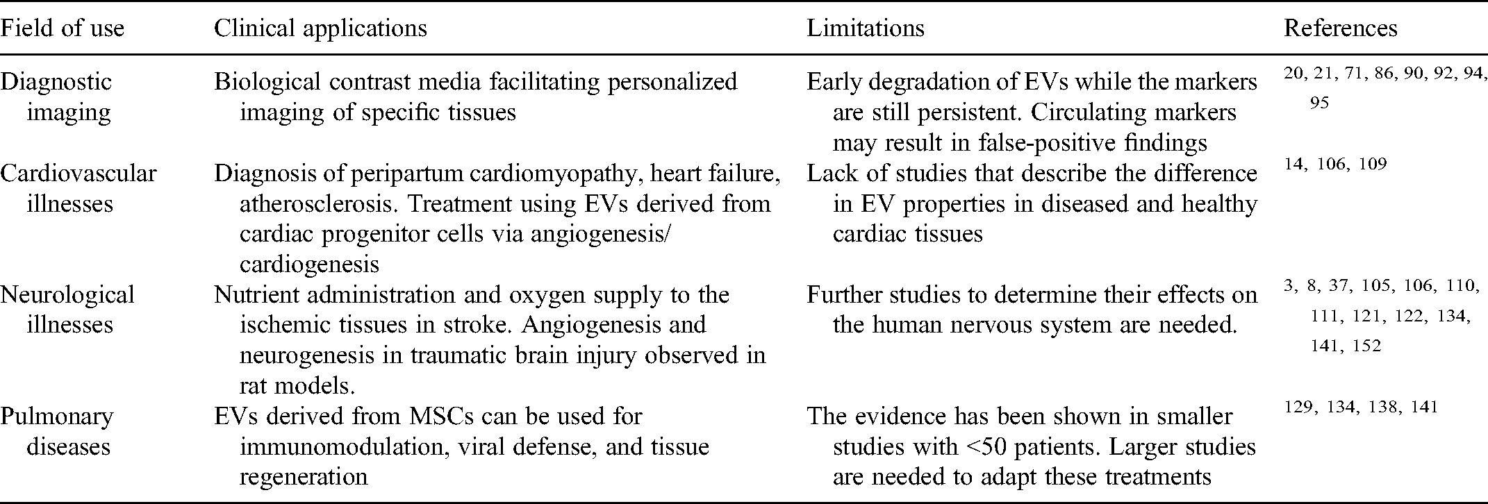
Surface protein bioengineering, biosensor development, drug loading, coupled with placement of ligands on the surface of EVs reprogram EVs into smart multifunctional drug-loaded systems,83,84 Figure 1.

Schematic illustration showing the application of extracellular vesicles in different diseases.
EVs in Diagnostic Imaging
EVs are also used in imaging studies.71,85 The 2 major methods used in detecting EVs in the visible light spectrum are Bioluminescence imaging and fluorescence imaging.86,87 Bioluminescence imaging involves protein-based labeling and has a high signal-to-noise ratio as it is transmitted without any light source.87,88 An ultra-sensitive camera is needed to detect the EVs. 89 Fluorescence imaging uses organic dyes or protein to emit signals under excitation with an external light source. 90 Fluorescence is more easily detected by a charge coupled device camera.91,92 Imaging tools commonly used are single-photon emission computed tomography, positron emission tomography, and magnetic resonance imaging.92-94 It should be remembered that the labeling compounds have a longer half-life than EVs and continue to be detected by imaging even after EVs are degraded.54,95 Bioengineering the EV membranes with inbuilt markers that degrade simultaneously with the EVs will be useful to avoid false-positive findings Table 2.
The protein and RNA cargo in the EVs associated with their cell of origin serve as molecular markers for screening a disease and to assess the progression.40,49,96 The assays using highly purified EV samples will provide relevant clinical information.40,49,96 Micro/nanofluidic technology with affinity capture for precise enrichment of EVs results in EVs that are highly purified compared to the conventional methods that isolate EVs irrespective of the cell of origin.97,98
Appropriate storage of EVs to maintain the stability of nucleic acids is essential.37,51 If these storage conditions are available, the EVs can most likely be used to diagnose gene mutations.37,51 Previous studies have proven that EVs and EV nucleic acids are stable in multiple environments.37,51,74 The EVs and nucleic acids were relatively more stable at 4 degrees centigrade (up to 168 h and 1 week) compared to room temperature (up to 48 h and 1 day).51,74,98
More studies to increase the shelf life of EVs in vitro and in vivo will surpass the current limitations to their use. 59 Preserving them with curcumin has been shown to increase the stability of EVs and potentiate the efficacy of the cargo.58,99,100 Experimentation on additive substances or bioengineered isotopes to increase the stability can widen the diagnostic opportunities of EVs. Tables 1 and 2.
EVs in the Treatment of Cardiovascular Illnesses
EVs containing miRNAs and proteins perform a multitude of functions in target tissues.101,102 The structure, contents, and profiles of the cargo change with tissue damage to produce tissue regenerating substances.57,103 In heart failure, they mediate communication between cardiomyocytes and fibroblasts. 104 For example, MiR-146a-loaded EVs are released by endothelial cells during the development of peripartum cardiomyopathy.105,106 These MiR-146a-loaded EVs serve as biomarkers for diagnosis.105,106 Therefore, they can be used as tissue biomarkers and diagnostic tools in heart failure. 104
EVs are natural carriers of signaling molecules involved in atherosclerosis.104,107 Determining the types and their specific functional properties will be of relevance to discover biomarkers and alter the pathophysiology to control atherosclerosis.
Cardiac progenitor cells (CPCs) and cardiosphere derived cells have shown promising results as useful tools in restoring heart function. 107 Unlike the EVs secreted from cardiomyocytes that have deleterious effects, EVs produced by CPCs are cardioprotective and mediate cardiogenesis.107-109 They induce angiogenesis to reduce ischemic injury, infarct size, and inhibit apoptosis to prevent undesired remodeling.110,111 However, little is known how the EVs are produced in diseased and non-diseased cardiac tissues and the interaction of the EVs with the surrounding and distant tissues. 110 Further studies in these areas would be greatly beneficial to add to the current management of cardiovascular diseases.
In mice, adipocyte-derived EVs in obese mice have been shown to activate differentiation of monocytes into macrophages, inducing insulin resistance.112,113 The same was not seen in lean mice. 114 Similarly, MiR and protein profiles of EVs from hypertensive rats were different from the normotensive rats.115,116 Further research to understand these differences can unfold the dynamic nature of EVs, Figure 1.
EVs in the Treatment of Neurological Illnesses
EVs in the neurons are the typical mode of communication between neurons and neuroglial cells.117,118 This interaction is important for the structural and functional integrity of the nervous system. 118 A lot of research has been done over the years to understand the molecular mechanisms involving the pathophysiology in neurodegenerative studies like Alzheimer's disease,119,120 Parkinson's disease, Huntington's disease, and other dementia disorders.120-122 But the treatment modalities are still very limited. Research is underway to expand this in terms of newer medications, cell therapy, and gene therapy. As the EVs can cross blood–brain barrier, and are less immunogenic, studies increasingly suggest EVs as preferred vehicles for intracranial medication administration. 123
There are previous studies that show there is a high probability of association between EVs and Alzheimer's disease.101,117 Multiple studies have proven that the therapeutic effect of mesenchymal stem cells and adult stem cells is mostly due to their paracrine effect and not the direct action of these cells.101,124 Paracrine factors released by stem cells induce angiogenesis and modulate the immune response to exert the therapeutic effects.125-127 Notably, the properties of purified mesenchymal stromal cells (MSC) derived EVs include most of the properties of mesenchymal cells itself. This provides an understanding that EVs from these cells are the major components involved in the disease process.102,127,128 This property has been exploited to use the extracted EVs in multiple treatments Figure 1.
MSC-derived EVs in rat models with traumatic brain injury and ischemic brain injury has been shown to increase angiogenesis and neurogenesis, thus improving functional recovery.129,130 Katsuda et al. suggested a new treatment possibility using EVs that carry neprilysin, an amyloid degrading enzyme as a cell-free drug therapy for Alzheimer's disease,131,132 Figure 1.
EVs in the Treatment of Lung Diseases Including Severe Acute Respiratory Syndrome Coronavirus 2 (SARS-CoV-2) Infection
EVs are secreted by the lining of the respiratory tract similar to any other cell in the body and their role in the pathogenesis of lung diseases has been established in previous studies.133,134 Recent studies show that EVs and viruses are interdependent on each other for cellular entry and exit owing to their similar physicochemical properties, for example, size and heterogeneous size distribution. 134 Therefore, trials using EVs loaded with antiviral medications to block viral intracellular entry can be explored.
In the past 1 year, mesenchymal stem cell (MSCs) therapy for SARS-CoV-2 infection is being explored in multiple studies.135,136 MSCs have unique potential for immunomodulation, antiviral defense, and tissue regeneration. Umbilical cord-derived MSCs have shown promising results in smaller studies.137,138 They induce the production of cytokines, paracrine factors that interact with immune cells to stimulate vascular endothelial growth factor, epidermal growth factor, transforming growth factor release mediated tissue regeneration.139,140 The promising tissue regenerative properties of MSC-derived EVs support pulmonary tissue regeneration post coronavirus 2019 (COVID-19) illness.
The limitations with MSC use are shorter half-life, lasting a few minutes in the blood stream. MSCs derived from embryogenic tissue can be mutagenic and tumorgenic.141,142 Intravenous administration of MSCs can cause platelet aggregation and thrombogenesis leading to emboli. 142
EVs derived from MSCs can be administered via intranasal and inhalational route, contributing to lesser immunogenic or thrombogenic side effects.143,144 They do not enter the circulation where they can be lost in a few minutes and do not cause thrombus formation.145-147 They do not self-replicate like MSCs, therefore are less tumorigenic.148,149
EVs can be administered along with antiviral drugs.150,151 Preliminary studies suggest that co-treatment with EVs obtained from mesenchymal cell secretomes is effective in treating COVID-19 patients.135,136,151 Further preclinical data considering safety, ethics, and practicality is needed to adapt this regimen for COVID-19 treatment.
Summary of Clinical Applications
Conclusion and Future Directions
EVs show great promise as diagnostic and therapeutic markers in many diseases. There is a dire need to work on standardization of isolation and storage methods of EVs. This will enable a wider application of EVs in large-scale treatment trials. Several studies have shown that the current methods available have limitations like contamination of samples, cost issues, instability issues, among others. Ultracentrifugation with immunoprecipitation has been a promising method. If the preprocessing methods are standardized and ways to curb the cost factor are explored. This method can be adapted for therapeutic EV production as it produces highly selective EVs. Further studies are urgently needed to explore these methods which can pave the path for the clinical application of EVs.
The pathological processes in which the EVs are being applied show great benefits with minimal side effects. As EVs are derived from human cells, there is always the benefit of biocompatibility compared to other modalities of treatment. In addition to cardiovascular and neurological diseases, the need of the hour is the treatment of SARS-CoV-2 infection. Though the studies so far for EVs derived from MSCs in COVID illness have been small, there is a great scope as they offer a combination of benefits providing immunomodulatory function, viral defense, and tissue regeneration. This can decrease the severity and duration of COVID-19 illness. Further studies in this area are needed. The regenerative property can also be utilized for cardiac remodeling in post-MI patients, neuroregeneration in stroke patients, thus decreasing the morbidity and mortality.
Footnotes
Declaration of Conflicting Interests
The authors declared no potential conflicts of interest with respect to the research, authorship, and/or publication of this article.
Funding
The authors received no financial support for the research, authorship and/or publication of this article.