
Abstract
Background and Aims:
This study evaluated the prognostic value of 18F-fluorodeoxyglucose positron emission tomography with integrated computed tomography (18F-FDG PET/CT) performed before and after concurrent chemoradiotherapy (CCRT) in esophageal cancer.
Methods:
We analyzed the prognosis of 50 non-metastatic squamous cell esophageal cancer (T1-4N0-2) patients who underwent CCRT with curative intent at Inje University Busan Paik Hospital and Haeundae Paik Hospital from 2009 to 2019. Median total radiation dose was 54 Gy (range 34-66 Gy). Our aim was to investigate the relationship between PET/CT values and prognosis. The primary end point was progression-free survival (PFS).
Results:
The median follow-up period was 9.9 months (range 1.7-85.7). Median baseline maximum standard uptake value (SUVmax) was 14.2 (range 3.2-27.7). After treatment, 29 patients (58%) showed disease progression. The 3-year PFS and overall survival (OS) were 24.2% and 54.5%, respectively. PFS was significantly lower (P = 0.015) when SUVmax of initial PET/CT exceeded 10 (n = 22). However, OS did not reach a significant difference based on maximum SUV (P = 0.282). Small metabolic tumor volume (≤14.1) was related with good PFS (P = 0.002) and OS (P = 0.001). Small total lesion of glycolysis (≤107.3) also had a significant good prognostic effect on PFS (P = 0.009) and OS (P = 0.025). In a subgroup analysis of 18 patients with follow-up PET/CT, the patients with SUV max ≤3.5 in follow-up PET/CT showed longer PFS (P = 0.028) than those with a maximum SUV >3.5.
Conclusion:
Maximum SUV of PET/CT is useful in predicting prognosis of esophageal cancer patients treated with CCRT. Efforts to find more effective treatments for patients at high risk of progression are still warranted.
Introduction
18F-fluorodeoxyglucose positron emission tomography with integrated computed tomography (18F-FDG PET/CT) is important for the diagnosis, staging, and radiation therapy treatment planning of esophageal cancer. 1,2 The maximum standard uptake value (SUVmax) is widely used as a surrogate marker for tumor metabolic status. 3,4 In addition, metabolic tumor volume (MTV) has been shown to be a good prognostic marker for esophageal cancer. 5,6 Further, total lesion of glycolysis (TLG), defined as the product of mean SUV and MTV, has been recently introduced as a new metabolic parameter of esophageal cancer treated with concurrent chemoradiotherapy (CCRT). 7,8
Previous studies have shown that PET/CT can be helpful for the therapeutic monitoring of esophageal cancer. 5 Especially, SUVmax at diagnosis or tumor volume on PET/CT has been reported to provide valuable information for the prognosis of esophageal cancer. 9 Recent studies have shown that cancer prognosis tended to be determined by the degree of SUVmax decrease after treatment. 10,11 Although a consensus has not been reached on the appropriate cut-off value for SUV, Higuchi et al 12 reported a prognostic difference in esophageal cancer based on SUVmax values of 2.5 after CCRT. Conversely, Brown et al 13 suggested a cut-off point of SUVmax 5 for determining prognosis. Furthermore, Kim et al 14 reported that serial PET/CT was meaningful in predicting pathologic response of esophageal cancer after neoadjuvant chemoradiotherapy.
The aim of this study was to investigate whether the SUVmax and other parameters of PET/CT at diagnosis were helpful for predicting the prognosis of esophageal cancer patients after chemoradiotherapy. The main goal of this study was to assess the feasibility of PET/CT parameters in prognosis prediction in esophageal cancer.
Materials and Methods
Patients
We analyzed a total of 50 esophageal cancer patients who were treated with CCRT from December 2009 to December 2019 at Inje University Busan Paik Hospital and Haeundae Paik Hospital. During screening, patients who had distant metastasis at the initial diagnosis or who had no abnormal hypermetabolism in the primary tumor were excluded from further analysis. All patients in this study underwent PET/CT prior to treatment, and the presence of squamous cell cancer was confirmed by biopsy. This retrospective study was examined and approved by the Institutional Review Board in Inje University Busan Hospital (IRB No. 18-0046).
Patients’ clinical information, collected from the Electronic Medical Record (EMR), and PET/CT parameters were used for analysis. Evaluation of the relation between SUVmax and prognosis was prioritized. At the initial diagnosis, patients were divided into 2 groups based on SUVmax 10, which is strongly indicative of a malignant tumor. 15 Besides SUVmax, additional PET/CT parameters, MTV and TLG, were available for evaluation in 42 patients. Thus, further analyses on the prognostic effect of MTV and TLG, obtained from initial PET/CT, were performed in those patients.
Tumor location was classified into 3 levels: upper thoracic esophagus (20-25 cm from upper incisors), middle thoracic esophagus (25-30 cm from upper incisors) and lower thoracic esophagus (30-50 cm from upper incisors).
This study was based on the assumption that a higher SUVmax value and a greater tumor volume are associated with a worse prognosis. The primary end point of this study was progression-free survival (PFS) and the secondary end point was overall survival (OS). Failure pattern was also observed and evaluated. PFS was calculated from the start of radiotherapy (RT) until recurrence, and OS was calculated from the start of RT until the date of death or last follow-up.
Image Acquisition and Analysis
All patients fasted for >6 h before undergoing PET/CT, and the blood glucose level was <180 mg/dl. Images from the mid skull to the upper thigh were taken approximately 60 min after intravenous administration of 370 MBq F-18 FDG. The Discovery™ PET/CT tomograph (GE Healthcare, Milwaukee, WI, USA) and Biograph Truepoint 64™ PET/CT (Siemens Healthineers) were used. All PET images were reconstructed using an iterative algorithm and attenuation correction with CT images.
Two nuclear medicine specialists independently reviewed the images. For semiquantitative analysis, all images were reviewed on a designated workstation. SUVmax was measured within a designated region of interest (ROI) and defined as the highest SUV of pixel. MTV, which was determined by measuring the volume of the lesion above the 3.0 SUV value and the TLG was determined by the MTV × SUVmean. MTV and TLG were calculated as the summation of individual MTV and TLG within the field of image.
Treatment
All patients underwent cisplatin and 5-fluorouracil chemotherapy after diagnosis, and RT was concurrently administered with a total dose of 39.6 Gy-66 Gy (1.8-2.0 Gy per fraction).
Among the patients in this study, 18 patients underwent follow-up PET/CT at 2-7 months after initiation of RT. Forty-two patients (84%) were irradiated with over 50 Gy of radiation. Gross tumor volume on simulation CT of RT planning was also measured and used for further analyses.
Statistical Methods
MedCalc statistical software (MedCalc Software version 19.2.0 bv, Ostend, Belgium) was used for statistical evaluation. Paired-T test was used to compare initial SUV and follow-up SUV values. Chi-square test or Fisher’s exact test was performed to find clinical factors associated with disease progression. In addition, Kaplan-Meier test was performed to estimate PFS and OS, and log-rank test was used to identify factors related with prognosis. For the test with PET/CT parameters (continuous variables), median values were used to classify patients into 2 groups. Cox-regression test was used to identify independent prognostic factors for PFS and OS.
Results
Patients Characteristics
Table 1 shows patient characteristics. All patients were pathologically diagnosed with squamous cell esophageal cancer. The median age at diagnosis was 66.5 years (range 45-86 years). Thirteen patients (26.0%) had active smoking history. Two patients (4.0%) were female. In regard to T and N stage, the number of patients diagnosed with T1, T2, T3, and T4 stages were 7, 13, 22, and 8, respectively, and N0, N1, and N2 were 18, 17, and 15, respectively. No patients had distant metastases upon initial diagnosis. Regarding tumor sites, 13 were upper-thorax (26%), 23 middle-thorax (46%), and 14 lower-thorax (28%) esophageal cancers. The median SUV max of initial PET/CT was 14.2 (range 3.2-27.7). When the SUVmax of baseline PET/CT exceeded 10, the clinical T stage was higher (T1-2 vs. T3-4, P = 0.007). Additionally, positive N stage was also related with the value of baseline SUVmax (P = 0.022).
Patient Characteristics.

Abbreviations: PET/CT, positron emission tomography with integrated computed tomography; SUVmax, maximum standard uptake value.
Patterns of Failure
Median follow-up was 9.9 months (range 1.7-85.7). During the follow-up period, 29 patients (58%) experienced progression. The 2 female patients experienced progression, and both died eventually because of cancer progression. However, progression rate was not significantly different by gender (P = 0.503). Progression occurred in 38.5% (5/13) of smoking patients, and there was no statistically significant correlation between past smoking history and progression (P = 0.116). More specifically, treatment failure was due to local failure in 10 patients, regional failure in 16 patients, and distant failure in 11 patients (there were a number of patients with multiple sites metastases). Lung was the most common distant metastases site (n = 4). One patient was cured following esophagectomy after progression. Of the 4 patients with complete response in the follow-up PET/CT, 50% (n = 2) did not develop relapse permanently.
Survival Analysis
A total of 16 (32%) patients died during the follow-up period. Deaths after CCRT were commonly caused by pneumonia (n = 4) or heart failure (n = 3). Overall, the 3-year PFS and OS were 24.2% and 54.5%, respectively (Figure 1).
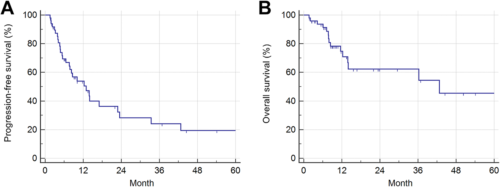
(A) PFS and (B) OS (n = 50).
Table 2 shows the clinical factors affecting PFS and OS. When the SUVmax of initial PET/CT exceeded 10, which was a strong indicator of malignancy (n = 22), PFS was significantly lower (P = 0.015, Figure 2A, Table 2). However, initial PET/CT SUVmax did not significantly influence OS (P = 0.282, Figure 2B, Table 2). T stage (T1-2 vs. T3-4, P = 0.008) and N stage (N0 vs. N1-2, P = 0.002) significantly affected PFS. Regarding tumor location, even though it did not reach statistical significance, PFS tended to be better when the tumors were located in the lower esophagus (n = 14) than when they were located in the upper or middle esophagus (n = 36, P = 0.091). Further, PFS was significantly improved when the total RT dose exceeded 50 Gy (P < 0.009). Another prognostic factor significantly affecting OS was a high T stage (T3-T4, P = 0.002). In multivariate analyses, initial total LN metastases (P = 0.049, hazard ratio [HR] 2.499, 95% confidential interval [CI] 1.002-6.180) and RT total dose above 50 Gy (P = 0.036, HR 0.333, 95% CI 0.119-0.932) were independent prognostic factors for PFS (Table 2). OS was apparently affected by T stage in multivariate analysis (P = 0.006, HR 6.039, 95% CI 1.312-27.796).
Prognostic Factors for Progression-Free Survival and Overall Survival.

Abbreviations: PET/CT, positron emission tomography with integrated computed tomography; SUVmax, maximum standard uptake value.
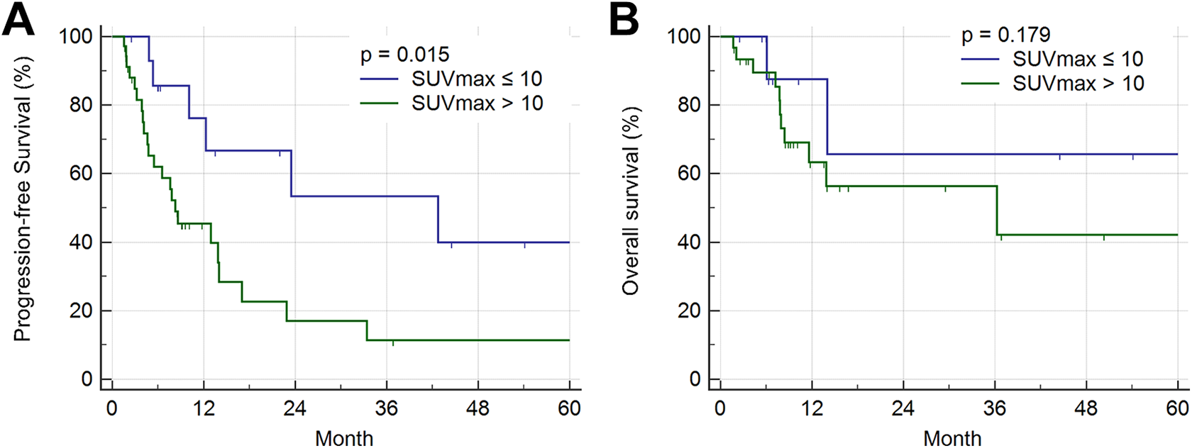
Dichotomous (A) PFS and (B) OS by baseline SUVmax 10 (n = 50).
Metabolic Tumor Volume and Total Lesions of Glycolysis
Subgroup analysis was performed for patients with further PET/CT related parameters, such as MTV and TLG (n = 42). Their median MTV was 14.1 (range 4.0-102.2) and their median TLG was 107.3 (range 9.6-687.5). A small MTV was significantly correlated with a high PFS (P = 0.002) and OS (P = 0.001).
Also, TLG had a significant effect on PFS and OS. Patients whose TLG was greater than 107.3 showed poor PFS (P = 0.009, Figure 3). TLG also had a significant effect on OS (P = 0.025, Figure 3).

(A) PFS and (B) OS by size of metabolic tumor volume. (C) PFS and (D) OS by size of total lesions of TLG (n = 42).
Prognostic Value of Follow-Up PET/CT
In 18 patients who underwent follow-up PET/CT, the SUVmax trend of baseline PET/CT had a significant effect on the results of SUVmax value of follow-up PET/CT (P < 0.001). The median SUVmax of follow-up PET/CT (n = 18) was 3.8 (range 0-7.3). Among patients who underwent follow-up PET/CT (n = 18), those with a follow-up SUVmax ≤3.5 had a longer PFS than patients with maximum SUVmax >3.5 (P = 0.028, Figure 4). However, there was no gain in OS in low SUVmax (≤3.5) group of follow-up PET/CT (P = 0.132).

Subgroup analysis: PFS by post-treatment PET/CT SUVmax 3.5 in patients with follow-up PET/CT (n = 18).
A case, described in Supplement 1 and 2, shows good treatment effect of CCRT; PET/CT before (Supplement 1) and after CCRT (Supplement 2) treatment. After 3 months of treatment, the maximum SUV of the lower cervical esophagus significantly declined. This patient did not experience progression afterward.
Discussion
In summary, PFS was short after esophageal cancer CCRT when PET/CT SUV max was more than 10 at diagnosis. In cases of a PET/CT SUVmax of 3.5 or higher after CCRT, PFS, or OS was lower. When RT was given with a total of 50 Gy or more, PFS was significantly improved.
PET/CT parameters are useful for predicting cancer prognosis 16 as well as checking current disease status. 17 Consistent with previous studies, follow-up PET/CT SUV values were helpful for the prediction of prognosis in terms of PFS and OS. 18 -20 Previous studies have reported that SUV values of 2.5-5 after CCRT are adequate thresholds for prognosis. 13 In this study, PFS was different when patients were divided according to follow-up PET/CT SUV 3.5. Despite the multivariate analyses not demonstrating significance, PET/CT parameters are meaningful prognostic predictors. This study also showed that initial tumor volume and TLG are also important predictors of PFS.
In this study, tumor volume, which represents disease burden, was important for prognosis. Similar to a previous study by Yin et al, 21 T stage was an important prognostic factor for survival. Metabolic tumor volume, guided by PET/CT, was also an important factor in predicting PFS and OS. TLG, which reflects both SUV value and tumor volume, was also helpful for the prediction of PFS and OS. Both TLG and MTV can predict survival, so they can be considered to have strong prognosis prediction abilities. Moreover, both PET/CT parameters could also be used for treatment response evaluation. In fact, a recent study by Borggreve et al 22 showed the possibility of using TLG for RT response prediction.
RT also had a great impact on prognosis. According to our data, RT should be administered with at least 50 Gy to improve PFS. In particular, a sufficient radiation dose was shown to help prevent progression in patients with a high SUVmax. Further, according to this study, there were many patients with regional failure after CCRT. Therefore, it is worth trying elective nodal irradiation to improve treatment results. In addition, the fact that heart failure and pneumonia were the cause of death in a substantial number of cases suggests that efforts should be continued with the aim of minimizing the dose of radiation affecting the lungs and heart during RT. 23
This study has some limitations. This study was a retrospective study and analyzed patients from 2 institutions. The total number of patients was not sufficient. Thus, the results of this study require caution in interpretation, with a small number of patients. Moreover, despite the fact that TLG and MTV were predictors of survival, the analysis of such parameters was not possible in all of the patients. Plus, for it was a retrospective study, the difference in gender distribution was unavoidably biased, so only a small portion of the patients were female (n = 2%, 4%). However, we don’t think that the gender bias causes a problem in trusting the entire study. Therefore, it should be noted that there may be a selection bias in the interpretation of the study results. Other institutions with a large number of patients may be able to obtain more reliable data if they conduct studies related to this subject. In this study, SUVmax 3.5 in follow-up PET/CT was proposed as the cut-off value for discerning treatment outcomes. However, a consensus has not been reached on the appropriate cut-off value of SUVmax, and so further research is still warranted. Recent studies have been conducted on the relationship between metabolic tumor volume on PET/CT and tumor volume on diffusion-weighted magnetic resonance imaging (DW-MRI), 22,24 and thus further studies related to DW-MRI may be useful in this field.
In conclusion, the SUVmax or tumor volume of PET/CT parameters seems to be useful for predicting prognosis of esophageal cancer patients treated with CCRT or treatment response evaluation after CCRT. Since initial disease status is an important factor in prognosis, it will be necessary to find an effective method for the early detection of esophageal cancer. A sufficient dose of radiation in patients with esophageal cancer is also essential to improve prognosis. Overall, the prognosis of esophageal cancer is still not good enough and thus, efforts to find more effective treatments are warranted in the future.
Supplemental Material
Supplemental Material, sj-tif-1-tct-10.1177_15330338211024655 - 18F-FDG PET/CT Parameters for Predicting Prognosis in Esophageal Cancer Patients Treated With Concurrent Chemoradiotherapy
Supplemental Material, sj-tif-1-tct-10.1177_15330338211024655 for 18F-FDG PET/CT Parameters for Predicting Prognosis in Esophageal Cancer Patients Treated With Concurrent Chemoradiotherapy by Seokmo Lee, Yunseon Choi, Geumju Park, Sunmi Jo, Sun Seong Lee, Jisun Park and Hye-Kyung Shim in Technology in Cancer Research & Treatment
Supplemental Material
Supplemental Material, sj-tif-2-tct-10.1177_15330338211024655 - 18F-FDG PET/CT Parameters for Predicting Prognosis in Esophageal Cancer Patients Treated With Concurrent Chemoradiotherapy
Supplemental Material, sj-tif-2-tct-10.1177_15330338211024655 for 18F-FDG PET/CT Parameters for Predicting Prognosis in Esophageal Cancer Patients Treated With Concurrent Chemoradiotherapy by Seokmo Lee, Yunseon Choi, Geumju Park, Sunmi Jo, Sun Seong Lee, Jisun Park and Hye-Kyung Shim in Technology in Cancer Research & Treatment
Footnotes
Authors’ Note
It was a retrospective study by EMR review. The potential risk for patients is very low. So, we got a waive of patients’ consent by Inje University Busan Paik Hospital IRB.
Declaration of Conflicting Interests
The author(s) declared no potential conflicts of interest with respect to the research, authorship, and/or publication of this article.
Funding
The author(s) disclosed receipt of the following financial support for the research, authorship, and/or publication of this article: For Yunseon Choi, this work was supported by a grant from Research Year of Inje University in 20180016.
Supplemental Material
Supplemental material for this article is available online.
References
Supplementary Material
Please find the following supplemental material available below.
For Open Access articles published under a Creative Commons License, all supplemental material carries the same license as the article it is associated with.
For non-Open Access articles published, all supplemental material carries a non-exclusive license, and permission requests for re-use of supplemental material or any part of supplemental material shall be sent directly to the copyright owner as specified in the copyright notice associated with the article.