
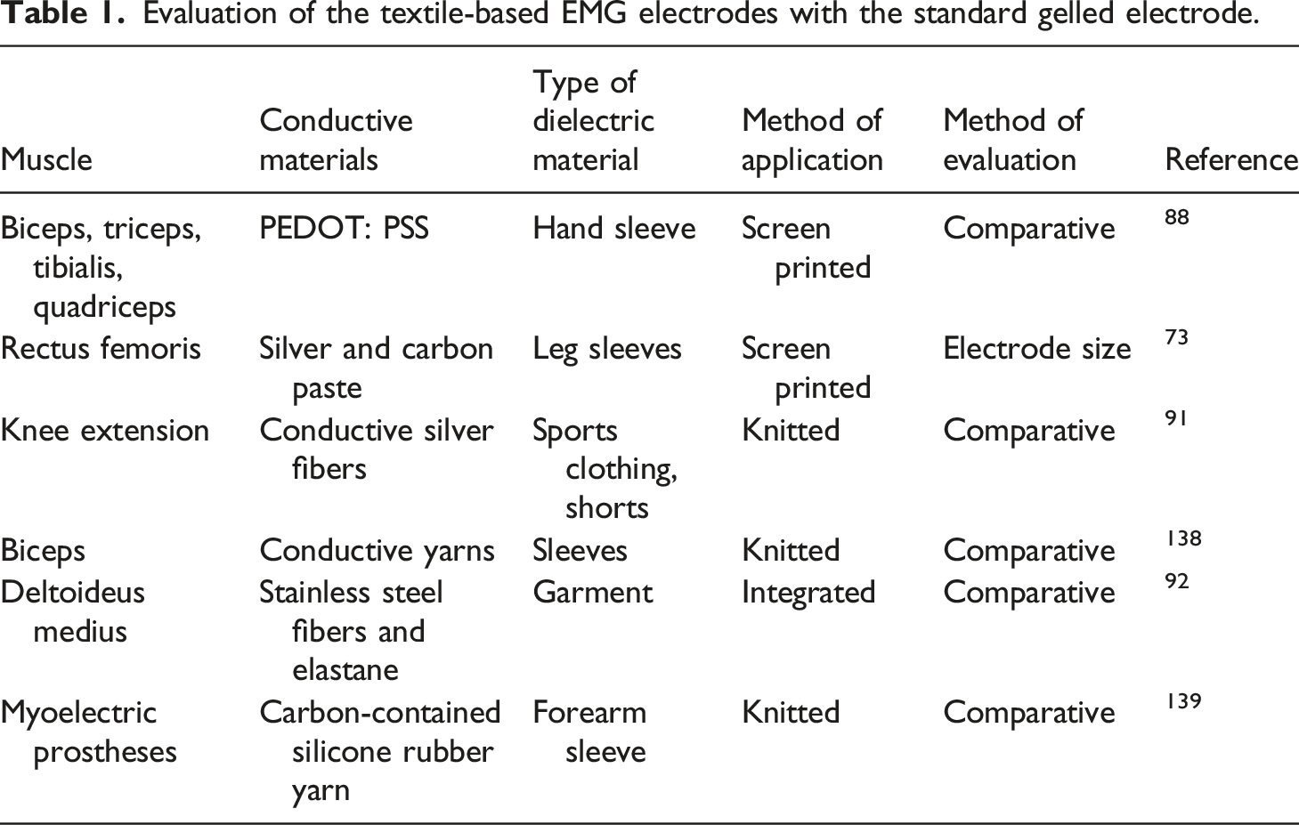
Abstract
Due to recent developments in wearable sensor technology, textile electrodes are routinely being employed in electromyography (EMG) for continuous monitoring of the biosignals from the muscles. However, the performance of such smart textile-based health monitoring devices depends on several factors such as, the sensitivity (impedance), durability (reusable/washable), users' comfort ability, integrability, and automatability. In this article we review the characteristics and the performance of the EMG textile electrodes, in the context of functional textile materials, smart textile materials, and smart textile systems for biosignals monitoring. The functional textile materials are confined to signal transmission alone, whereas, the smart textile materials include signal transducers and sensors. The more advanced smart textile systems include signal conditioning circuits with displays. Nowadays, textile-based sensors embedded in garments are becoming a part of users' normal life, in particular, the textile systems that continuously monitor the vital physiological signals from muscles are being sought after in healthcare settings.
Keywords
Introduction
Smart textiles are categorized into functional textile materials (passive), smart textile materials (active), and smart textile systems (STS, ultra smart).1–4 Electrically conductive textile materials are often used to connect the various components in a smart textile system, such as the batteries, the data processing units and the sensors. Though they are essential in smart textiles by being electrically conductive, they cannot be considered as “smart”. Functional textiles are the fabric materials that not only possess signal transmission properties (e.g. optical fibers or electrically conductive wires) but also include antimicrobial, fire resistance, UV protection, crease recovery and others.5,6 Smart textiles materials on the other-hand incorporates signal transducing sensors and respond to an external stimulus such as thermal, optical, mechanical or electrical.
2
A chromic pigment composed of strontium aluminate printed on to a cellulosic fabric has been shown to change its color on exposure to UV light
7
Figure 1. Other examples of smart textile materials include phase change materials or piezoelectric materials.
8
Sensors are components that detect changes in physical properties; whereas, the ‘transducers’, not only sense but also convert a signal from one energy form (mechanical/pressure, thermal, optical etc.) to another, especially, electrical. To measure electromyography there are specific sensors available in the market.9–12 An important component in the smart textiles is the electrode designed using appropriate conductive material such as metallic fibers or fibers coated with conductive polymers. Conducting Electro active Polymers (CEP) of pyrrole coated on silk or cotton fibres using continuous vapor phase polymerization enabled production of uniformly coated yarns with low electrical resistivity and good conductivity.13,14 However, the stability of such conductive polymers in wearable sensor technology during its manufacturing processes at high temperatures still remains a challenge. The photograph on the left shows a screen-printed cotton fabric (0.08 wt%) before illumination (red color) and the right one shows the same fabric after illumination (patterned blue and green substrate) with UV (λ = 365 nm). The irradiation was carried out at room temperature for 1 min.
7
Reproduced with the permission of Copyright Clearance Center, Inc. (CCC).
In a smart textile system, an energy component or power supply is required to supply power or energy to its sensors and actuator components. In smart textiles design, efficient and compact lithium polymer (Lipo) batteries or solar cells.15–17 coupled with capacitors 18 that store harvested energy are used.19–21 Developments in textiles technology has facilitated both the sensing capabilities such as measuring touch, movement, stretch, sound and light; and actuation capabilities such as changing shape, color, emit heat, light, etc. 22 In human activity monitoring technology, STS is deployed to acquire and classify body signals with respect to the activity being performed. Human activity monitoring technology is concerned with facilitating lifestyle through digital means at home or in the office. 23 For example, providing security through surveillance, improving fitness and sports activities, monitoring health of older people. Sometimes, STS is used to detect occupational related physical activities to make accurate assessment of the hazards in the work place.24,25 Since STS can be programmed, it makes use of the latest communication technologies such as Internet, aeronautical networks, IoT and mobile networks. 26 One of the concerns of using smart textile sensing is the decreased quality of the monitored signal governed by the less adherent interface present between the textile electrode and the skin. 27 Another major challenge of wearable textile systems is the occurrence of resistance and heating at the electro-conductive contacts (EC-contacts). Electro-conductive contacts occurs when two electro-conductive yarns intersect perpendicularly or woven into a hybrid fabric. 28
Biopotential monitoring
EMG is a Greek word that stands for the recording or measurement of the biological electrical activity of the muscle.29,30 EMG is an analytic technique that monitors the health condition of the muscle and its associated nerve cells, by measuring the electrical signal transmitted by the contraction and relaxation of the associated muscle fibers.
31
The EMG signals can be measured using either the intramuscular or extra muscular electrodes 31–33 as shown in Figure 2 The measurement using intramuscular electrodes is an invasive method, because the needle electrodes are inserted (or ‘deep seated’) into the intramuscular cells. The latter type of electrodes referred to as surface electromyography (sEMG) are non-invasive in nature. The deployment of any type of electrode, i.e., intramuscular or surface electrodes, require careful consideration of the size of muscle under investigation. For example, to monitor a large superficial surface, the surface electrode is preferred, whereas, to study a small region of the muscle, intramuscular electrodes are sufficient. Intramuscular (left) electrode
136
and extra muscular surface electrode (right) reproduced from
31
under Creative Commons Attribution License (http://creativecommons.org/licenses/by/3.0).
Intramuscular needle electrode contains a shielded wire placed inside a cannula (or needle shaft) and its tip remains unshielded or bare. Such types of electrodes provide improved signal compared to other available electrode types. The size of these electrode allow detection of individual Motor Unit Action Potentials (MUAPs) and sense new tissue territories within the muscle through relatively low force contractions, and repositioning. 31 But the insertion of the needle electrode cause discomfort and muscle soreness; hence, its usage is limited to inpatient services and requires medical supervision.
Unlike the needle electrodes, used by medical professionals in the clinics, the surface electrodes can be used outside of clinics such as in sports with less medical professionals’ assistance. sEMG provides an easy and straightforward way to measure muscle contraction under the influence of different variables such as loads/weights, skin contact pressure and exercise. There are two types of sEMG electrodes available namely, gelled and dry EMG electrodes. The gelled EMG electrodes require a gel based electrolytic substance applied at the interface between the skin and the electrode, whereas, the dry EMG electrodes are devoid of gel layer interface. One of the most commonly used surface electrode is the metallic silver-silver chloride (Ag/AgCl) electrode which show several favorable characteristics such as stable behavior, microcidal activity, non-toxic composition, low frequency noise, and low cost.34,35 They can be used either in dry form or in wet form with an electrolytic gel applied on to the skin surface.
sEMG’s are susceptible to electrical and mechanical artifacts compared to intramuscular EMG.
36
Since EMG measurements are affected by physiological and anatomical characteristics of the muscles under consideration, we will briefly explain the structural features of the general muscle fiber and the biosignals generated by it.
37
A set of muscle fibers innervated by a single neuron constitutes the spindle shaped functional unit called the ‘motor unit’ (MU), see Figure 3. The MU innervated by a motor neuron forms a junction called motor endplates. The motor neurons have their axons extended from the spinal cord to the skeletal muscle fibers of a motor unit; with its cell body residing in the spinal cord. Since the control of muscle is realized at the level of the motor unit (MU), it seems important to consider the physiological properties of motor units when attempting to understand and predict muscle fatigue.
38
A typical. α− motor neuron extends from the spinal cords and ends at the motor end-plates in the skeletal muscle fiber, adapted from
137
under CC BY-NC-ND 3.0.
On receiving the signal from the α-motor neuron, the muscle fiber gets depolarized. This depolarization also called action potential (AP) propagates along the muscle fiber in both directions from its origin/source or innervation zone (IZ), Generally, the action potential, (which generates a potential difference along the length of the muscle fiber) originates as a dipole at the IZ (or motor endplate-muscle fiber junction), and propagates as a ‘tripole’ (because of the generation of a secondary action potential that follows the primary action potential). The tripole terminates at the ‘tendon zone’ (TZ) present at the end of fiber and give rise to the ‘end fiber action potential’. The potential difference originating from each MU is defined as the MUAP. The EMG signal is the sum of the MUAP’s originating from several activated MU’s and the electrodes measure the sum of MUAPs. When EMG is recorded using surface electrodes, the amplitude of the signal gets attenuated by the connective tissues, subcutaneous adipose layer and skin layers that is present in between the source of MUAPS and the skin surface. 39 Hence, when simulating an EMG signal, a model for the signal attenuation in terms of ‘volume conductor’ is considered.
Currently, researches are focused towards evolution of the surface electrode into textile electrodes for improved acquisition and processing of electromyography (EMG), electrocardiography (ECG), electroencephalography (EEG), phonocardiography (PCG), and other biosignals in clinical settings as well as other domains. Development of compatible textile electrodes for biosignal acquisition requires careful consideration for materials selection, substrate deposition (materials coated on textile), and functional efficiency in terms of signal amplitude, signal to noise ratio (SNR), root mean square (RMS), average rectified value (ARV) and impedance characteristics when compared to conventional electrodes. In this review, we provide an overview of the previous and the current materials, methods and techniques used in EMG measurements with relevance to surface textile electrodes. We have also outlined the challenges faced during the usage of the textile electrodes in clinical setting and measures to overcome them.
Basics of EMG Electrodes
The Electrochemical Cell and the Equivalent Electronic Circuit
To develop a basic understanding of the electrode properties, the theoretical basis for the transfer of the electrical charge from the source (body) to the electrode is necessary. Consider a gelled electrode setup Figure 4(b), in which a metal electrode disc is placed on a cleaned skin surface with a pre-gelled electrolyte material. This arrangement leads to the formation of two unique interfaces. The first interface is seen between the metal electrode and the gel electrolyte (top part), the second interface is seen between the gel-electrolyte and the skin (bottom part). When a metal electrode comes in contact with an electrolyte, the metal atom ionizes into its cationic form by losing an electron to the electrode and the cation is released into the electrolyte solution. The high concentration of cations near the electrode attracts more anion from the electrolyte solution and form a charge barrier with an associated potential called ‘Helmholtz double layer’ ionic potential. This potential is also referred to as the, ‘half-cell potential’, and is characteristic of the different metal-electrolytes being used.33,40 For example, Ag metal dipped in NaCl electrolyte, also referred to as Ag/AgCl, has a half-cell (Ehc) potential of −0.69 V. Half-cell potential of a metal-electrolyte can be experimentally determined by connecting it to a reference half-cell, realized by dipping a platinum metal electrode into an acidic solution bubbled with hydrogen gas. The potential of the reference half-cell is considered to be 0 V. The second interface is present in between the electrolyte and the skin, which also generates a potential difference referred to as the ‘contact potential’ that is significantly greater than the ‘double layer potential’. The ‘contact potential’ is more important than the ‘Helmholtz double layer’ and it contributes significantly to bio-impedances. Both of the above-mentioned interfaces are modelled using an equivalent electrical circuit
41
as shown in Figure 4(a) (a) The electrode model showing the metal-electrolyte interface and electrolyte-skin interface. (b) The physical representation of the model.
41
Adapted under CC by 4.0.
In Figure 4(a), the interface potential (Helmholtz or contact potential) is represented as
Impedance property of biopotential electrodes
The skin-electrode impedance is an essential parameter that influences the overall performance of electrodes in biosignal monitoring. It implies, if the skin-electrode impedance is high, the electrodes cannot be used for biosignal recording. Impedance, is represented as
The impedance is made proportional to the voltage difference across an interface and is quantitatively expressed using Ohms law as,
Another important aspect of the impedance, Bode plots for measuring the impedance of screen-printed electrodes using conductive ink 3 (left) and conductive ink 4 (right) and Nyquist plots of conductive ink three and four on polyester substrate (bottom center).
46
The commercially available silver ink used in screen printing is composed of 1.5 µm silver flake suspended in butyl carbitol at a concentration of 75%wt (Metalon® HPS-FG32, www.novacentrix.com). Reproduced under CC by 4.0.
Influence of the human skin on biopotential measurements
As mentioned earlier, the skin-electrode impedance is a critical parameter for measuring biosignals. The resistance and reactance to alternating current (AC) affects the quality of signal directly while recording the biopotential. Electrodes placed on the skin surface record the muscle’s biopotential transmitted through the skin’s outer layer mediated by the electrochemical activity occurring at the interface of electrode and the skin. The outer layer of the skin contains three principal sublayers: stratum germinatium, which is the internal part of the epidermis that contain hair follicles and sweat ducts, stratum granulosum, the middle layer and stratum corneum (SC), which is the outer most layer of the skin. The SC layer is the most resistive and protective dielectric layer that contains dead cells.
48
The high electrode-skin impedance of SC layer results in low mobility of ionic current from the internal region of skin epidermis to the outer layer with diminished electron transfer at the skin-electrode interface. The resultant weak conductivity path between the skin and electrodes leads to low SNR and low signal amplitude.48,49 To decrease the impedance of the SC layer and to allow better flow of current, the hairs grown at the region of interest should be shaved, and the skin is gently scraped to remove the dead cells. Though it is painful and may increase the risk of infection, the preparation of the skin surface is important in order to improve the skin electrode interfacial contact.
50
Additionally, the standard disposable electrodes require application of gel to moisturize the skin and maintain good contact between the electrodes and skin, for minimizing the skin-electrode impedance. Gel layer also creates an ionic path between the metal part of the electrode and the skin underlying the SC layer, allowing easy conversion of the ionic currents to electrical current.
51
Though the skin preparation and the use of gel in conventional Ag/AgCl electrodes result in high quality signal,
52
the impedance increases over time due to the drying property of the gel. Oliveira et al.,
43
measured the electrode-skin impedance and evaluated the quality of the EMG signal for long-term monitoring purposes and have addressed this issue.
43
The skin-electrode interface circuit containing the resistance of the underlying layers (
Types of EMG Electrodes
Rigid gelled electrodes
Conventional Ag/AgCl gelled electrodes are the commonly used electrodes for recording biopotential in medical and other applications as described above. 40 They consists of a metal part, often with Ag/AgCl coating and embedded in a package containing the electrolyte that can be glued on to the skin for optimal contact. A snap button connector is present on the back to attach the wires for readout. For EMG, the packaging can be premade with 2–three electrodes, see Figure 2.
Rigid dry electrodes
The widely used Ag/AgCl electrodes in biopotential monitoring provide excellent signal quality, however, with prolonged usage signal degradation and possible skin allergies are observed because of its dependency on gel. Dry or gel free electrodes are considered as alternatives for recording biosignals to replace gelled electrodes. It can be made from different metals like stainless-steel, gold, silver or some polymers, and are available as flat disc or as surface microstructure electrodes.53,54 Rigid metal disks-based electrodes show higher contact impedance, because of the non-flexibility issue when applied onto the curvilinear surface such as the skin. Also the ‘slippage effect’ in the contact area during measurements give rise to motion artifacts in the signal.
In micro and nano spiked electrodes, the electrode penetration into the SC layer causes the impedance to be lowered, but on the downside, they are very expensive and might cause irritation and infections.55,56 Lopez et al., characterized the impedance of micro needle electrodes with two electrode measurement technique and studied the effects of the contact pressure, electrode size, and frequency on the impedance. 54
Textile electrodes
Recently, flexible dry electrodes are given more attention to monitor biosignal; among these the textile electrodes are the most prominent. Mostly textile electrodes are embedded inside stretchable clothes or elastic straps in order to enhance skin contact and lower the skin-electrode impedance. The tightness level of the cloth or strap do affect the impedance property of the textrode. Taji, B and co-workers
57
measured the skin-electrode impedance of silver coated nylon textile electrodes for frequencies ranging between 1 Hz to 1 MHz with the textile electrodes placed on the left bicep at an inter electrode distance of 10 cm. Starting from 0 mmHg, the tightness level of the cuff was increased by 40 mmHg unit until a maximum value of 120 mmHg. They reported that the impedance decreased significantly at high tightness level. The size of the electrode and the position where the electrodes were placed also affected the impedance property. Skin-electrode impedance decreases as the size of the electrode increases, due to larger contact area enabling the acquisition of better signal quality.
58
The precise and repeatable characterization of textile electrodes based on the effects of textile structure (woven/knitted, non-woven, massive material), sweat content, sensor size, and long-term stability using electrochemical impedance spectroscopy was studied by Priniotakis et al.
59
This study shows that even for a corrosion resistant stainless steel textrode, the resistance increase rapidly for the first few days (∼5) mediated by electrochemical corrosion rather than surface oxidation. By simulating the human skin layers as an electrochemical cell similar to the model shown in Figure 5, but with an additional capacitor component, the impedance characteristics of the textile electrode-skin interface was studied for different textile substrates screen printed with silver inks.
46
Based on Nyquist plot, it was found that both the textile substrate and the silver ink formulations had direct influence on the impedance properties, see Figure 6. The silver inks printed on all studied substrates were identified to be suitable for external applications, because its impedance values were found to be less than 2500 Ω, compared to the maximum acceptable limit of 50 kΩ, for the frequencies ranging between 0.5 Hz to 500 kHz. Several studies carried out on the performance of the textile electrodes in comparison with the conventional gel electrode are tabulated in Table 1. The diagram of an electrochemical cell (left), and customized prototype of a textile electrode similar to an electrochemical cell (right).
46
Reproduced under CC by 4.0. The artificial sweat used in the vitro model contained, 20 g/L of NaCl, 1 g/L Urea, and 500 mg/L of other salts, with NaOH or HCl used for adjusting pH value to 5.8.
59
Evaluation of the textile-based EMG electrodes with the standard gelled electrode.
Evolution of the textile electrodes for EMG measurement
EMG Smart textile system
The EMG smart textile system (STS) is comprised of sensors, actuators, and a data processing unit. They are wearable and have the capacity to sense, actuate, adopt, communicate, self-power, and perform as an effective tool for monitoring and treating patients outside the hospital environment. It is also used in guided effective therapeutics (iSMD), robotic interfacing and improving prosthetics.60–62 Textile EMG integrated systems are tested practically by contracting and relaxing the muscle. An example given by D.Farinal et al., proposes the use of smart fabric and interactive textile for EMG measurements based prostheses control. 63
There are several terminologies related to textiles used in literature such as, fibre, yarns, fabric/textiles. Fibres are the fundamental structural unit of a textile.64,65 The fiber could be made out of a conductive or non-conductive material. On the other-hand, the yarns or threads are made by twining two or more fibers together. The textile or fabric are composed of either fibre or yarns and form a 2D sheet of material. Closely related terms such as weaving, knitting, and embroidery, are concerned with how the fabrics are constructed out of the fiber or yarn. Woven textiles are developed by interweaving two sets of yarns in perpendicular manner, whereas, knitting uses a needle and a single continuous yarn/thread to connect chains of yarn together. Embroidery is carried out for decorations or creating conductive fabric on the surface of an already finished textile.66,67 Research shows that embroidery provides repeatable and highly accurate results in manufacturing e-textile EMG Electrode.
43
When textile EMG electrodes are integrated into clothing, close fit of the clothing on to the body determines the signal quality and therefore the ‘cut and pattern’ become important factors. Fit has a direct influence on the contact pressure between the skin and e-textile electrode, which in turn improves the SNR and decreases the resistivity present at skin-electrode interface. The effect of clothing design patterns on the performance of embroidered EMG electrodes’ during arm movement has been compared with the conventional hydrogel Ag/AgCl electrodes.
68
When additional electronic components are integrated into the textiles, the textiles are termed as ‘smart textiles’ or ‘e-textiles’.1,69 The process of imparting specific functionality through electronic integration is referred to as the ‘functionalization’ of the textiles. The degree of electronic integration into textiles varies depending on the application and utility (see Degree of integration of textile electrode in smart textiles). Based on the degree of its functionality, they are classified as passive (containing only sensors), active (containing both sensors and actuators) and ultra-smart textiles (adaptively respond in addition to it sensing and actuation functions.2,70,71 At fundamental level, the functionalization could be realized by embedding yarns or fibers made out of conductive materials such as carbon, copper, nickel, or silver into the fabrics (conductive textiles). The conductive textiles are homogenous compared to the standard incorporation of metal wire in to the textiles, because the thickness and shape of the embedded wire varies spatially. On the other-hand, the use of either a single conductive yarn/fiber or several conductive yarns knitted together, ensures less variability in conductive textiles.
72
Integration of the electro conductive yarns in the form of patch electrodes sewed or embroidered on to the fabric is often observed. Alternatively, fabric coated with electrically conductive materials were also used for electrode construction. The influence of the electrode shape was investigated by Hyelim Kim et al.,
66
who prepared circle and wave shape embroidered electrodes for bipolar EMG measurement made from silver conductive yarn. They were worn as a leg sleeve on the rectus femoris target muscle as shown in Figure 7. Their tests showed that the wave-type electrodes were morphologically more stable than the circle-type electrode, and could withstand up to a strain of 30%. During the strain test, the shape of the electrode region was not affected significantly despite an increase in the area of the embroidered region. However, the validity of textile electrodes, such as durability, repeatability, and inter electrode distance (IED) effects, on the EMG monitoring system were not taken into consideration in their investigations. Different shapes of the embroidery electrode used for sEMG signal acquisition (left). (Right) (a) designed leg sleeve; (b) testing postures; (c) sEMG signal testing.
66
Originally published under a CC-BY 4.0.
In another study, Kim et al. prepared round bipolar EMG electrodes from carbon and silver paste which can be worn as leg sleeves,
73
Figure 8. They tested for different clothing strain between 0 and 30 mm, with the pressure assessed indirectly through pattern reduction rates (PRRs). PRR is obtained from the changes in the width of the sleeve and not from its length. They found a significant effect of the clothing pressure on the performance of the textile electrodes and concluded that a minimum pressure of 10 mmHg is required for its performance to be comparable to that of the commercial Ag/AgCl electrodes. In addition, electrode diameter of 20 mm was found to be optimum, with reduced baseline noise and good signal-to-noise ratio. The authors also mentioned that these results may differ for textile electrodes prepared with different materials and methods. Electrode preparation in varied (a) electrode diameters and varied (b) pattern reduction rate (PRR). (c) Positioning of the electrode on the rectus femoris. From reference
73
published under a CC-BY 4.0.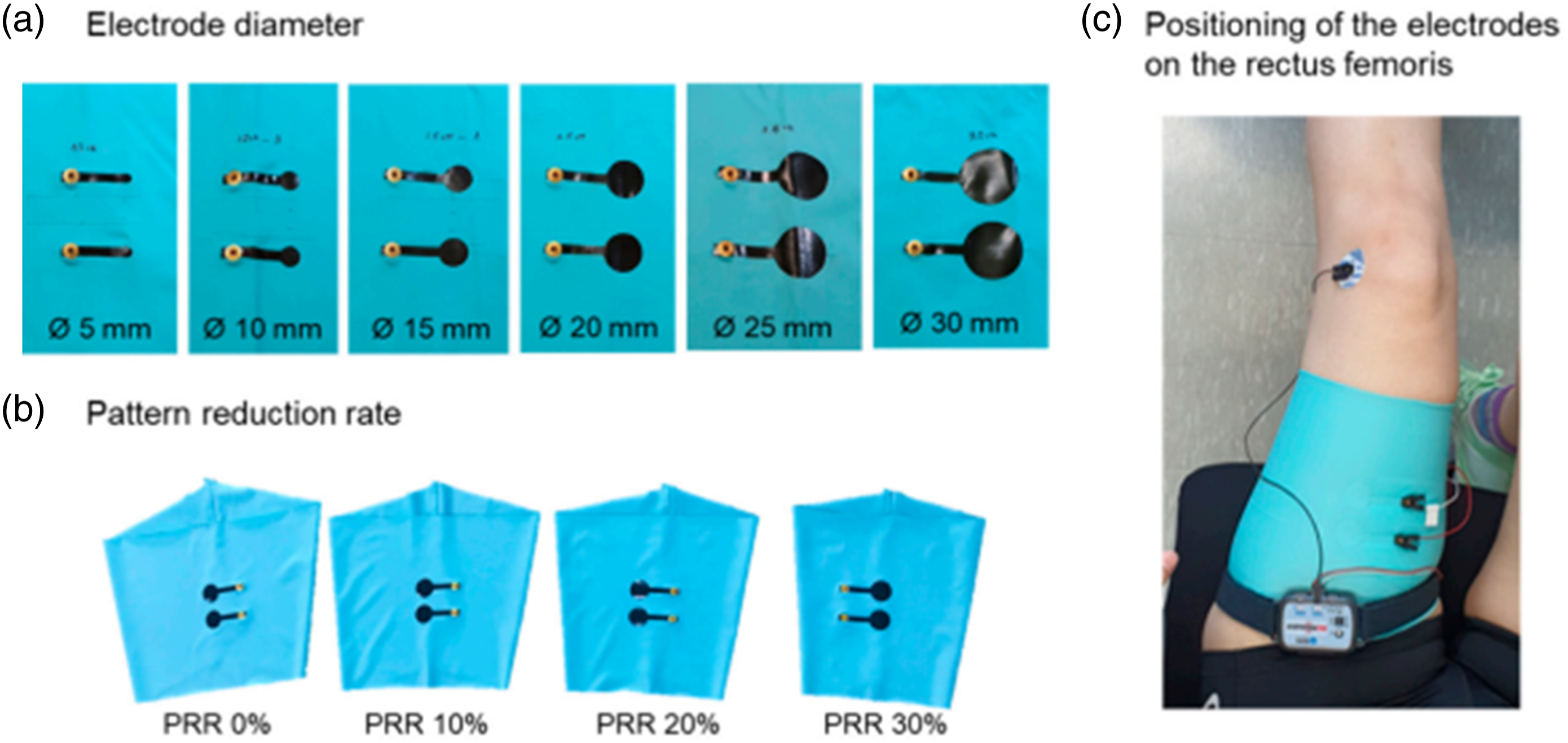
The influence of body motion on wearable EMG monitoring was investigated by Gozde Goncu-Berk et al., who prepared an embroidered textile electrode using t-shirt prototypes constructed with set-in and raglan sleeve styles.
74
Comparison of these two types reveal that within the same clothing size range, the raglan-sleeve performed better than set-in sleeves at custom fit as well as slightly snug or slightly loose conditions,
75
see Figure 9. Ohiri et al. compared screen printed, stretchable silver ink, and a silver plated lycra based commercial conductive stretch material (CCSM) for the electrode assembly in an athletic compression garment.
76
Their sEMG suit comprised of arm sleeves, shorts, and calf sleeves, for different muscle types such as bicep/tricep, quadriceps/hamstring, and tibialis anterior/gastrocnemius. In addition they also studied the performance of CCSM and PEDOT:PSS for the skin contact impedance at 1 kHz input signal and showed its impedances to be similar to silver–silver chloride electrode under damp condition. Left: (a) Set-in and Raglan sleeves with embroidered electrodes, (b) The t-shirt prototypes constructed with set-in and raglan sleeve styles, Right: Acquired sEMGs.
75
Originally published under a CC-BY 4.0.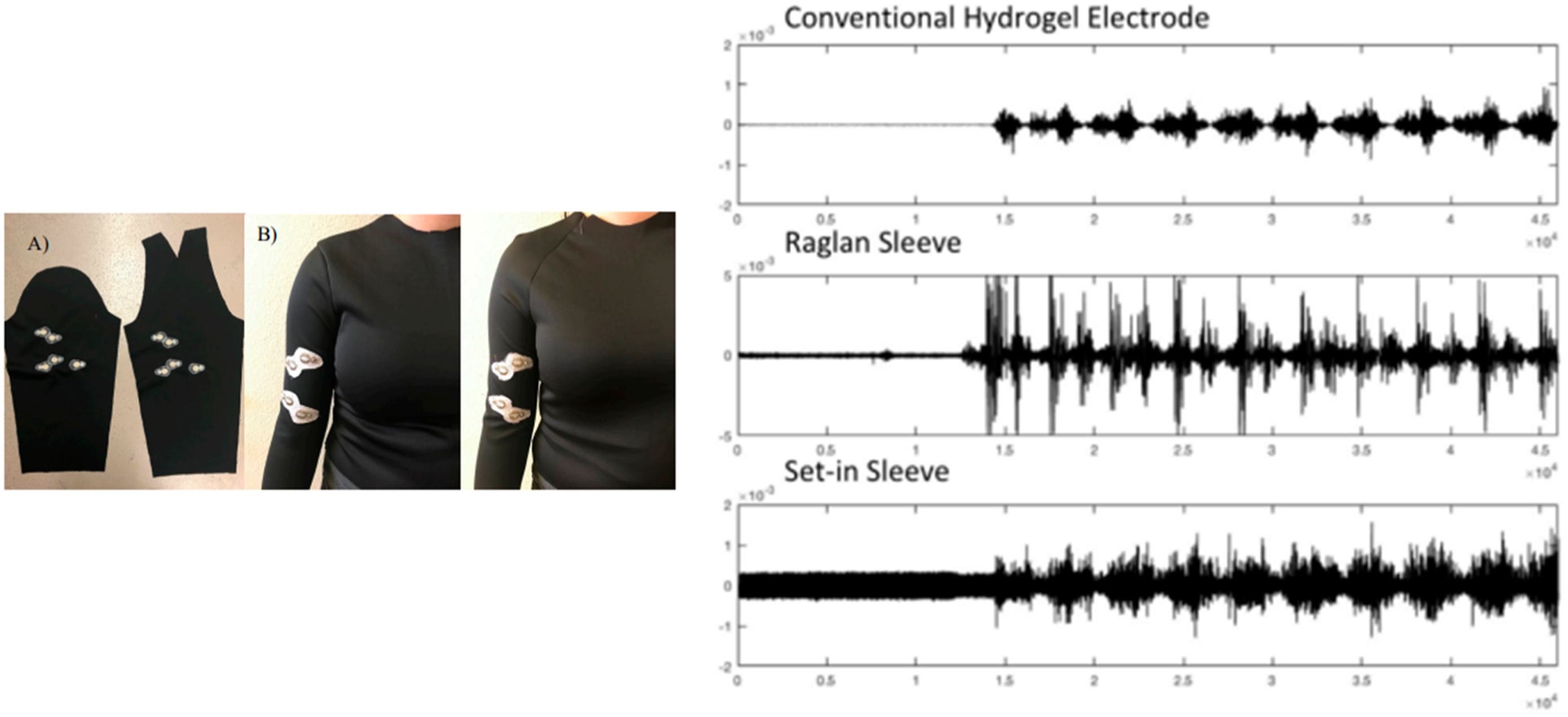
Jingjing Zhao et al., developed 3D textile electrode inspired by the embroidery technique for EMG, and ECG, from 1D composite yarns containing a mixture of reduced graphene oxide (RGO), sericin and water retention polymer,
77
Figure 10. T.E.peterson et al., also developed 3D printable bioelectronic sensors using graphene and PDMS composite material, targeting home based monitoring of hand function, and for potential applications in neurological and musculoskeletal conditions.
78
They tested those electrodes for gripping force and compared its performance with the functional electrode at different strain and pressure levels. The textile electrode constructed from composite yarns (a-j right) and schematic illustration of the 3D textile electrode fabrication process structural characterization for ECG and EMG applications,
77
With permission of Copyright Clearance Center, Inc. (CCC).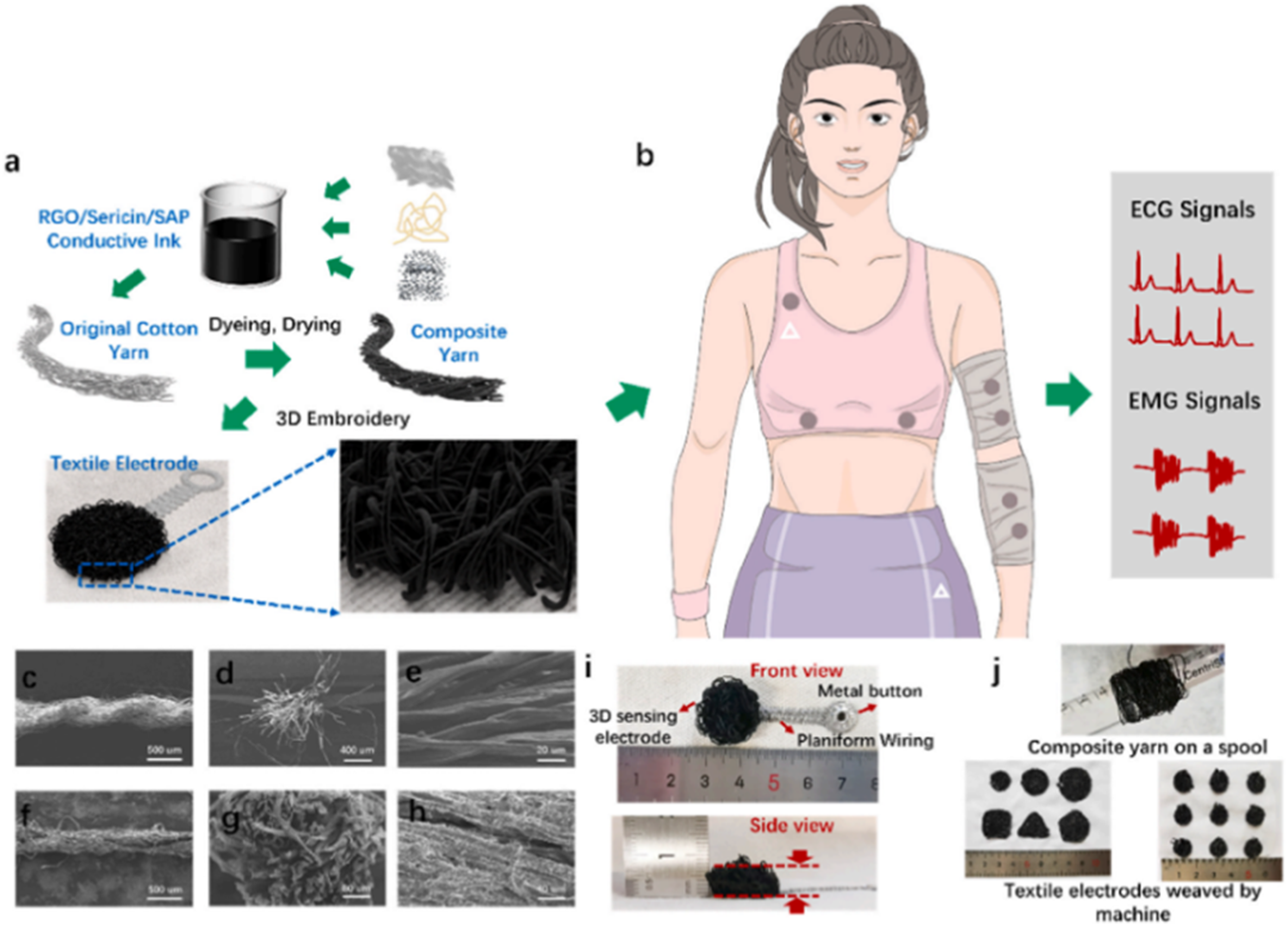
Degree of integration of textile electrode in smart textiles
The design of ‘smart textiles’, requires consideration for three main components namely, (1) the materials and the design of the substrate used, (2) the design of the electronic components such as the sensors, actuator and electrode, and (3) the technique used to integrate the electronics into the substrate to realize the textile system. Here, we will be focusing on the materials (first point) and the degree of integration of the ‘textile electrode’ into the textile substrate (third point); but will exclude the designing aspect of the electronic components such as sensors, actuators, etc. Integrating the conductive yarns alongside the electrodes imparts a functional aspect to the textile. Functionalization of the textile into a smart textile system is influenced by techniques such as knitting, weaving, embroidery, or by coating, and materials such as conductive fibers, yarns, and patch fabrics as shown in Figure 11. The four levels of degree of integration of the ‘textile electrodes’ into the smart textile are as follows. Different levels of electrode integration level in textiles
79
under CC BY-NC4.0.
In Level 1 – the conductive patch that serves as the electrode, are ‘added on’ or affixed onto the surface of the supporting textile substrate materials using the cut and sew method. 79 The textile substrate by itself has no functionality other than physically supporting the textile electrode attached to it. The sewing/embroidery method requires fewer production facilities and less space, thus making it far more flexible than coating or printing processes. A study carried out in Sweden showed that the wearable textile electrodes ensured high user comfortability and quality ECG signal measurement. 80 In another study an electrically conductive textile electrode was used to measure EEG in neonates. 81 Recently, a multilayer sweat absorbable textile electrode has been developed with a usability duration of more than a month. 82 One study showed the viability of recording EMG using electrodes made up of stainless-steel conductive threads sewn in to a pair of leggings. 83 Similarly, several such dry textile electrodes for EMG measurement have been reported.
Level 2 – The integration of electrodes ‘onto’ the textile substrate using coating and printing techniques are classified under ‘integrate onto’ techniques.79–85 In this technique, partial modification of the textile substrate takes place due to the coating/printing processes. Different types of textile electrodes are designed by coating or printing the electrode upon the textile substrate.79,86 The drawbacks of the printing technique are associated with the ink properties such as viscosity, curing temperature, and surface properties such as surface roughness and surface energy, that affect the electrode’s performance accuracy. Dip coating is one of the simplest methods to coat yarns or fabrics, and the process consists of immersing the substrate in a solution containing conductive materials such as metallic particles and carbon nanotubes. Though carbon nanotubes are being widely used, its application in biomedical domain is limited due to its toxic properties on the skin. Among metals, silver is the preferred materials due to its biocompatibility, stability and conductivity. Pani et al., made comparative characterization of functional electrodes and textile electrodes using screen printed conductive polymer (PEDOT: PSS) for detecting sEMG.
87
Sinha et al., compared hydrogel based commercial Ag/AgCl electrode with a screen printed dry PEDOT:PSS electrodes on textiles substrate
88
as shown in Figure 12. PEDOT:PSS based EMG electrodes screen printed on a commercially sold spandex arm sleeve was used in this study to record EMG signals originating from four different muscles. It was shown that the screen printed electrodes performed marginally better than Ag/AgCl electrodes in terms of SNR and signal amplitude. Scotchgard coating of PEDOT:PSS electrodes made it more hydrophobic and was shown to be stable over 10 cycles of accelerated wash and dry phases. Additionally, PEDOT:PSS electrodes have also been shown to record electro-dermal activity (EDA) and ECG signals from the wrist. Finally, a prototype t-shirt was fabricated which recorded ECG, EMG and EDA together under simultaneous cognitive stress and exercise. This easy-to-fabricate metal-free skin conformable electrodes could find application in continuous health monitoring as well as physiotherapy.
88
However in their study, the sustainable factors of conductive polymer such as environmental stability and repeated wash ability for long term continuous monitoring were not considered. (a) molecular complexation of PEDOT:PSS; (b) DMSO molecule; (c) Electrodes printed on a sleeve; (d) Biceps with the sleeve placed on it; (e) Comparative EMG signal recorded from PEDOT:PSS electrode (red) and Ag/AgCl electrode (blue).
88
Under CC BY 4.0.
In level 3 – ‘integrate into’, the electrodes are incorporated into the wearable textile.89,90 Here, the miniaturized sensors and actuators are embedded in to the fabric/textile alongside the conductive yarns. In different studies, silver was incorporated into fabrics made out of cotton, nylon, or polyester threads. The other type of metal commonly used for developing conductive textiles is stainless steel. Similarly, copper is also a good candidate metal for producing conductive textile because of its high electrical conductivity, but it is prone to oxidation. 84 Another study carried out on 50 healthy subjects validated the use of embedded textile electrodes in shorts. 91 In this study, the woven textile material consisted of both the non-conductive synthetic yarn and the electro-conductive silver yarns. A study conducted on the deltoid muscle (which is located on the upper arm) of six healthy people concluded that the performance of the conventional electrode was similar to that of an electrode made form conductive stainless steel yarn and non-conductive elastane fabric. 92 Another study assessed the performance of EMG sensors integrated with soft fabric as an exoskeleton for robotic hand. 93 Here, three electrodes made of a silver knit fabric were worn on the upper arm and served as a sensor to measure the bio potential endogenously generated within the tissues.
Finally, in level 4, the complex pattern design resulting from ‘seamless integration’ is made possible through computerized knitting. 79 In this technique, the electronic components itself is designed during the knitting processes, eliminating the presence of any joints. The functionalized sensor/actuator are fabricated at desired regions by adopting specific patterning of conductive yarns at such locations during the process of fabric knitting. With the integration of digital knitting technology, specialized design options and textile-based sensor construction is easily realizable. This strategy open up new possibilities for design freedom, quick prototype development, inclusive of green production procedure. This production technology enable incorporation of right materials into a variety of wearable electronic textile systems in a systematic manner. In an application study, the sensors were seamlessly integrated into the glove which enabled them to detect the motion of hand or robotic fingers. 94 In another study, the generation of seamless glove with pneumatic actuation for applications in soft robotics showed better scalability and customizability compared to the conventional ‘cut-sew’ method. 95 They realized several modes of actuation with better grip property, by incorporating different degree of extensible lycra yarn with less extensible ‘plied’ yarn in three layers (top, middle and bottom). All the methods from level 1 to 3 necessitate the post-processing step of attaching the electrodes into/onto the wearable system, whereas, in seamless integration this step is not required. 79
The demand for smart textiles with integrated sensors is increasing in various healthcare applications, in line with significant technological improvements. Development of these textiles requires expertise from various domains of science (materials), technology (information) and engineering (textile, chemical, biomedical chemical, electrical & electronics).79,96 Of increasing interest are the wearable sensors that utilize embedded electronic systems, e.g., thermally and electrically insulated sensors embedded within a textile to measure specific spatiotemporal signals originating from the human body. Electrical circuits and circuit elements built on a textile material using stainless steel, tin, titanium, silver or gold, conductive yarns, are already available in the market.
97
In general, functional textiles have become a crucial base for different applications and its utility has been increasing steadily till 2021, as shown in Figure 13. Further, an increasing number of conferences are being organized with focus on functional textiles and related material science fields.98,99 Analysis of Research Fronts in Scientific articles on Smart/Functional Textiles”, between 2009–2021, shows there are 1633 published articles. Bargraphs indicate the dynamic of research activity over the last 12 years.
Among the flexible substrates, textile by itself can be used as an electronic system through modifications; however, even better would be a wearable electronic system that is flexible and can be shaped or modified according to the changes in the environment or personal preferences. In the future, wearable computing systems which also assist with smart ‘decision making’ aspects are being considered to improve the quality of life of the patients. 71 Embedding textile electrodes (textrode) in clothing have significant benefits in diverse fields, including healthcare, athletic performance, military training, and restorative therapies. Textrode applications mainly depend on smart textiles' core properties: comfort, flexibility, and lightness. 100 In general, textile electrodes incorporated into the garment measure physiological biosignals and monitor vital signs. The main disadvantage of the textrode is the lack of good skin-electrode interface which causes noise disturbances and low sensitivity of the measured signal. The signal disturbances can be mitigated by proper choice of the electrode material and ergonomic design of the garments which ensure effective and stable contact with the skin. 101 Notably, Acar et al., evaluated the overall performance of the electrode primarily based on comfort level, 84 SNR, skin-electrode impedance, and other electrical characteristics. On comparing the ECG signal measured with the standard electrode, Ag/AgCl, the textile electrodes were found to be flexible and matched in line with the surface topography. It provides close contact with the pores and skin, thereby enhancing the wearable comfort and also prevented the skin infection in long-term monitoring cases. 74 The density of the fabric determines the sheet resistance by controlling the effective conductive thread count per fabric unit area. Hence, in addition to obtaining high-quality ECG signals, optimized fabric density provides the wearer with a degree of comfort. The signal quality of the e-textile based EMG measurements is determined by the cut and pattern of the material. The performance parameters such as electrical resistivity, SNR, are determined by the close fit of the wearable sensor to the skin, even in the presence of any physical activity such as arm movements. 74
Acquisitions, processing and analysis of sEMG signals
Preparation and acquisition of sEMG signal
In order to record high quality sEMG signal using gelled electrodes, pretreatment of skin such as hair removal, application of gel and prevention of sweat accumulation, are necessary. The proper electrode positioning and alignment of the sensor with the muscle also significantly affect the performance of the signal acquisition. On the other hand, dry electrodes do not require skin preparation, but only require the electrode to be positioned stably. However, they are usually bulky and heavier when compared to gelled electrodes.102,103 Such increased inertial mass required for the electrode fixation in turn lead to discomfort problems. EMG signal acquisition is the process of recording and measuring time domain signal from different states such as resting or exercise conditions.
sEMG Signal processing
The signal processing helps to get rid of the artifacts present within the raw signal. 36 sEMG signal is highly variable in terms of intensity and frequency and has a large-frequency range. It is highly susceptible to noises originating from electronics equipment’s, motion artifact, electrode contact etc. In general, the sEMG signals are denoised (noise reduced through low pass and high pass filters), rectified (the negative signal is made positive) and smoothened (which brings out the slowly varying ‘envelop’ profile concerned with the muscle contraction episodes through its low pass filtering effect). 104 Feature extraction is the process of transforming the input EMG surface signals and extracting a set of parameters (features) for further interpretation.
sEMG Signal analysis
In general, features in the sEMG signal analysis can be categorized into three categories such as time or signal domain, frequency or spectral domain, and time-frequency domain.105,106 Important parameters such as, RMS, ARV, statistical moments/parameters (coefficient of variation (CV), standard deviation (SD)) are calculated from the processed signal. Some parameters are derived from time domain signal, e.g. RMS, integrated EMG (iEMG), ARV. Some are derived from frequency domain spectra, e.g. median frequency (MDF), mean power frequency (MPF), entropy and recurrence quantification analysis (RQA). In a recent study, ARV was used to assess the subcutaneous fat content of four different muscle. 107 The derived parameters are sometimes subjected to multivariate analysis such as, principal component analysis (PCA) to obtain temporal patterns present across a large time-series dataset.
Application of sEMG based textile electrodes
Application of sEMG based textile electrodes.

Application of sEMG based textile electrodes in diagnosis
sEMG is one of the best diagnostic device for estimating, capturing, and analyzing muscle electrical activity. A sequence of myoelectrical potential fluctuations in sEMG is caused by cycles of voluntary or involuntary muscular contraction and relaxation.84,109,110 Several sEMG application based case studies are available in the literature and some of these are discussed here.
A sEMG comparative study of the healthy and neuropathic individuals, based on the RMS and ARV, concluded that the peak amplitude of different frequency components of the neuropathic subjects are significantly higher than that of the normal subjects. 111 A systematic review on the treatment of temporo mandibular disorder (TMD) patients in orthodontics and maxillofacial orthopedics wards describes the use of sEMG for diagnosis purpose. 112 In another case, electromyography-based diagnosis and recovery monitoring for masticatory muscle injury (zygomatico maxillary complex fractures) has been studied. sEMG has also been used for monitoring the activity of the masseter muscles in patients with facial bone fractures. 113 There are many related works reported in previous literature concerning biosensors that have been embedded into smart watches, clothes, and accessories to collect various bioelectric signals in real time for sports and fitness exercise (wearable sensors).114,115 Hand-sewn electrodes for surface electromyography are widely deployed in hospitals especially across countries with low income like Ethiopia. 116
A recent study investigated the efficacy and feasibility of dry-contact electrode in smart textiles and compared it with conventional gel. 117 The noninvasive flexible sensor was successfully used to monitor patients having limb fractures. 118 In the same study, EMG was recorded on animals like rats using flexible wearable sensors. This work used machine learning based on various parameters such as, RMS, iEMG, ARV in time-domain features, and MDF, MPF, entropy and recurrence quantification analysis (RQA), in frequency-domain. The studies relied on the prior knowledge that muscle strength has a strong correlation with RMS and iEMG, and muscle fatigue is correlated with MDF and MPF. 118 Azadeh Soroudi et al., developed an elastic paste that is electro-conductive, compatible with skin and are applied onto textile electrodes. The elastic paste enhanced the stability of the electrode surface in the presence of external touch, uniformity and smoothness. It also reduced the side effects of pore size and humidity in the signal. Such modifications enabled them to monitor Parkinson and epilepsy disease conditions for a long duration. 80
Application of sEMG based textile electrodes in sports and rehabilitation
sEMG techniques are widely used in the sport sciences, rehabilitation research, kinesiology, and ergonomics. 119 Fitness training and diagnosis in health monitoring are among the most popular applications, where, sEMG has been applied. Liu Shing et al. developed a sEMG patch to detect real-time muscle fatigue during exercise. These patches could be worn on the gastrocnemius muscle or the lower leg. 120 In another screen-printing technology-based study, the sEMG of the trans-radial amputees and healthy subjects were recorded using textile electrode for myoelectric prostheses control. 121 Giorgio Biagetti et al., designed a wearable wireless system at low cost to monitor sports and fitness activities using an accelerometer and a sEMG sensor. 122
Advantages, disadvantages and challenges in using textile electrodes for EMG applications
Different sources of interference noise are observed when conventional electrodes are used for sEMG measurements. The interaction among different muscles during their activation is also a major problem leading to cross signal contamination. Furthermore, the electrode should be placed stably on the skin, else the signal will be distorted. For untrained medical personnel, the correct placement of the electrodes will be challenging because the inter electrode spacing and its precise location significantly affect the quality of the measured sEMG signal. Hence, to circumvent these drawbacks, the sEMG sensors are nowadays integrated into a wearable clothing or textile, upon wearing it, the sensors are localized to its correct positions. In biomedical field, most of the electrodes require electrolytic gel to be applied at the skin-electrode interface to reduce the impedance present at the interface. However, such electrolyte applications are suitable only for short-term monitoring. In long-term monitoring, the gel will dehydrate or cause skin irritation, allergies or even bacterial growth. In addition to this, the life-span of such electrodes does not exceed 1 year even when properly stored. Overall, the Ag/AgCl electrodes are susceptible to drying, and its successive usage results in noisy and error prone signals, thus making it a one-time use device. Often in clinics, the dead skin cells on the surface of the skin are abrased to reduce the contact impedance. However, in long-term monitoring, the abrased skin surface will be renewed within a short period of time and repeated abrasion can cause discomfort to the user. Additionally, motional errors or ‘slippage’ effects caused by loosely attached electrodes also lead to noise/errors in measurements. In textile electrodes, sometimes, hydrogel membranes are used in the place of gelled electrolytes, to minimize the noise artifacts.123,124 In addition to the common factors that affect sEMG signal, the “Non-Invasive Assessment of Muscles” (SENIAM), a European Union project has set the standards for sensor design, placement, and data analysis. It recommends the following features to be considered during the acquisition such as: frequency ranges, inter electrode spacing, reference selection, and electrode design depending on application.32,83
Conventional Ag/AgCl electrodes through their conductive electrolytic medium exhibits enhanced polarization. The electrolytic conductor increases the charges at the interface of the skin-electrode, and reduces the impedance of the electrode polarization. However, in the case of textile electrodes increased total impedance is observed, because it does not contain any such electrolytic medium.
58
The area of the skin–electrode interface also plays an important role in affecting the skin-electrode impedance, because impedance is inversely proportional to the contact area of the electrode.
84
The skin-electrode impedance is affected by different sewing patterns which in turn affects the contact areas. In practice, it is difficult to compare different electrodes based on their impedance property as the skin characteristics of an individual varies within a population as well as with time. Several studies indicated that the electrode area of sEMG significantly affect the sEMG amplitude.
125
Amplitude is measured from peak to peak and is proportional to the distance between the recording electrode at the surface and the muscle fiber within the motor unit. The signal mostly represents few of the depolarized fibers that are adjacent to the recording electrode. It is suggested to place the electrode between the innervation zone and the tendon zone,
31
so that its location is confined to the center of the muscle belly, see Figure 14. Placing the sensor in an inappropriate position will decrease the intensity, quality of the EMG signal due to less motor units being observed and the interference/crosstalk effect prevalent among the muscle fibers. Electrode placement
31
under CC-BY_SA 3.0.
Challenges also come from the ‘electrode imbalance’ caused by loose attachment of one electrode with respect to the other or the presence of hair beneath each electrode or differences in the holding pressure applied to each electrode. 44 It is also necessary to give some time (at least 3–5 min) for the electrode to stabilize and reach an equilibrium state prior to any signal measurement. 58 Hence, a systematic evaluation of textile electrode should consider, seamless integration, 79 surface texture, porosity of textile fabric, knowledge of ink chemistry low impedance, and powering unit for continuous and long-term monitoring applications.43,126,127
Conclusion and future work
Since textiles cover the entire body from head to toes and is proximal to the skin, the sensing capacity of textile electrodes is significantly higher than those of individual biosensors/equipment. For example, by covering the head with a cap, signals from body parts such as the nose, ears, and eyes could be recorded; the whole of chest area and the upper body that include vital organs such as heart and lungs can be monitored through smart textile-based shirts. Thus, STS based textile enable continuous monitoring of vital physiological signals of a patient on a daily basis and create personalized medical database. Novel smart materials are being integrated with the textile structure through various technologies such as embroidering, sewing, nonwovens, knitting, weaving, spinning, braiding, coating/laminating, printing, and chemical treatments. Recent advancements in STS has enabled, flexible, stretchable fabrics which generate energy via triboelectrification mechanism to power up its components. 128 Further, stretchable fabric that reduces electrode slippage effects in textrodes have been developed and commercialized. The miniaturization of textrodes using nanomaterials (nanofibres, nanowires, nanosol) and nano techniques (nanomaterial coating) is been actively pursued. On the downside, the embedded nanoparticles within polymer matrix has durability issues, hence functionalizing the fabric surface by physical or chemical methods prior to nano-coating is considered as a viable option. 129 From the sustainability perspective, the recycling the Micro/Nano electromechanical systems (MEMS/NEMS) incorporated in to the fabric is challenging and is under review.130,131 Dry textile electrodes sometimes perform less than the gel electrodes especially, when motion artifact are present. Researchers have suggested applying sufficient quantity of water or saline solution onto the dry electrodes to achieve performance comparable to that of the conventional gel electrodes.132,133 However, in the case of the textile electrodes, to remain wet for a prolonged period of time, high compression textile fabric with improved moisture retention property are being considered. When it comes to modern applications in health monitoring devices, pressure sensing system combined with a flexible textile-based sensor array with real-time readout during cardiac operation has already been devised. 134 A wearable health-shirt with e-textile materials for the continuous and cuffless monitoring of arterial blood pressure has been developed. Textile electrode integrated with air mattresses to measure the interfacial pressure of high-risk patients during surgical operations are being developed so as to minimize the pressure injury. 135
Footnotes
Declaration of conflicting interests
The author(s) declared no potential conflicts of interest with respect to the research, authorship, and/or publication of this article.
Funding
The author(s) disclosed receipt of the following financial support for the research, authorship, and/or publication of this article: This work was supported by Prof. Lievavan Langenhove (mounascere20212026).