
Abstract
Recently, nanosized cellulose materials preparation has been extensively increased from the sources of sustainable materials and utilization in various functional applications as nanofillers. Cellulose nanofibrils (CNF) extraction through green bio-based materials featured as promising interest in the field of science. In this research, cellulose nanofibrils were extracted from a cellulose rich biomass Ficus natalensis barkcloth. Furthermore, extracted cellulose nanofibrils and Zinc oxide nanoparticles (ZnONPs) were mixed and casted to posterior analysis of formed sodium carboxymethyl cellulose (CMC) film at various concentrations of CNF (1 wt%, 5 wt%, 8 wt%, and 12 wt%) and fixed amount of ZnONPs (0.5wt%) based on CMC weight. Results revealed that CNF was smoothly distributed in the polymer matrix to form even and flexible films indicating the cellulose nanofibrils and zinc oxide are highly compatible with the CMC. Similarly, the water solubility percentage (WVP) of CMC film was low at lower content of CNF, and increased with the increase of CNF percentage. Addition of CNF and ZnONPs in nanocomposite films improved the thermal stability values and antibacterial activities of CMC films. Thus on the basis of various tremendous performance, this study showed that F.natalensis barkcloth could be considered as an alternative source of cellulose nanofibrils. Similarly, the prepared nanocomposite films can have potential application in packaging films for the extension of shelf life of fresh and minimally processed fruits and vegetables.
Keywords
Introduction
Environmental pollution concerns are extensively increasing because of petroleum based materials used for various applications, accelerating the growth of ecofriendly biodegradable polymers especially edible and non-edible for packaging fields. 1 Accordingly, carbohydrates and proteins or a hybrid composite of both, mixed with lipids molecules often used as the sources of edible biopolymers are minimizing the pollution generation issues. Various polysaccharides are evaluated to prepare bio-degradable film used for food packaging such as thermoplastic starch, 2 carrageenan, 3 chitosan, 4 agar, 5 and carboxymethyl cellulose. 6 However, due to weak mechanical and blocking characteristics, the industrial application of biodegradable-based bioactive polymer packing films has not been practiced. 7 Generally, biopolymers owing to weak thermo-physical and barrier properties, the practice of carbohydrates proves to be relatively excellent film making properties in packaging materials as compared to proteins and lipids. Carboxymethyl cellulose is an abundant carbohydrate biopolymer in food packaging materials because of excellent film-forming potential, inherent renewability, sustainable, biocompatible, excellent mechanical, chemical and physical properties, as well as suitable barrier and transparent activities of its films.6,8 To expand the diversity of food packaging films manufactured by natural bio-resource materials to attain essential process requirements, enhance their dimensional and mechanical properties, certain limitations have been overcome with the addition of nano-scaled materials, biodegradable polymers, lipids, plasticizers, as well as cross-linker and nanofillers.
Recently, biopolymer film properties are reported to be improved by using nanofillers like nanometals, nanoclays, and cellulosic nanofibers which induce excellent ultraviolet, antibacterial, and antioxidant properties in nanocomposite films. 9 Generally nanocomposite film properties are dependent upon the nanofillers type, shape, and size, as well as their interfacial characteristics. 10 Similarly, it has been noted that single type of nanofiller used for incorporation may not enhance enough the film characteristics for industrial-based applications. 11 Recently, studies show that the nanocomposite films have been prepared by combining various polymer nanofillers due to renewable, sustainable, and exhibit low abrasive nature as reported by Susan Azizi. 12 Combine merits of the individual components in nanocomposite materials like multifunctional and high-performance properties of two or more nanofillers have been reported. 13 In nanofillers, cellulose nanofibrils extracted from natural resources such as plant wood or stems like jute, ramie, cotton, as well as agricultural residue are getting a tremendous level of attention. 14 This is mainly due to the most abundant biopolymers on the earth with unique and excellent features such as inexpensive, abundance, renewable, non-toxic, high aspect ratio, and lightweight natural biopolymers. 15 CNF has been reported as an excellent nanofiller improving various properties of nanocomposites such as mechanical, structural, and blocking properties.16,17 CNFs are reported to enhance the physical characteristics of biopolymeric membranes, 18 additionally, the thermal, physical, and blocking properties of chitosan, 19 starch derivatives, 20 agar, 17 poly(lactic acid), 21 and alginate 21 films.
Similarly, Zinc oxide (ZnO) is receiving considerable attention to be used in hybrid nanocomposite films because of excellent functional properties such as photocatalytic and antimicrobial activities. 22 ZnO is a FDA (U.S. Food and Drug Administration) approved (21CFR182.8991) nanomaterial against bacterial strains and fungi. 23 ZnO and nanocellulose acting as nanofillers have been used to enhance the mechanical properties and provide the antibacterial activities of poly(vinyl alcohol)/chitosan blend films.12,24
Thus, in this study, extraction of cellulose nanofibrils from a novel material Ficus natalensis barkcloth is done and utilized in preparation of antibacterial functional film by combining with CMC and ZnONPs.
Materials and Methods
Materials
F.natalensis barkcloth was purchased from Uganda. F.natalensis barkcloth is a biosource material contains a rich amount of cellulose, which can be used for cellulose and cellulose nanofibrils extraction for further applications. Generally, F.natalensis trees are 20m in height and considered a part of Moracseae family, usually local people called it as Natal fig, or Mutuba.25,26 F.natalensis barkcloth is a naturally biodegradable material generated from the tree bark of F.natalensis. This tree is speckled in most of the regions of Zambia, Malawi, South Africa, Zimbabwe, Kenya, Mozambique, and surroundings. 27 Obtained raw material was washed thoroughly with the distilled water. Then it was fully dried in an oven at 40°C to eliminate the maximum moisture. Zinc oxide nanoparticles (ZnO, 40 nm) and sodium carboxymethyl cellulose having a molecular weight of 40,000 kDa were obtained from Aladdin Chemistry Co. Ltd., Shanghai, China. Similarly, sodium hydroxide (NaOH, 98%), sodium chlorite (NaClO2, 80%), sodium hypochlorite (NaClO, 13%), hydrochloric acid (HCl, 37%), and sodium bromide (NaBr, 99%) were purchased from Sino-pharm Chemical Reagent Co., Ltd., Shanghai, China. The other remaining chemicals including glycerol, and 2,2,6,6,-tetramethylpiperidin-1-oxyl (TEMPO, 98%) were obtained from Aladdin Chemistry Co. Ltd., Shanghai, China.
Extraction of cellulose nanofibrils and composite film preparation
Figure 1 shows the graphical illustration of the chemical process performed in this manuscript. Cellulose nanomaterials were extracted from F.natalensis barkcloth by a method mentioned in our previous study.
28
In short, barkcloth was cut into small pieces, washed thoroughly with distilled water for about 1 hour, and then oven dried until the maximum moisture elimination was done at 40°C for 20h. At the end after fully drying the barkcloth small pieces, residue was grounded into powder form by using a small crusher machine named as DJ-04 grinder (Shanghai Dianjiu Traditional Medicine Machinery Manufacture Co., Ltd, Shanghai, China). Powder barkcloth was passed through 60-mesh sieve to get a confined form of the F.natalensis barkcloth. After that, 50 g/L dried powder residue was weighted and mixed with 1.0 M NaOH solution for about 2 h at 80°C; meanwhile, stirring of the solution was done constantly using a magnetic stirrer. Later on, residue was washed various times with deionized water to remove the alkali chemicals on the samples, filtered, and then oven dried for 6h at 40°C. Graphical abstract
Later on alkaline treated dried barkcloth residue was bleached by using 2.5% w/v sodium chlorite through material to liquor ratio of 1:20 maintaining pH of the solution as 4–5 for 1 h using continuous stirring. After the bleaching process, mixture was carefully washed with deionized water using a glass beaker and oven dried at 60°C until the constant weight was achieved. At the end, obtained material was cellulose and then it was ground into powder form.
Later on cellulose was converted into cellulose nanofibrils by applying TEMPO oxidation technique. For this purpose, powder cellulose samples (10g) were mixed with 190g of water for 6h. After that, a fixed amount of NaBr (0.20 g) and TEMPO (0.02 g) was distributed into the solution. Additionally, 12% NaClO (18 g) was added drop wise into the solution to start the oxidation process. The pH of the solution was maintained at 10–10.5 using 0.5 mol/L sodium hydroxide aqueous solution. At the end, ethanol (5 mL) was carefully added into the mixture and stirring of the solution was done to stop the oxidation reaction. After that the washing of the obtained process was done to extract the chemicals by several wash cycles using deionized water by centrifugation process at 1960g for 15 min. Similarly, sonication at 10,000 r/min for 10 min was done by placing the solution in an ice bath; solution was prepared using 1.0 g CNF slurry in 100g water. Cellulose nanofibrils were separated from the solution by using centrifugation process to use it for further applications.
Various percentage of carboxymethyl cellulose, Cellulose nanofibrils, and ZnO in 50g film forming solution.

CMC: carboxymethyl cellulose.
Film solutions were prepared by dissolving 2 g CMC powder in 100 mL water at 900 r/min stirring for 30 min at 90°C. After that the solution was added slowly with 1.5 mL of glycerol (75% based on CMC weight) as a plasticizer at 40°C. Meanwhile stirring of the solution was done vigorously using a magnetic stirrer for 10 min at 700 r/min. Later on, the solution was cooled at room temperature to remove the apparent air bubbles. After pouring onto a 15 mm leveled glass plate, the film solution was dried by using circulator- equipped incubator at 35°C for 24 h. The resultant film was peeled from the casting surface and stored in a closed tight bag before the tests. Obtained film was named as CMCc and considered as control film. In order to prepare CNF nanocomposites films, process is same as CMCc but there is a little modification, such as precisely weighed ZnO nanoparticles (at a level of 0.5% (w/w) per film forming solution) was dispersed and homogenized using a high shear mixer Miccra D9 (ART Prozess and Labortechnik GmbH, Mullheim Germany) at 16,000 r/min for 10 min. After that, CMC (2g) was added slowly at 50°C; meanwhile, a magnetic stirrer was used to stimulate the solution carefully in order to obtain a fine solution. By using solution interaction method, CMC nanocomposite films having various contents of cellulose nanofibrils (1 wt%, 5 wt%, 8 wt%, and 12 wt% based on CMC weight) were prepared prior to glycerol addition. The obtained film solutions were centrifuged for 5 min at 5000 r/min, and then 1.5 mL of glycerol (75% based on CMC weight) was added. Thereafter, the film solution was poured and placed for 30 min into a leveled glass plate to remove the air bubbles. The glass plates were then dried for 24 h at 40°C. Thus, the films were pealed out from the glass plates and before characterization, nanocomposite films were placed in a humidity chamber at 25 °C/50 RH for 4 days. At the end, obtained film samples were entitled as CMCc, CMC1, CMC2 CMC3, and CMC4 according to CNF concentration as 0, 1 wt%, 5 wt%, 8 wt%, and 12 wt%, respectively.
Physical appearance of raw material during cellulose extraction
Various content present in barkcloth were evaluated and noted as cellulose, hemicellulose, and lignin were determined as 43.5%, 24.5%, and 19.5% using different standard methods.
30
During cellulose production, barkcloth material was processed in different stages such as chopping into small pieces (Figure 2 (a)), conversion of raw material into powder form (Figure 2 (b)), after initial washing, cleaning and chemical treatment, and finally cellulose extraction was done as shown in the Figure 2(c). Figure 2(c) shows that the sample were light yellow in pretreated process but very fine and white after chemical treatment indicating the cellulose extraction as massive fraction of non-cellulosic components were removed during the whole process.
31
Barkcloth fibers cut pieces (a), barkcloth powder (b), and extracted cellulose samples (c).
Characterization
Surface morphology and thickness measurements
The morphology of cellulose nanofibrils and nanocomposite films were analyzed by using scanning electron microscope (SEM.) JSM-6360LA, instrument (JEOL Ltd, Tokyo, Japan) and transmission electron microscope (TEM) JEM-2100 TEM (JEOL Ltd, Tokyo, Japan). Samples were mounted on the stabs separately and firmly coated with a thin film of gold by sputtering method using an accelerating voltage of 30 kV. Cellulose nanofibrils dimensions were analyzed using TEM images by a drop of each sample with ethanol dispersion solution at a concentration of 0.5 wt% using an accelerating voltage of 120 kV. Similarly, the thickness of every nanocomposite film was calculated by using a digital micrometer (model QLR digit-IP54, Qinghai, China) to the nearest 0.0001 mm. So measurements were taken at ten random places of the films. Thus, the average thickness of each film was used.
Fourier-transformed infrared spectroscopy of nanocomposite films
Fourier-transformed infrared spectroscopy (FTIR) spectra were examined in transmission mode (T%). Spectrograms of all the samples were recorded from 400 to 4000 cm−1, with 32 scans at a resolution of about 8 cm−1 on Nicolet 8700 FTIR spectrometer (Thermo Fisher Scientific Co., Ltd, Waltham, MA, USA).
Film solubility (%) in water
The nanocomposite film solubility was checked by using the specific technique named as “cup method” (ASTM 1993) as mentioned in.
32
First of all, the dry weight of every film sample was determined by thermal processing at 100°C to obtain a constant weight and was named as initial dry weight. Then small pieces of each film sample were cut (2 × 2 cm2) and weighed to the nearest as 0.0001 g. Water solubility was determined by soaking of every film sample in 30 mL water at 25°C. Later on, centrifugation of the solution was done at 3000 r/min for 15 min to separate the insoluble film from the supernatant. After this, insoluble film was extracted and subjected to drying process at 100°C in an oven until the weight was constant and considered as final dry weight. The solubility percentage (%) of every film was calculated through the following equation
Antibacterial activity of the films
Disk diffusion technique was applied to examine the antimicrobial activity of the prepared films against Staphylococcus aureus (S. aureus) ATCC 29213 (as gram positive) and Escherichia coli (E. coli) ATCC 25922 (as gram negative) bacteria by a method mentioned in. 33 These two species were nominated due to their importance to human health. In order to assess the antimicrobial activity of each film, every bacterial culture was diluted to the final concentration of 104 cells/ml and about 700 μL of that solution was spread onto new specific medium agar plates. Each film was cut into disc shaped sample having 10 mm diameter and placed on ager petri dishes which were already inoculated with the corresponding bacteria. Petri dishes were later on incubated at 35–37°C for 12h. The bacterial activity was examined by the inhibition diameter on the film disc and results were expressed as diameter of growth inhibition (mm).
Results and discussion
Morphological analysis of raw material and CNF samples
Barkcloth in raw material shape without any treatment as well as morphology of cellulose and cellulose nanofibrils extraction after various processes is exhibited in the Figure 3. Figures 3(a) and (b) show the physical appearance of barkcloth raw material in chopped and powder form, respectively. Figure 3(c) exhibiting the SEM images of barkcloth showing an uneven arrangement of natural fibers connecting and supporting each other at different angles, might be due to presence of non-cellulosic impurities.
34
Figure 3(d) exhibited the SEM scans of the samples in powder form; exhibiting that the fibers are separated but unaligned. Similarly, Figure 3(e) shows the cellulose samples having a fibrous shape, indicating the removal of non-cellulosic components including lignin, pectin, and hemicellulose during chemical purification as alkali-treatment helped to eliminate the non-cellulosic impurities, leading to reduced dispersion of micro-fibrils.31,35 Figure 3(f) is exhibiting the TEM images of cellulose nanofibrils. Images of Ficus natalensis barkcloth in shopped form (a), powder form (b), morphological analysis by scanning electron microscopy (SEM) of shopped raw material (c), milled powder form (d), SEM scan of cellulose (e), and transmission electron microscope image of cellulose nanofibrils (f).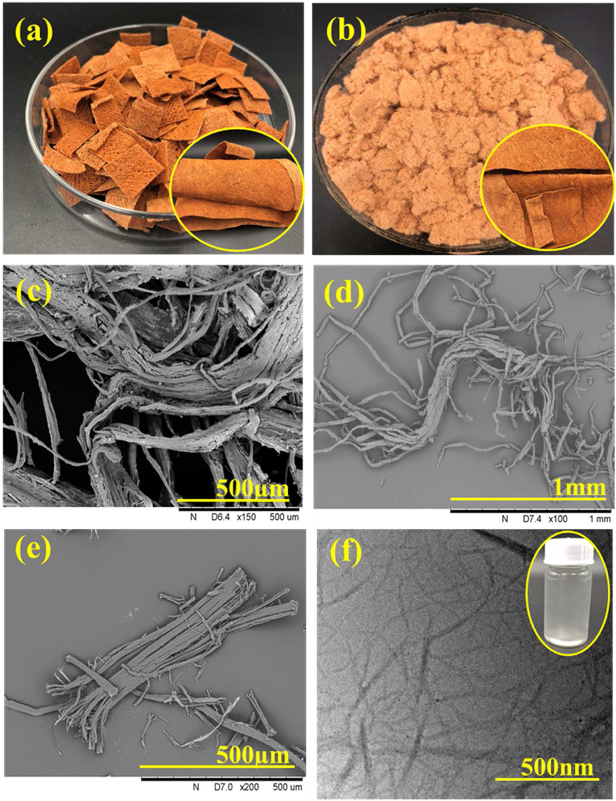
Similarly, the diameter distributions of cellulose nanofirbils was taken by using image J. software of nearly 100 samples. Figure 1 supplementary file (SF) shows the diameter and frequency histogram, exhibiting that CNF had a narrower distribution range where mostly nanoparticles diameters were in the range of 12–40 nm.
Microstructural characteristics and film thickness of nanocomposite films
In order to observe the degree of cellulose nanofibrils as well as zinc oxide nanoparticles distribution and the proper interface between CNF and polymer matric scanning electron microscope (SEM) was used as shown in Figure 4. Figure 4(a) shows the image of CMC nanocomposite film. SEM technique (Figure 4(b)) shows that CMC surface was smooth and compact with no obvious pore or crack on the surface of the film as acknowledged in, Ref [36] and the surface of the films were continuing to be smooth until CNF percentage was 5% as shown in Figures 4(c) and (d). This shows that cellulose nanofibrils are excellent compatible with polymer matrix outstandingly dispersed throughout the CMC polymer to procedure a well-developed nanocomposite films. But the nanocomposite films surface show less uniformity with the increase of CNF concentration (Figures 4(e) and (f)) because cellulose nanofibrils exhibit extensive accumulation at higher concentration.
37
The similar results were reported for the effect of adding GTE to chitosan.
38
No agglomeration of cellulose nanofibrils and any crack were observed in Figure 4(b) containing CNF (1%); similarly, well separation of cellulose nanofibrils (Figure 4(c)) chains were witnessed because of uniform distribution of cellulose nanofirbils and zinc oxide. However, with the increase of CNF accumulation cracks were observed in nanocomposite films as depicted from the Figures 4(e) and (f). Film thickness was noted between 60 ± 4.52 and 76 77 ± 5.45 μm (Table 1, SF). Pristine CMC film shows minimum thickness, on incorporation with ZnO and cellulose nanofibrils, the thickness of the films increased. As shown in Table 1 (SF), the thickness of CMC4 film was greater because of higher concentration of cellulose nanofibrils. Physical appearance of CMC film (a), SEM analysis of CMCc (b), CMC1 (c), CMC2 (d), CMC3 (e), and CMC4 (f). CMC: carboxymethyl cellulose.
Thermal stability of CMC and CMC/CNF composite Films
Carboxymethyl cellulose and CNF/CMC nanocomposite films were evaluated to examine the thermal stability values by using TGA techniques. Figure 5 exhibits the TGA and DTG curves of CMC and cellulose nanofibrils (5%) and CMC nanocomposite films. As from the Figure 5(a) it is obvious that TGA curves are displaying a pattern of weight decreasing for each sample during the thermal degradation process; however, the DTG curves exhibit the highest decomposition temperature at every stage of thermal degradation.
39
Generally, CMC and CMC/CNF films exhibited lower thermal stability values as compared to other biopolymer-based films because of weak thermal stability properties of CMC.
40
In this study, thermo gravimetric analysis showed three distinct stages of weight loss for all the samples. The first weight loss of every film was noted about 3.4 and 3.2% for CMC and CMC2 films, respectively, at temperature between 40 and110°C, second weight loss of CMCc and CMC/CNF 5% was nearly 31 and 29%, respectively, showing temperature between 110 and 235°C, the final degradation was noted at 235–350°C and the final weight after thermal degradation at 600°C was noted as 25 and 29% for CMCc and CMC2 films, respectively. The first weight loss is attributed to moisture,
41
second is due to glycerol evaporation,
42
and the third weight loss was because of carbohydrate polymer decomposition.
39
The CMC2 film shows higher thermal stability values as compared to controlled CMC film as in every phase of thermal degradation CMC2 shows less weight loss as compared to CMCc film which is apparently due to higher thermal stability values of cellulose nanofibrils as compared to CMC polymer matrix as well as zinc oxide nanoparticles caused to increase the thermal stabilities of polymer films as Kim et al. confirmed the increase of thermal stability values with the addition of zinc oxide nanoparticles in polymer films.
43
TGA (a) and DTG (b) of CMCc and CMC2 films. CMC: carboxymethyl cellulose.
Water solubility Percentage (WSP)
Water solubility (WS) of biopolymer films is an important factor for many applications which may require insolubility of the films to enhance the product shelf life and moisture blocking properties. Results (Table 1 SF) showed that water solubility percentage (%) of the CMC films was high as compared to nanocomposite films containing cellulose nanofibrils, CMC, and zinc oxide nanoparticles. The solubility rate of neat CMC film (CMCc) was noted higher than those of the composite films; with the increase of content of nanomaterials, the fragmentation and solubilization rates of the nanocomposites was decreased. The higher water solubility behavior of CMCc film could be because of more hydrophilic structure of CMC molecule causing the film matrix more accessible to water. In previous studies, similar findings have been reported that on addition of CMC the solubility of final composite films was increased.44,45 Additionally, WS decreased by adding a little amount of zinc oxide nanoparticles (as shown in Table 1 SF) indicating the increase of cohesiveness of the biopolymer matrix. Synthetic polymers generally possess hydrophobic nature and biopolymers are supposed to hydrophilic in nature. Film water solubility is advantageous in some situations such as composite films will be consumed with a product that is heated prior to consumption. 46 Similarly, it has been proved that the water solubility of nanocomposite films assist the biodegradability of them. 47 As shown in the Table 1 (SF) that the CMCc film was totally water soluble and WS decreased with the addition of nanomaterials in CMC matrix (from 100.00 ± 00.001 to 37.20 ± 2.6, respectively), which is similar with the previous findings.48–50 The WS of CMC films could be due to more hydrophilic structure of carboxymethyl cellulose molecule making the film matrix more open to water. In a similar finding, CMC was added in a composite film to increase the solubility of the end product. 45 The water solubility (%) of nanocomposite films decreased with the addition of zinc oxide nanoparticles which is a clear indication of cohesiveness of the biopolymer matrix and zinc oxide, and thus decreasing WS values. Even with the addition of cellulose nanofibrils (1%) and constant amount of ZnO (0.5%), the water solubility of obtained nanocomposite film was lower than CMCc film (100.00a ±00.00). Furthermore, with the increase of CNF (12%) and ZnO (0.5%) the water solubility (%) decreased significantly.
FTIR analysis
Figure 2 (SF) shows the FTIR spectra of various film samples using FTIR analysis to examine the interactions between polymer and zinc oxide nanoparticles. The FTIR spectra of CMC film displayed characteristic peaks within the range of 3360–1038 cm−1. The peak at about 3330–3360 cm−1 was due to hydroxyl group (O–H) stretching. 51 The peak at 2828–2918 cm−1 was appeared due to C–H stretching connected with the rings of methane hydrogen atoms. 52 The controlled CMC film (CMCc) displayed two peaks at 1416 and 1520 cm−1, indicating the symmetrical and asymmetrical stretching vibrations of the carboxylate group. 53 The characteristic absorption peaks at 1038–1200 cm−1 indicated C–O stretching of controlled CMC film. Similar kind of peaks with higher intensity can be seen in the FTIR spectra of the all nanocomposite films. Nevertheless, some peaks were moved to higher and lower wavenumbers with the addition of zinc oxide nanoparticles. Such as controlled CMC FTIR spectra having peaks at 3197, 2918, 1606, 1415, 1323, and 1038 cm−1 were shifted to 3285, 2898, 1602,1418, 1326, and 1038 cm−1, respectively, in case of various nanocomposite films. The moving of absorption peaks showed that the specific interactions between zinc oxide nanoparticles and biopolymer matrix were happened. 54
Antibacterial Activity
Antibacterial activities of all the film samples were measured against two foodborne pathogens groups by using zone inhibition method or disc diffusion method. Figure 6 shows that the controlled CMC film did not exhibit any antimicrobial activity; however, with the addition of zinc oxide nanoparticles, films showed excellent reduction in cell viability of both S. aureus and E. coli. Results in Figure 6 show the antibacterial activity against S. aureus, revealing that controlled CMC exhibited minimum inhibition zone diameter (Figure 6(a)), but the nanocomposite films containing zinc oxide were showing excellent antibacterial activities as shown in Figure 6(b), (c), (d), and (e). Nearly, an average inhibition zone diameter of 1.4 mm was noted as shown in Table 2 (SF) for various samples against two models S. aureus and E. coli. Results showed that more reduction in cell viability was noticed against Gram positive S. aureus as compared to Gram negative E. coli (Table 2 SF). This shows that zinc oxide nanoparticles were active against Gram positive than the Gram negative bacteria. The ZnO nanoparticles release Zn2+ ions which can easily penetrate in the bacteria’s cell wall to react with the cytoplasmic content and kill the bacteria. It is believed that zinc oxide nanoparticles are known to create a highly oxidizing agent named as hydrogen peroxide (H2O2) to destroy the cell membrane of bacteria.
55
Antimicrobial characteristics of controlled CMCc film (a), CMC1 (b), CMC2 (c), CMC3 (d), and CMC4 (e) against S. aureus. CMC: carboxymethyl cellulose.
Similar findings reported that the nanocomposite films containing ZnO nanoparticles indicated strong antibacterial activities against Gram positive bacteria.
56
Additionally, Anitha, Brabu, Thiruvadigal, Gopalakrishnan, and Natarajan (2012) studied that Gram positive S. aureus was highly inhibited by zinc oxide nanoparticles as compared to Gram negative E. coli.
54
It is reported that the antibacterial activity of zinc oxide nanoparticles is dependent on the bacteria’s structure of cell wall. Gram positive cell wall are reported as more thick and made of multilayer peptidoglycan; however, the Gram negative bacteria’s cell wall is a complex structure and comparatively composed of thin peptidoglycan layers.
57
Figure 7 shows graph of antibacterial activity for various samples. Antibacterial activity graph of various film samples.
Conclusion
In this study, cellulose nanofibrils have been produced from the novel material F.natalensis barkcloth and utilized in improving the various properties of CMC films by incorporating with zinc oxide and CMC nanomaterials. Results revealed that the extracted cellulose nanofibrils diameter was in the range of 12–40 nm. Similarly, CMC nanocomposite films exhibited excellent thermal properties, reduction in film solubility percentage, and excellent antibacterial properties with the addition of cellulose nanofibrils and zinc oxide nanoparticles. SEM images revealed that CNF was smoothly distributed in the polymer matrix to form even and flexible films, indicating the cellulose nanofibrils and zinc oxide are highly compatible with the CMC. Additionally, the water solubility percentage of CMC film was low at lower content of CNF, and increased with the increase of CNF percentage. The addition of cellulose nanofibrils and zinc oxide nanoparticles in nanocomposite films improved the thermal stability values and antibacterial activities of CMC composite films. Thus, the F.natalensis barkcloth appeared to be excellent raw material for cellulose nanofibrils extraction and CMC film making. Similarly, this research can lead to the application of barkcloth in cellulose nanofibrils extraction and then using them as value addition products in developing aerogels, composites, bioactive materials, and inorganic/organic hybrid materials.
Supplemental Material
sj-pdf-1-jit-10.1177_15280837221082036 – Supplemental Material for Extraction of cellulose nanofirbils from Ficus natalensis barkcloth and utilization in preparation of antimicrobial bio-nanocomposite films for possible food packaging applications
Supplemental Material, sj-pdf-1-jit-10.1177_15280837221082036 for Extraction of cellulose nanofirbils from Ficus natalensis barkcloth and utilization in preparation of antimicrobial bio-nanocomposite films for possible food packaging applications by Amjad Farooq, Mohammed K Patoary, Azmat Hussain, Syed Rashedul Islam, and Lifang Liu in Journal of Industrial Textiles
Footnotes
Declaration of conflicting interests
The author(s) declared no potential conflicts of interest with respect to the research, authorship, and/or publication of this article.
Funding
The author(s) disclosed receipt of the following financial support for the research, authorship, and/or publication of this article: This work was supported by the National Key Laboratory Foundation of China (2018YFC2000900).
Supplemental Material
Supplemental material for this article is available online.
References
Supplementary Material
Please find the following supplemental material available below.
For Open Access articles published under a Creative Commons License, all supplemental material carries the same license as the article it is associated with.
For non-Open Access articles published, all supplemental material carries a non-exclusive license, and permission requests for re-use of supplemental material or any part of supplemental material shall be sent directly to the copyright owner as specified in the copyright notice associated with the article.