
Abstract
Expandable graphite was applied to an upholstery polyester textile material as an eco-friendly flame-retardant alternative to halogenated compounds. Fabrics treated with the flame retardant were evaluated by flammability tests. Besides, thermal analysis was carried out by Differential Scanning Calorimetry (DSC) and Thermogravimetric Analysis (TGA). Moreover, a percutaneous absorption study was conducted to verify the safety in terms of dermal penetration of the flame-retardant treated upholstery fabric for human use.
The upholstery fabric treated with expandable graphite successfully passed the flammability tests. Thermogravimetric analyses with TGA and DSC showed that the flame-retardant application slightly decreased the initial decomposition temperatures regardless of the atmosphere but increased the final residue at 600°C (35.7% in O2 and 44.5% in N2) compared to non-treated upholstered fabrics. Dermal permeation of expandable graphite applied on the polyester fabric showed no penetration after 24 h of fabric exposure time. This result demonstrated that graphite-treated polyester fabric is dermatologically and toxicologically safe for use in upholstery.
Applied expandable graphite proved an efficient and safe flame-retardant alternative to conventional flame retardants for eco-friendly flame resistance applications in upholstery fabrics.
Introduction
Many accidental fires in dwellings involve upholstered furniture. One of the main requirements for contract upholstery fabrics is their flame retardancy. Contract upholstery fabrics in Europe are legislated according to fire safety by cigarette and match tests, and EN standard regulations such as EN 597-1 and 2: 20151,2 and EN 1021-1 and 2:2015.3,4 There are two ways to chemically treat upholstery textiles to make them flame retardant: 1) coating technique and 2) dipping method. 5 For substrates made of natural fibres or blended fabrics (with a high percentage of natural fibres), dipping is the most common fire-treatment method. This method consists of dipping the textile substrate into a chemical solution, which is absorbed by the textile fibres. With the coating technique, a flame retardant (FR) is applied on the back side of the upholstered fabric, which does not come into contact with the end-user. Coating with different FRs have been used to prepare flame-retardant coating polyester (PET) fabrics.6,7 The flame-retardant coating provides protection against fire, but it also braces and stiffens the textile, therefore improving fabric characteristics for upholstery usage. 8
Common FRs used for upholstery textile applications are based on halogenated compounds due to their high efficiency and the low dosages of FR needed in the formulation. 9 They are commonly used in combination with antimony trioxide to provide a synergistic effect. 10 However, halogenated compounds have been shown to exhibit ecotoxicological issues and persistence and toxicity related to dioxins, and furans are produced during their incineration.11,12 The presence of antimony trioxide also leads to health concerns since it is considered to be a carcinogenic compound. This type of FR also has potentially harmful effects on human health. Therefore, research on alternatives, such as those based on phosphorus and nitrogen, are finding acceptance in the market.13–15 Phosphorus-containing flame retardants cover a wide range of inorganic and organic compounds and include both reactive products which are chemically bound to the polymer material as well as additive products which are integrated into the material by physical mixing only. Nitrogen compounds are mostly used in combination with phosphorous-based flame retardants due to their synergistic effect. N-based flame retardants alone have a lower intrinsic fire-retardant performance. On the other hand, expandable graphite (EG) is used in a growing number of flame-retardant applications as a blowing agent and suppressor for smoke. 16 EG absorbs heat, which leads to the reduction of heat release rate value and exhibited an increased flame resistance. 17 Meanwhile, the thermal conductivity and thermal stability 18 give EG an edge in preparing functional composites. 19 However, little has been reported on flame-retardant efficiency in EG coatings.20,21
The FLAREX project has conducted work on the use and development of FR alternatives to halogenated compounds, antimony trioxide and formaldehyde releasing related compounds. 22 Expandable graphite (EG) was selected in the project as a promising alternative FR material for upholstery application to commonly used halogenated compounds.23,24 EG is usually integrated into polyurethane foams in the automotive industry and as a back-side coating in carpets for aviation applications to achieve high thermal stability and flame retardancy. EG is considered a convenient FR compound for eco-friendly flame resistance applications because of its low cost (easy to prepare) and low smoke production as well as its anti-dripping and halogen-free characteristics, and then the coating technique could be adequate because these textiles are usually thick (hardly dried when soaked) and their surface relief could be impacted by squeezing between rollers during the impregnation. 25 To assess the properties of the FR materials applied in textiles, thermal and calorimetric techniques, such as Thermogravimetric Analysis (TGA) and Differential Scanning Calorimetry (DSC), can be applied to provide information on the formation of volatiles and decomposition temperatures.
Since upholstery fabrics could be in contact with the skin, it is important to evaluate the possible dermal toxicity of the EG flame retardant. Therefore, the dermal toxic potential of compounds applied to PET has also been studied, using the percutaneous absorption technique. So far, no studies have been conducted on the FR migration from a textile to the skin upon their contact during service life. However, increasing evidence suggests that dermal absorption is a potentially significant pathway of human exposure to FR25.16,26 Efforts to fill this gap are limited by the difficulties related to ethical problems with human studies. The procedure for assessing the risks to human health of a product (in our case, an FR product) is to compare the exposure levels to which populations are or may be exposed with the exposure level at which no toxic effects are expected.
The dermal toxicological potential of FR applied to polyester has also been studied using the percutaneous absorption technique in Franz cells according to the OECD methodology. 27 In this case, FR analyses were performed by confocal Raman microscopy (CRM). Confocal Raman spectroscopy is useful for investigating the penetration of graphite into the skin due to the sensitivity of the technique to the graphite structure and ease of tracking the graphite signal. 28
The procedure for evaluating the risks of a product to human health (in our case, a fire-retardant product) consists of comparing the level to which the customer could be exposed with the exposure level where toxic effects are not expected to occur. The risk evaluation of a chemical consists of four basic steps: hazard identification, hazard characterization (commonly used is the non-observed adverse effect level (NOAEL)), exposure assessment, and the risk characterization (margin of safety (MoS)). 29
Therefore, the main aim of this work is to determine the feasibility of the use of EG expandable graphite as an alternative flame retardant for PET upholstery fabrics. The flammable properties of the fabric were evaluated, and the thermal properties were assessed by means of DSC and TGA analyses. Safety of the fabric for human use was also determined through a percutaneous absorption and risk assessment studies. These safety studies are not usually carried out; however, we believe they are essential for tissues that may be in contact with the skin.
Materials and methods
Materials
A polyester woven upholstered fabric with 320 g/m2 of weight per square meter was supplied by ATEVAL (Ontinyent, Valencia). The PET fabric has two different sides: the front with an aesthetic red, white and black pattern, and the back with a grey pattern.
Expandable graphite (EG) is a commercial product ‘FRC 827’ supplied by the EOC Group (Oudenaarde, Belgium). It is a grey-coloured product. It is a halogen-free flame retardant and a ready-to-use compound. It is based on a waterborne polymer dispersion containing expandable graphite and calcium carbonate. This compound has a 54.0–56.0% solid content, pH: 7.0–9.0, viscosity (BV RV ¾): 14.000–16.000 cPs and density of 1.00–1.40 kg/L.
Fabric treatment and soaking procedures
The PET upholstery fabric was treated with EG by the back-coating technique at laboratory scale. This technique consists of the application of the FR coating on the back side of the upholstery fabric. The fabric was mounted in a stenter frame, and the FR was applied along the textile with a blade-knife with 1.5 mm of gap. The coating thickness was adjusted by varying the gap between the blade-knife and the roll (knife-over-roll coating). The treated PET fabric was then dried and cured in a stenter at 150°C for 3 min. Several applications were performed, and different FR add-ons, 50%, 60% and 70% (over dry weight), were deposited on the PET fabric by depositing single-layer and double-layer coatings onto the back side of the textile. Treated fabrics had A4-sample dimensions.
Before the ignitability tests were carried out, the fabrics were subjected to water-soaking treatment according to UNE-EN 1021-1:2015. 3 The coated fabrics were immersed in a water bath (water hardness ranging from 8dH to 10dH, bath ratio of 1:20, 1 g PET fabric per 20 ml of bath) at a temperature of 40°C for 30 min and were folded so that the reverse side was exposed to water. After water treatment, the coated fabrics were rinsed for 2 min.
Flammability tests
Flammability tests were performed like UNE-EN 1021-2:2015 4. (Part 2), where a match flame was used as the ignition source.
Flammability tests were conducted with fabric samples at the laboratory scale. The treated fabrics were mounted on the top of two flexible foams of 75 mm thickness (MR086) according to normative conditions. The frame had the shape of a sofa onto which the foams were fastened. The frame had a height of 35 mm. The test consisted of placing a match-flame near the fabric testing area for 15 s. Following these 15 s, the flame was removed, and the flaming behaviour of the textile was tested over time. Samples pass the fire test if the flame causes no flaming or progressive smouldering (<15 min ±1) of the material. The test time should be shortened than 2 min. The test was performed on three different test areas. If the first two areas did not pass the test, the third area was not tested.
Thermal Analysis: DSC and TGA analysis
Characterization of the thermal and physical properties of FR fabric was conducted by DSC analysis using a Mettler-Toledo DSC-823 apparatus. Approximately 3 mg of fabric was placed in microperforated aluminum pans for internal pressure control to ensure the complete removal of water and other volatiles from the testing pans. Tests were conducted from 30°C to 500°C at a scan rate of 10°C/min under 50 mL/min of N2 flux 28 because at 500°C, the thermal decomposition of samples were completed.
The air-oxidation behaviour of the FR-textile sample was determined by TGA analysis using Mettler-Toledo TGA/SDTA 840 equipment. Approximately 10 mg samples were placed in non-sealed aluminum pans, not to exceed their melting temperature, and heated from 25°C to 600°C at a scan rate of 10°C/min under N2 flux and O2 flux, both of which were 60 mL/min. 30
The analysis were performed in the Thermal Analysis and Calorimetry Laboratory “Josep Carilla” from IQAC-CSIC.
In vitro dermal permeation
The skin permeation assay was performed using an in vitro methodology using Franz vertical diffusion cells (3 mL, 1.86 cm2, Lara-Spiral, Courtenon, France). The OECD guidelines 31 and the published recommendations of the Scientific Committee on Cosmetic Products and Non-Food Products (SCCNFP) 32 were followed throughout the test.
The skin permeation assay has been described in previous studies.33,34 In the test, the skin is placed on both parts of the Franz cell. In this case, the skin used was the porcine skin, which shows the same morphological and histological properties as the human skin.35,36 The unboiled porcine skin was obtained from the dorsal area of female white/Landrace pigs weighing 40 kg. The skin was obtained from the department of cardiology from the Clinic Hospital of Barcelona. Animal handling was approved by the Institutional Review Board and Ethics Committee of Institut d’Investigacions Biomèdiques August Pi i Sunyer (IDIBAPS), and the management of the animals conformed to the Guide for the Care and Use of Laboratory Animals. 37 The skin was shaved and dermatomed at 500 ± 50 μm with a dermatome (Aesculap, Germany). The dermatomed skin was cut into pieces and stored at −20°C until further use.
The skin was placed so that its surface faced the donor compartment of the cell. The non-coated side of the EG-treated fabric was placed in contact with the skin surface. Twenty microlitres of distilled water was added between the textile and the skin to ensure close contact. The test was performed in triplicate. Moreover, a pressure was applied with a steel cylinder at a constant and standard pressure of 125 g/cm2 (ISO 105-E04). 38 The cells were placed in a thermostated water bath assuring skin hydration and a skin surface temperature of 32°C. An additional Franz cell treated with fabric, without EG application was used as a control. The receptor compartment was filled with phosphate-buffered saline (Sigma, USA) at pH 7.6.
After 24 h of exposure, fabric textiles and the skin were removed from the Franz cell. The superficial stratum corneum was removed by a series of stripping because the compounds in the stratum corneum layer are considered not-penetrated due to peeling of the skin. 26 The rest of the skin (inner stratum corneum, viable epidermis and dermis) was analysed directly by confocal Raman microscopy to detect graphite. Layer separation was not necessary as the confocal Raman microscopy images yield information about depth in the skin sample. The experimental conditions are described in the section Confocal Raman Microscopy Analysis.
The in vitro permeation of substance permits to know its systemic exposure dosage. The safety evaluation can be obtained for a substance by the calculation of the margin of safety (MoS)
29
with the following formula

Confocal Raman microscopy analysis
PET fabric without treatment (control fabric), fabric treated with EG and samples of the skin used after the permeation assay were analysed by CRM. Raman spectra were obtained by using the confocal Raman microscopy (Witec alpha-300 R-AFM, WITec GmbH, Ulm, Germany), with a 532 nm excitation laser and a ×100 objective lens (NA = 0.95). The incident laser power was 0.5, 1 and 5 mW. The resolution of the recorded Raman spectra was 0.02 cm-1. Raman spectra were recorded over a spectral range from 100 to 3700 cm-1. The scan area was 100 × 30 μm, the parameter of the Raman image was 100 × 30 pixels, and the integration time per pixel was 0.5 s. Slices at depths up to 30 μm below the surface of the skin were examined. The collected Raman spectra were analysed by using Witec Control Plus Software (Witec, Ulm, Germany).
Results and discussion
Fabric treatment
Applications of expandable graphite on the PET upholstery fabric conducted at the laboratory scale.
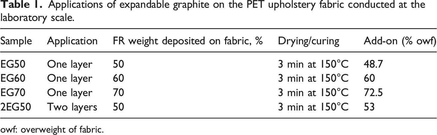
owf: overweight of fabric.
Application of the EG resulted in hardening of the upholstered textile. An increase in the FR amount on the upholstered fabric led to an increase in the addition of FR coating on the fabric. An increase of the weight deposited on the textile to 60% and higher (EG60 and EG70) led to a better coating uniformity. The application of two layers of EG (2EG50) also led to a more uniform coating compared to the fabric coated with a single layer of EG (EG50).
The reverse side of the upholstery fabric adopted a grey colour due to the inherent colour of the EG, which did not present an aesthetic problem because the colour modification only occurred on the back side of the textile and not on the front, visible side of the upholstery fabric. Functional properties, such as elasticity, also changed, and fabric stiffening was observed after the coating treatment due to the high add-on, higher than 50% of the weight of the fabric. This effect was more apparent in the upholstery fabric samples treated with 60 and especially with 70% of weight deposited, which showed very rigid properties, and therefore, a modification of the functional properties is needed for an upholstery fabric. EG fixation happens through mechanical fixation and adhesion between the polymer dispersion containing expandable graphite and calcium carbonate particles and the porous structure (pores, fibre interstices, etc.) of the PET upholstered textiles.
Flammability tests
Fire test results of UNE-EN 1021-2:2015 of upholstered fabrics treated with expandable graphite EG at the laboratory scale.

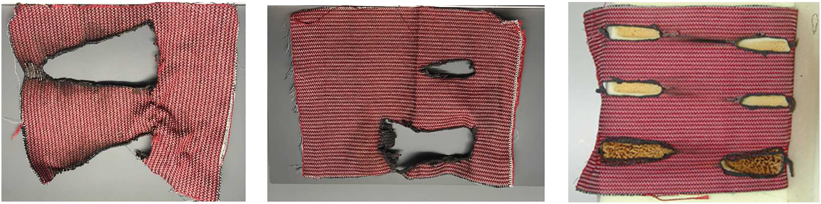
a) Untreated PET upholstered fabric, b) treated upholstered PET fabric with a single layer of 50% of weight deposited of expandable graphite FR and c) treated upholstered PET fabric with two layers and 50% of weight of EG.
The outstanding fire-resistant behaviour of EG is due to its structure. EG adopts a stack-like structure of parallel planes or carbon atoms with intercalation agents inserted between the planes and positioned between the graphite lattices. When graphite is exposed to heat or flame, the molecules of the intercalation agents decompose and release gases. The graphite layers are then forced apart by the gas, and the graphite consequently expands, creating a protective and insulating layer that inhibits flame propagation and further ignition. The outstanding behaviour of EG is due to its fire resistance mechanism and because it can expand to a greater degree than traditional intumescent coatings thus providing better insulation properties.
Given the favourable results of the FR fabric of 2EG50 in the inflammation tests, this fabric was chosen to perform the thermal tests and percutaneous absorption.
Thermal analysis
The application of thermal analysis techniques, such as DSC and TGA, enables the assessment of the effect of FRs on the thermal stability and final residue at 600°C in N2 and O2 atmospheres. The results are consistent with those of the heat release rate given by a micro-scale combustion calorimeter MCC related by Qin Chen and Tao Zhao. 39
Figure 2 shows the DSC and TGA plots of the PET upholstery fabric conducted in N2 and O2 atmospheres. The DSC plot enables the identification of the peak of PET melting at 255.6°C followed by an endo-exo transition attributed to PET decomposition. DSC (in blue) and TGA (O2 atmosphere in red and N2 in black) plots of the original PET fabric for upholstery.
Summary of the TGA results up to 600°C of the original PET upholstery fabric and that treated with EG in N2 and O2 atmosphere: T5 temperature of 5% of mass loss, Tend temperature of PET decomposition determined by DTGA, loss of mass of these steps and final residue.

When EG was used as ecological intumescent FR, there was an effect on the thermal behaviour which is shown in Figure 3. Its application modifies the T5 temperature depending on the atmosphere. In N2 atmosphere,T5 is increased by 18°C, while in O2 atmosphere, a decrease in 36°C is observed. The main protective effect of EG occurred at 313°C, at which the DSC thermogram showed a wide endotherm attributed to the graphite expansion, which protects PET and greatly reduces the loss of mass due to decomposition. As shown in Table 3, the temperature at which the decomposition of PET, determined by the DTGA, ends, and Tend ascends in 7°C in N2 atmosphere up to 484.4°C and decreases in 18°C in O2 atmosphere to 444.3°C. The loss of mass from T5 to Tend is greatly reduced (48.1% in N2 and 58.2% in O2). The additional loss of mass observed after PET decomposition of the untreated fabric in O2 atmosphere that accounts for a loss of mass of 12.4%, probably caused by the oxidation of the formed char products, is also observed at the end of EG-treated PET decomposition, accounting for a 13.2% of mass loss that can also be attributed to a sudden oxidation of the derived char products formed during PET decomposition. Table 3 summarizes the results including the temperature at which a loss of mass of 5% is attained, T5; the temperature at which the decomposition of polyester, determined by DTGA, ends Tend; the mass losses from both T5 to Tend and from Tend to 600°C; and the final residue at 600°C. The residue is greatly increased by the presence of EG (35.7% in O2 and 44.5% in N2). The application of alternative FR limits the emission of volatiles and generates a higher volume of final residue than the conventional FR.22,41 DSC (in blue) and TGA (O2 atmosphere in red and N2 in black) thermographs of the PET upholstery fabric treated with EG as ecological intumescent FR.
In vitro dermal permeation
Confocal Raman microscopy analysis was first optimized by analysing the textiles. A control sample of non-treated PET fabric and samples of PET treated with expandable graphite were analysed by confocal Raman microscopy prior to its application on the skin. A piece of each fabric was placed on a glass slide, and a selected area was imaged. Large domains of graphite, approximately 10 micrometres in size, were perfectly distributed in the treated fabric covering all parts of the surface, as shown by the optical microscopy images obtained with the confocal Raman (Figure 4). Graphite scales on graphite applied on PET fabric observed by optical microscopy.
Then, Raman mapping of a selected area of 80 μm x 80 μm was carried out in the graphite domains. Three compounds were detected: graphite, an organic compound and microparticles of a carbonate (possibly calcium carbonate). The Raman spectrum and the microscopy mapping image of the textile treated with EG are shown in Figure 5. As shown in the image (Figure 5), the main component detected was graphite (red-coloured areas in the image). Spherical particles were found on top of the graphite identified as calcium carbonate (green-coloured areas in the image). Raman spectrum (left) and microscopy mapping image of the fabric face treated with EG (right): graphite (red-coloured), organic substance (blue-coloured) and calcium carbonate (green-coulored) were identified.
The characteristic spectrum of graphite (Figure 5 left in red) was observed. Three constituent bands (D, G and 2D) were separated from the Raman spectra using the Lorentzian peak fitting function (Figure 6). The disorder-induced defect D-band was located at approximately 1360 cm-1, the structural order-induced graphite G-band was located at approximately 1580 cm-1, and the secondary D-peak of 2D was located at approximately 2700 cm-1.42,43 Raman spectrum of graphite with three constituent peaks, D, G and 2D, identified.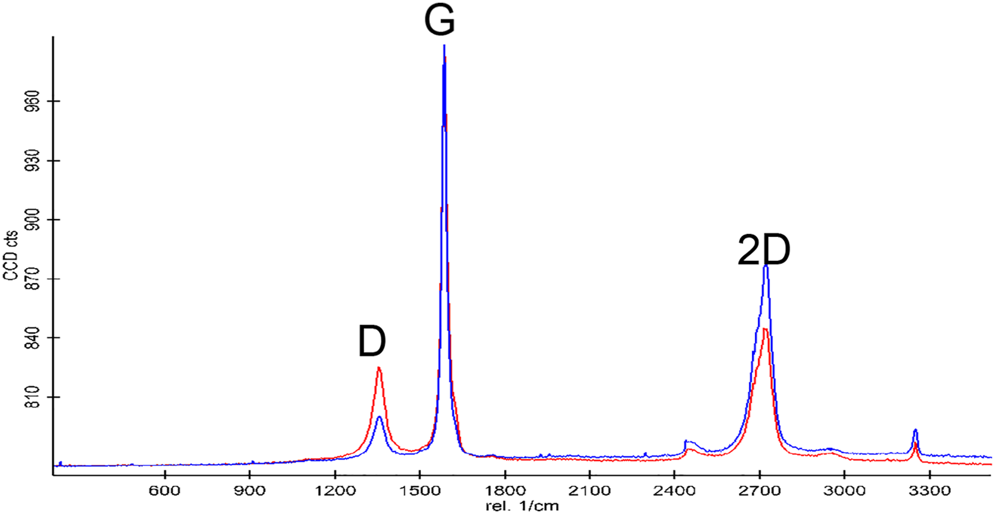
Next, the percutaneous absorption assay was performed. Three samples of treated textile were applied on the skin surface (via the non-coated side of fabric, which was brought into contact with the skin) deposited in the Franz diffusion cells. One sample of PET fabric without the FR compound was used as a control. After 24 h of exposure time, the fabrics were recovered, and a series of stripping was carried out to remove the superficial stratum corneum. The rest of the skin (inner stratum corneum, viable epidermis and dermis) was analysed directly by using confocal Raman microscopy following the protocol described in the section In vitro Dermal Permeation.
The typical Raman signal of the skin was obtained from the control skin (Figure 7).
44
Raman spectra at the skin surface showed a residual stratum corneum layer after the stripping step (the red spectrum). Viable epidermis Raman spectra were obtained at 20 μm depth (blue line) with a higher water signal at 3300–3600 cm-.
1
Raman spectrum of the control dried and hydrated skin and of the possible compounds which could be present due to the EG-treated textile.
Next, skin samples treated with EG fabric were analysed. Raman mappings of 100 μm length and 30 μm depth into the skin were carried out with a ×100 objective and radiation of 5 mW for 0.5 s (an example is shown in Figure 8). As expected, the residual SC signal (in red) was observed, showing that the skin is drier on the surface. With increasing depth into the viable epidermis, the skin becomes more hydrated (higher intensity of the water signal at 3300–3600 cm-1), as indicated by the blue colour at a depth of 30 μm. Microscopy image and Raman mapping 30 μm deth into the skin applied with the EG50 textile fabric during 24h (residual SC in red, viable epidermis and dermis in blue).
Additionally, XY mapping at approximately 20 μm depth with dimensions of 150x150 μm was obtained (Figure 9). In the mapping, it was possible to detect mostly viable epidermis (blue area): the observed small areas corresponded to the residual SC (red areas). Microscopy image and XY Raman mapping at 20 μm depth into the skin applied with EG50 textile fabric during 24h (residual SC in red, viable epidermis in blue).
The results obtained from the skin, which was in contact with the treated textile, were equal to those obtained from the control textile. No EG (green spectrum in Figure 7) was detected in any of the samples (Figures 8 and 9), and only the typical Raman signal of the skin was obtained. The optical microscope images indicated that there was also no graphite attached to the skin surface. As a result, the amount of FR compound absorbed to the skin was zero.
Therefore, the dermal permeation of graphite applied in polyester fabric indicated no penetration after 24 h of fabric exposure time. Therefore, in this case, the exposure assessment was done by the limit of detection equipment. The risk characterization (margin of safety (MoS)) 18 was higher than 1032. This result is consistently higher than the target of 100 and demonstrated that the polyester fabric coated with graphite is toxicologically safe for use in upholstery. Therefore, the application of expandable graphite was shown to be a good alternative to conventional flame retardants for eco-friendly flame resistance applications in upholstery fabrics.
Conclusions
Expandable graphite is a new type of intumescent coating, which exhibits a physical fire-retardant mechanism. EG was applied on the PET fabric by back-knife coating at different concentrations (between 50% and 70%) resulting in a single- or double-layer application. Fabric with single layer of FR (50%) and obtained add-on of 48% did not pass the flammability test. However, application of two layers allowed obtaining an add-on of 53% implying a pass for the flammability test. Higher concentrations of EG applied at 60% and 70% allowed deposition of higher amount of EG, 60 and 72%, respectively, and did pass the flammability test. Fabric coated with two layers of EG showed the best results in terms of flame retardancy properties with the lowest concentration of flame retardant applied and maintenance of the flexibility properties of the upholstery textile.
The application of both differential scanning calorimetry DSC and thermogravimetric analysis TGA in O2 and N2 atmospheres enabled us to evaluate the effect of the FRs on the thermal behaviour of the PET fabric. The effect of the ecological intumescent FR on PET fabric based on EG slightly decreased the initial temperatures of decomposition regardless of the atmosphere and resulted in the highest increase in the final residue at 600°C.
Dermal permeation of graphite applied to the PET fabric was evaluated by an in vitro methodology through a percutaneous absorption, detection with confocal Raman microscopy and risk assessment studies. After 24 h of fabric exposure onto skin biopsies, no graphite was detected in the skin by confocal Raman microscopy, and the margin of safety (MoS) couldn’t be calculated. This result demonstrated that the polyester fabric coated with graphite is dermatologically safe for use in upholstery.
Footnotes
Acknowledgements
The present work could not be performed without the collaboration and contribution of the Service of Dermocosmetic Assessment from IQAC-CSIC. The authors are also grateful to Montserrat Rigol Muxart and Núria Solanes from the Department of Cardiology (Institut d’Investigacions Biomèdiques August Pi i Sunyer (IDIBAPS) Hospital Clínic, Universitat de Barcelona, Spain) for supplying the porcine skin biopsies.
Declaration of conflicting interest
The authors declare that there are no conflicts of interest to disclose.
Funding
The author(s) disclosed receipt of the following financial support for the research, authorship, and/or publication of this article: This study is supported by Ministerio de Ciencia, Innovación y Universidades, España (CTQ2018-094014-B-100) and LIFE Environmental Policy and Governance program from the European Union (LIFE16 ENV/ES/000374).